Contrast-enhanced mammography-based interpretable machine learning model for the prediction of the molecular subtype breast cancers
- PMID: 40596940
- PMCID: PMC12220444
- DOI: 10.1186/s12880-025-01765-3
Contrast-enhanced mammography-based interpretable machine learning model for the prediction of the molecular subtype breast cancers
Abstract
Objective: This study aims to establish a machine learning prediction model to explore the correlation between contrast-enhanced mammography (CEM) imaging features and molecular subtypes of mass-type breast cancer.
Materials and methods: This retrospective study included women with breast cancer who underwent CEM preoperatively between 2018 and 2021. We included 241 patients, which were randomly assigned to either a training or a test set in a 7:3 ratio. Twenty-one features were visually described, including four clinical features and seventeen radiological features, these radiological features which extracted from the CEM. Three binary classifications of subtypes were performed: Luminal vs. non-Luminal, HER2-enriched vs. non-HER2-enriched, and triple-negative (TNBC) vs. non-triple-negative. A multinomial naive Bayes (MNB) machine learning scheme was employed for the classification, and the least absolute shrink age and selection operator method were used to select the most predictive features for the classifiers. The classification performance was evaluated using the area under the receiver operating characteristic curve. We also utilized SHapley Additive exPlanation (SHAP) values to explain the prediction model.
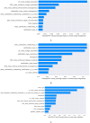
Results: The model that used a combination of low energy (LE) and dual-energy subtraction (DES) achieved the best performance compared to using either of the two images alone, yielding an area under the receiver operating characteristic curve of 0.798 for Luminal vs. non-Luminal subtypes, 0.695 for TNBC vs. non-TNBC, and 0.773 for HER2-enriched vs. non-HER2-enriched. The SHAP algorithm shows that "LE_mass_margin_spiculated," "DES_mass_enhanced_margin_spiculated," and "DES_mass_internal_enhancement_homogeneous" have the most significant impact on the model's performance in predicting Luminal and non-Luminal breast cancer. "mass_calcification_relationship_no," "calcification_ type_no," and "LE_mass_margin_spiculated" have a considerable impact on the model's performance in predicting HER2 and non-HER2 breast cancer.
Conclusions: The radiological characteristics of breast tumors extracted from CEM were found to be associated with breast cancer subtypes in our study. Future research is needed to validate these findings.
Keywords: Breast cancer; Contrast-enhanced mammography; Interpretable machine learning model; Molecular subtype; Multinomial naive Bayes; SHapley additive exPlanation.
© 2025. The Author(s).
Conflict of interest statement
Declarations. Ethical approval and consent to participate: The medical ethics committee of the Nanfang Hospital of Southern Medical University approved this retrospective study and waived the requirement for written informed consent. Authors confirm that all experiments were performed in accordance with relevant guidelines and regulations. Consent for publication: Not applicable. The images in this article are completely unidentifiable, and the manuscript contains no detailed information about the individual, so consent to publish the images is not required. Competing interest: The authors declare no competing interests.
Figures


Similar articles
-
Cost-effectiveness of using prognostic information to select women with breast cancer for adjuvant systemic therapy.Health Technol Assess. 2006 Sep;10(34):iii-iv, ix-xi, 1-204. doi: 10.3310/hta10340. Health Technol Assess. 2006. PMID: 16959170
-
Multiparametric MRI-based Interpretable Machine Learning Radiomics Model for Distinguishing Between Luminal and Non-luminal Tumors in Breast Cancer: A Multicenter Study.Acad Radiol. 2025 Jul;32(7):3801-3812. doi: 10.1016/j.acra.2025.03.010. Epub 2025 Apr 1. Acad Radiol. 2025. PMID: 40175203
-
Establishment of an interpretable MRI radiomics-based machine learning model capable of predicting axillary lymph node metastasis in invasive breast cancer.Sci Rep. 2025 Jul 18;15(1):26030. doi: 10.1038/s41598-025-10818-0. Sci Rep. 2025. PMID: 40676103 Free PMC article.
-
Peri-lesion regions in differentiating suspicious breast calcification-only lesions specifically on contrast enhanced mammography.J Xray Sci Technol. 2024;32(3):583-596. doi: 10.3233/XST-230332. J Xray Sci Technol. 2024. PMID: 38306089
-
Systemic pharmacological treatments for chronic plaque psoriasis: a network meta-analysis.Cochrane Database Syst Rev. 2021 Apr 19;4(4):CD011535. doi: 10.1002/14651858.CD011535.pub4. Cochrane Database Syst Rev. 2021. Update in: Cochrane Database Syst Rev. 2022 May 23;5:CD011535. doi: 10.1002/14651858.CD011535.pub5. PMID: 33871055 Free PMC article. Updated.
References
-
- Sung H, Ferlay J, Siegel R L, et al. Global cancer statistics 2020: GLOBOCAN estimates of incidence and mortality worldwide for 36 cancers in 185 countries[J]. CA: A Cancer J Clin. 2021;71(3):209–49. - PubMed
-
- Bomalaski J J, Tabano M, Hooper L, et al. Mammography[J]. Curr Opin Obstet Gynecol 2001;13(1):15–23. - PubMed
-
- Autier P, Boniol M. Mammography screening: a major issue in medicine[J]. Eur J Cancer. 2018;90:34–62. - PubMed
MeSH terms
Substances
LinkOut - more resources
Full Text Sources
Medical
Research Materials
Miscellaneous

