

Diagnostic value of preoperative advanced doppler imaging with cervical maneuvers in the detection of central cervical lymph node metastasis in papillary thyroid carcinoma
- PMID: 40596944
- PMCID: PMC12211922
- DOI: 10.1186/s12880-025-01750-w
Diagnostic value of preoperative advanced doppler imaging with cervical maneuvers in the detection of central cervical lymph node metastasis in papillary thyroid carcinoma
Abstract
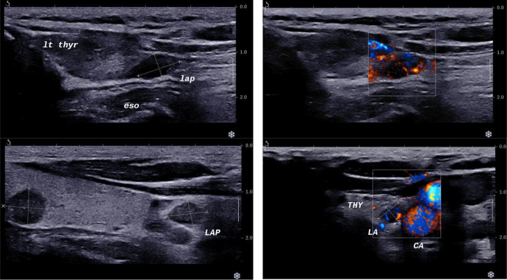
Objective: This study assesses the diagnostic value of preoperative greyscale and Doppler imaging and their combinative use with simultaneous advanced Doppler imaging and cervical maneuvers in detecting central cervical lymph node metastasis in papillary thyroid carcinoma patients.
Methods: In this cross-sectional survey, we included candidates for total or partial thyroidectomy with concomitant cervical lymph node dissection who referred to the TIRAD imaging center from February 2022 to September 2023 with papillary thyroid carcinoma diagnosis. Patients underwent preoperative ultrasonographic examination using the Aixplorer device (Supersonic Imagine, France) with a linear array transducer of 7.5-16 MHz to identify potential metastasis within the cervical lymph nodes. Ultrasonic assessments are presented using the totaling attributes such as sensitivity, specificity, positive and negative predictive values, and likelihood ratios.
Results: The post-operation pathology results showed metastasis in 85 (42.5%) patients. Standard imaging protocol without cervical approaches and advanced Doppler imaging capability detected metastatic involvement in 34 (17.0%) subjects. Meanwhile, the modified approach utilizing advanced Doppler imaging capability and cervical maneuvers identified metastatic involvement in 84 (42.0%) cases. The preoperative sensitivity without advanced Doppler imaging and maneuvers was 35.3%, specificity - 96.5%, positive predictive value - 88.2%, and negative predictive value - 66.9%. The introduction of advanced Doppler imaging and maneuvers yielded a sensitivity of 97.6%, specificity - of 99.1%, positive predictive value - 98.8%, and negative predictive value - 98.3%.
Conclusion: Advanced Doppler imaging can improve the visualization of the cervical areas, due to its ultrafast and ultrasensitive perception qualities, facilitating the early recognition of vascular pattern changes.
Keywords: Doppler ultrasonography; Lymph nodes; Neoplasm metastasis; Papillary thyroid carcinoma; Ultrasonography.
© 2025. The Author(s).
Conflict of interest statement
Declarations. Ethical approval: All procedures were conducted by the 1964 Declaration of Helsinki and its later extensions. Written informed consent was obtained from all participants. The ethics committee of AJA University of Medical Sciences reviewed and approved this study (IR.AJAUMS.REC.1402.036). Consent for publication: Consent for publication was obtained for every individual person’s data included in the study. Informed consent: Informed consent was obtained from all individual participants included in the study. Competing interests: The authors declare no competing interests.
Figures
References
-
- Surveillance E, and End Results Program Cancer Stat Facts: Thyroid Cancer: National Cancer Institute 2024 [Available from: https://seer.cancer.gov/statfacts/html/thyro.html]
-
- Iqbal MA, Wang X, Guoliang Z, Moazzam NF, Shahid AD, Qian X, Qian W. A comparison of the efficiency of diagnostic ultrasound and magnetic resonance imaging of cervical lymph nodes in papillary thyroid carcinoma. J Xray Sci Technol. 2021;29(6):1033–44. - PubMed
MeSH terms
LinkOut - more resources
Full Text Sources
Medical
