. 2025 Jul 1;25(1):1057.
doi: 10.1186/s12885-025-14470-9.

BAIAP2 as a driver of tumor progression in urothelial bladder cancer

Affiliations

BAIAP2 as a driver of tumor progression in urothelial bladder cancer

Long Huang et al. BMC Cancer. .

Abstract

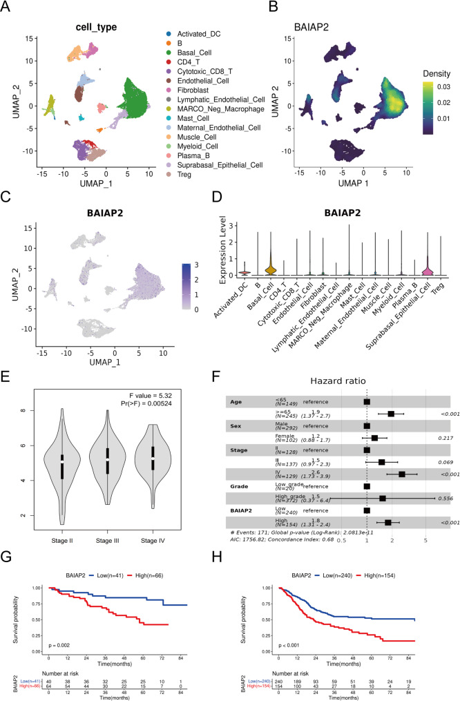
Background: Urothelial bladder cancer (UBC) is a highly heterogeneous malignancy with poor prognosis in muscle-invasive and high-grade subtypes. Epithelial-mesenchymal transition (EMT) drives tumor aggressiveness, yet its molecular mechanisms in UBC remain unclear. BAI1 associated protein 2 (BAIAP2) has been linked to cancer progression but remains unexplored in UBC. This study investigates the expression, functional role, and regulatory mechanisms of BAIAP2 in UBC, focusing on its contribution to tumor aggressiveness.

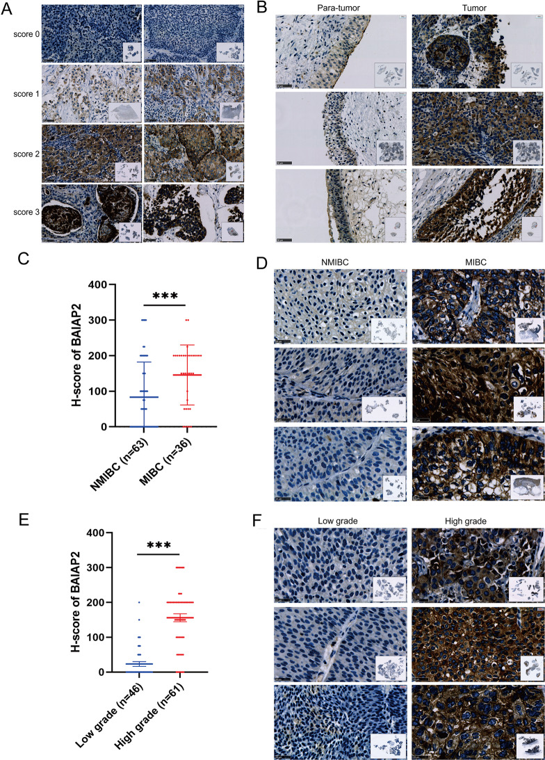
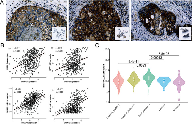
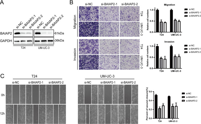
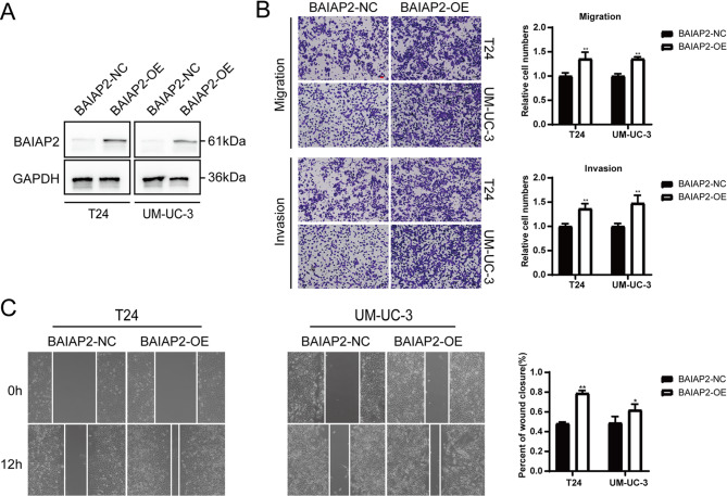
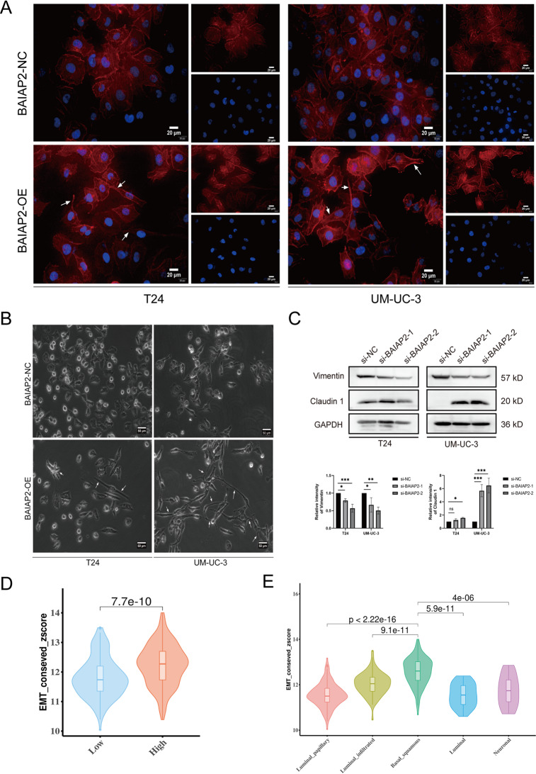
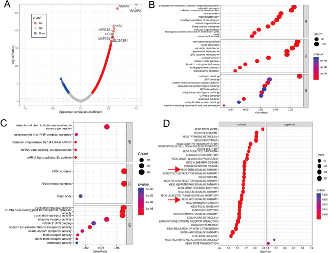
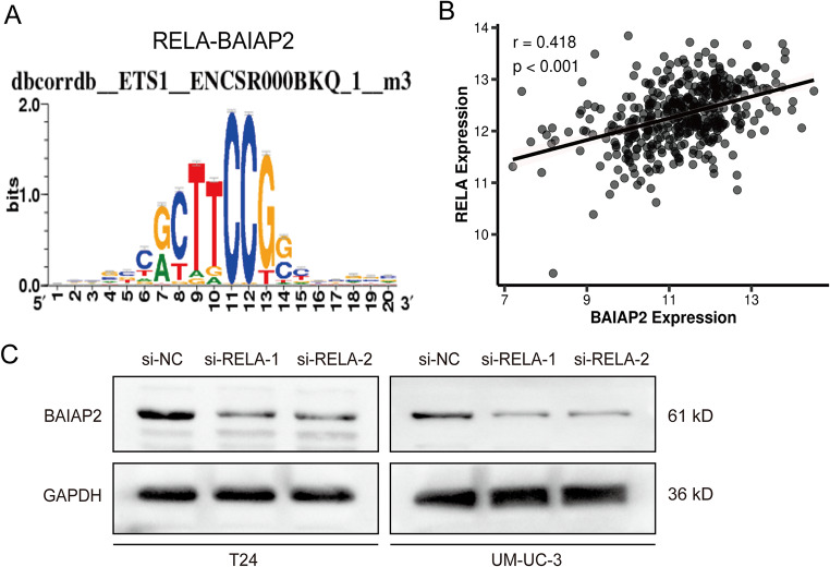
Methods: This study investigated the role of BAIAP2 in UBC using single-cell data analysis, bioinformatics, and functional assays. BAIAP2 expression was analyzed across UBC sub-populations, stages, and molecular subtypes via immunohistochemistry and quantitative methods. Transwell migration, invasion, and wound-healing assays were used to assess the impact of BAIAP2 knockdown and overexpression on cell behavior. EMT-like changes were examined through immunofluorescence and bright-field imaging. The roles of BAIAP2 in regulation of EMT pathways and its interaction with the transcription factor RELA were validated by Western blot analysis. Enrichment analysis of TCGA-BLCA datasets identified associated gene ontology terms and KEGG pathways.

Results: BAIAP2 was overexpressed in UBC, particularly in muscle-invasive and high-grade subtypes, and correlated with poor prognosis. Functional assays showed BAIAP2 promoted migration, invasion, and EMT-like changes, while its knockdown suppressed these behaviors. Bioinformatics analysis linked BAIAP2 to the transcription factor RELA, with RELA knockdown reducing BAIAP2 expression. Enrichment analysis implicated BAIAP2 in cytoskeletal reorganization and tumor progression, highlighting its role in UBC aggressiveness and potential for further therapeutic investigation.

Conclusions: BAIAP2 was highly expressed in muscle-invasive and high-grade tumors and was associated with poor prognosis. It promoted metastasis and EMT through activation of cytoskeletal remodeling. These findings identified BAIAP2 as a promising biomarker and a potential therapeutic target for the aggressive UBC.

Keywords: BAI1 associated protein 2 (BAIAP2); Epithelial-mesenchymal transition (EMT); Progression; Urothelial bladder cancer (UBC).


Conflict of interest statement

Declarations. Ethical approval: The study was approved by the Ethical Committee of the Second Affiliated Hospital and Yuying Children’s Hospital of Wenzhou Medical University (Approval No. 2021-K-101-01). Written informed consent was obtained from all participating patients. The consent process ensured that patients were fully informed about the study’s purpose, procedures, potential risks, and benefits, in accordance with the principles of the Declaration of Helsinki. All patient data were anonymized to protect confidentiality. Consent for publication: Not applicable. Competing interests: The authors declare no competing interests.

Figures

Fig. 1
Fig. 1
BAIAP2 expression in sub-populations of UBC tissues. (A) Clustering analysis of primary cell types. (B,C) BAIAP2 expression in the sub-populations, visualized in UMAP. (D) Violin plot shows expression level of BAIAP2 across major cell types. (E) Violin plots of BAIAP2 expression levels across three cancer stages: Stage II, Stage III, and Stage IV. (F) The forest plot illustrates the HR for various factors associated with BAIAP2 (G) Kaplan-Meier overall survival curves according to BAIAP2 expression levels in our in-house cohort of 107 UBC patients. The number at risk at each time point is shown below the survival curves (Note: Three cases were excluded due to missing event status). (H) High level of BAIAP2 predicted poor prognosis in TCGA-BLCA cohort. Log-rank P value < 0.05 was considered as statistically significant
Fig. 2
Fig. 2
The expression of BAIAP2 in UBC tissues. (A) Representative images showing the expression of BAIAP2 in 107 UBC tissues detected by IHC. Score from 0 to 3. (B) Representative images showing the expression of BAIAP2 in para-tumor and tumor tissues detected by IHC. (C) H-score of BAIAP2 in NMIBC (n = 63) and MIBC (n = 36) tissues (Note: Eight samples were excluded due to missing data). (D) Representative images showing the expression of BAIAP2 in NMIBC and MIBC tissues detected by IHC. (E) H-score of BAIAP2 in Low-grade (n = 46) and High-grade (n = 61) tissues. (F) Representative images showing the expression of BAIAP2 in Low-grade and High-grade tissues detected by IHC. Wilcox tests were used to evaluate the statistical significance of differences between two groups. ***P < 0.001
Fig. 3
Fig. 3
Association of BAIAP2 expression with Basal/Squamous features and molecular subtypes in UBC. (A) Representative images showing Basal/Squamous differentiation in tumors with high BAIAP2 expression. (B) Scatter plots to illustrate the correlation of BAIAP2 expression with Basal/Squamous cell markers. (C) Expression levels of BAIAP2 in different molecular subtypes of UBC (TCGA-BLCA)
Fig. 4
Fig. 4
Knockdown of BAIAP2 inhibited migration and invasion of UBC cells. (A) The protein abundance of BAIAP2 in UBC cells measured by western blot analysis. (B) Representative images and quantity of transwell migration and invasion assays. (C) Representative images and quantity of wound healing assays. (Data are presented as mean ± SD, n = 3. t-test, **P < 0.01)
Fig. 5
Fig. 5
Over expression of BAIAP2 promoted migration and invasion of UBC cells in vitro. (A) Western blot analysis showed overexpression of BAIAP2 in T24 and UM-UC-3 cells. (B) Representative images and quantity of transwell migration and invasion assays. (C) Representative images and quantity of wound healing assays. (Data are presented as mean ± SD, n = 3. t-test, *P < 0.05, **P < 0.01)
Fig. 6
Fig. 6
BAIAP2 promoted EMT-like changes of UBC cells in vitro. (A) T24 and UM-UC-3 cells transfected with BAIAP2 or control plasmid were stained with phalloidin (F-actin, red) and DAPI (nuclei, blue). White arrows indicate morphological changes. Scale bar: 20 μm. (B) Representative bright-field images of T24 and UM-UC-3 cells overexpressing BAIAP2 or transfected with control plasmid. Scale bar: 50 µm. White arrows indicate EMT-like changes. (C) Effects of BAIAP2 knockdown on EMT marker expression. (Top) Representative Western blots showing Vimentin and Claudin1 protein levels in T24 and UM-UC3 cells. (Bottom) Quantitative analysis of Vimentin and Claudin1 expression normalized to GAPDH (mean ± SD, n = 3. t-test, *P < 0.05, *** P < 0.001). (D) Violin plot showing the distribution of EMT scores in BAIAP2 high-expression and low-expression groups based on bioinformatics analysis of publicly available bladder cancer datasets (TCGA-BLCA). (E) Violin plot showing the distribution of EMT scores in different subtypes of UBC
Fig. 7
Fig. 7
Enrichment analysis of BAIAP2 gene co-expression network in UBC. (A) The volcano plot showed co-expression genes associated with BAIAP2 expression in the TCGA-BLCA datasets. (B) Enrichment analysis of GO terms for co-expression genes which were positively correlated with BAIAP2. (C) GO terms for co-expression genes which were negatively correlated with BAIAP2. (D) Enrichment analysis of KEGG terms for co-expression genes which were correlated with BAIAP2
Fig. 8
Fig. 8
Association of BAIAP2 with the transcription factor RELA and its functional validation. (A) RELA is one top associated transcription factor for BAIAP2. (B) Correlation between RELA and BAIAP2 expression in BLCA, based on data from the TCGA-BLCA database. (C) The expression of BAIAP2 was down-regulated in T24 and UM-UC-3 cells after siRELAs transfection

Similar articles

References

    1. Liang Y, et al. Single-cell atlases link macrophages and CD8(+) T-cell subpopulations to disease progression and immunotherapy response in urothelial carcinoma. Theranostics. 2022;12(18):7745–59. - PMC - PubMed
    1. Luo Y, et al. Single-Cell transcriptome comparison of bladder Cancer reveals its ecosystem. Front Oncol. 2022;12:818147. - PMC - PubMed
    1. Yu L, et al. Prognostic significance of lineage diversity in bladder Cancer revealed by Single-Cell sequencing. Front Genet. 2022;13:862634. - PMC - PubMed
    1. Zhou R, et al. Development and validation of an intra-tumor heterogeneity-related signature to predict prognosis of bladder cancer: a study based on single-cell RNA-seq. Aging. 2021;13(15):19415–41. - PMC - PubMed
    1. Bianchi M, et al. Age-stratified distribution of metastatic sites in bladder cancer: A population-based analysis. Can Urol Association J. 2014;8(3–4):148. - PMC - PubMed