. 2025 Jul 1;25(1):1029.
doi: 10.1186/s12885-025-14432-1.

Ex vivo engineering of phagocytic signals in breast cancer cells for a whole tumor cell-based vaccine

Affiliations

Ex vivo engineering of phagocytic signals in breast cancer cells for a whole tumor cell-based vaccine

Román Martí-Díaz et al. BMC Cancer. .

Abstract

Background: Today, cell therapies are constantly evolving and providing new options for cancer patients. These therapies are mostly based on the inoculation of immune cells extracted from a person's own tumor; however, some studies using whole tumor cell-based vaccines are approaching the level of maturity required for clinical use. Although these latest therapies will have to be developed further and adapted to overcome many ethical barriers, there is no doubt that therapeutic cancer vaccines are the next frontier of immunotherapy.

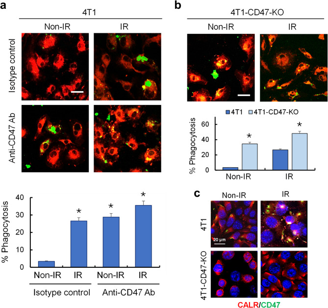
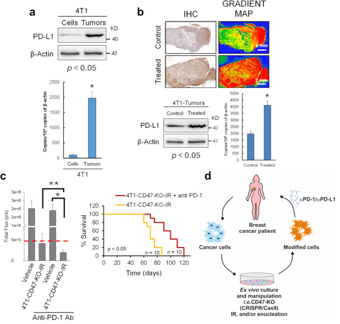
Methods: Ionizing radiation and CD47 knockout via CRISPR-Cas9 genome editing were used to optimize the macrophage-mediated phagocytosis of breast cancer cells. These cells were subsequently used in several mouse models to determine their potential as novel whole-cell-based vaccines to drive antitumor immunity. To improve the recognition of tumor cells by activated immune cells, this cellular therapy was combined with anti-PD-1 antibody treatments.

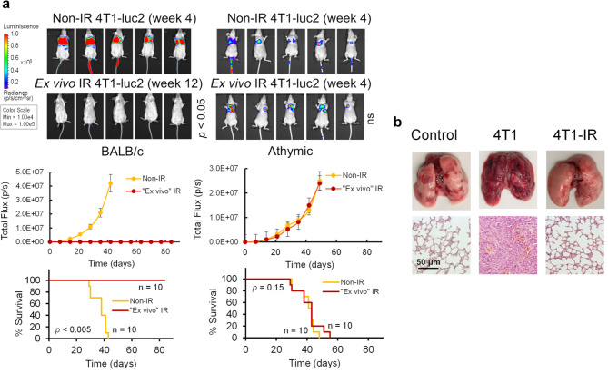
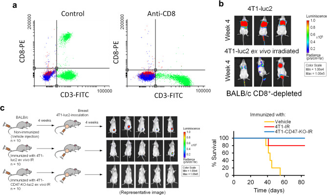
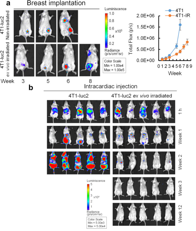
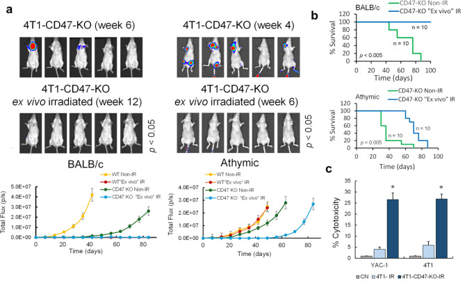
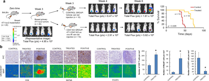
Results: Here, we showed that irradiation of 4T1 breast cancer cells increases their immunogenicity and, when injected into the blood of immunocompetent mice, elicits a complete antitumor immune response mediated, in part, by the adaptive immune system. Next, to improve the macrophage-mediated phagocytosis of breast cancer cells, we knocked out CD47 in 4T1 cells. When injected in the bloodstream, irradiated CD47 knockout cells activated both the adaptive and the innate immune systems. Therefore, we used these ex vivo engineered cells as a whole tumor cell-based vaccine to treat breast tumors in immunocompetent mice. A better response was obtained when these cells were combined with an anti-PD-1 antibody.

Conclusion: These results suggest that tumor cells obtained from surgical samples of a breast cancer patient could be engineered ex vivo and used as a novel cell therapy to drive antitumor immunity.

Keywords: Anti-tumor immunity; Breast cancer; CD47; Ionizing radiation; Whole tumor cell-based vaccines.


Conflict of interest statement

Declarations. Ethics approval and consent to participate: Animals were bred and maintained according to Spanish legislation on the ‘Protection of Animals used for Experimental and other Scientific Purposes’ and in accordance with the directives of the European Community. All animal procedures were approved by the Ethical Committee of the University of Murcia and the Direccion General de Ganaderia y Pesca, Comunidad Autonoma de Murcia (Project reference A13151101). Consent for publication: Not applicable. Competing interests: The authors declare no competing interests.

Figures

Fig. 1
Fig. 1
The effect of ex vivo irradiation on the immune response against breast cancer cells. (a) Luciferase imaging of BALB/c and athymic mice injected with 4T1-luc2 cells via the tail vein. When indicated, the cells were irradiated ex vivo (10 Gy; 24 h). The total flux (photons/s) was compared between the ex vivo irradiated and non-irradiated groups (ns, not significant) and the histogram compares tumor evolution in groups injected with non-irradiated 4T1-luc2 (Non-IR) and ex vivo irradiated 4T1-luc2 (“Ex vivo” IR) cells. The values (mean ± S.D.) are representative of two independent experiments showing similar results (n = 10 for each experimental condition). Survival data are presented in the graph. (b) Lungs from BALB/c mice injected via the tail vein with vehicle (control) or the indicated cells (4T1-IR, ex vivo irradiated 4T1 cells). A microscopic view of the lung tissue structure (H&E stain, ×100) is shown below
Fig. 2
Fig. 2
Ex vivo irradiation of breast cancer cells induces an adaptive immune response. (a) Depletion of CD8+ T lymphocytes. BALB/c mice were treated intraperitoneally with a dose (250 µg) of IgG isotype control (left panel) or anti-CD8-clone 2.43 (right panel) monoclonal antibody every 3 days. After four doses, peripheral blood was collected and stained to test for the presence of CD8+ cytotoxic T lymphocytes among lymphoid cells by flow cytometry. The data show the complete clearance of T CD8+ cells in a representative experiment out of two with similar results. The remaining blood cell counts (granulocyte, monocyte, NK and B lymphocyte counts) were unchanged (data not shown). (b) Inactivation of CD8+ T cells in immunocompetent BALB/c mice results in tumor growth after the injection of 4T1-luc2 cells. The differences were significant (p < 0.05) compared with those of the BALB/c mice inoculated with ex vivo-irradiated 4T1-luc2 cells, as shown in Fig. 1A. (c) Vaccination assay. Non-immunized animals were injected with vehicle, while the immunized groups were injected with ex vivo-irradiated 4T1-luc2 cells (4T1-IR) or ex vivo-irradiated 4T1-CD47-KO-luc2 cells (4T1-CD47-KO-IR). After 4 weeks, non-irradiated 4T1-luc2 cells were injected intradermally. Analysis of primary tumors was carried out 4 weeks after breast pad injection. Differences between control and both immunized groups were statistically significant (p < 0.05). Survival was examined (right panel)
Fig. 3
Fig. 3
The injection site of 4T1-luc2 cells determines the immune response in mice. (a) Luciferase imaging of a model where tumor cells were directly inoculated into the mammary path of BALB/c mice. 4T1-luc2 cells (both non-irradiated and ex vivo irradiated) induced primary tumor formation and metastasis in this mouse model. The histogram compares primary tumor evolution in groups injected with non-irradiated 4T1-luc2 (4T1) and ex vivo irradiated 4T1-luc2 (4T1-IR) cells. The values (mean ± S.D.) are representative of three independent experiments (n = 10 for each experimental condition). (b) Luciferase imaging of an intracardiac model where tumor cells were directly inoculated into the arterial blood supply of BALB/c mice. The images compare the evolution of tumors generated by injection of non-irradiated cells (4T1-luc2) versus those generated by injection of ex vivo irradiated cells (4T1-luc2 ex vivo irradiated)
Fig. 4
Fig. 4
Knocking down CD47 sensitizes breast cancer cells to macrophage phagocytosis. (a) CFSE-labeled 4T1-luc2 (4T1) irradiated (IR) or non-irradiated (non-IR) cells along with a blocking antibody against CD47 (B6H12.2, 10 µg/ml) were added to cultures of PKH26-labeled PBMC-derived macrophages. Two hours later, the cultures were washed and examined with an inverted confocal microscope. The data shown are representative microphotographs of three individual experiments. Green: CFSE-labeled melanoma cells; red: PKH26-labeled macrophages. Scale bar, 20 μm. The histogram shows the comparison of the phagocytosis index of macrophages against the indicated 4T1 cells. The phagocytosis index was calculated as the number of phagocytized CFSE+ cells per 100 macrophages (n = 3, mean ± S.D.; *p < 0.05 compared with the non-IR-isotope control group). (b) As in Fig. 4a, but CFSE-labeled 4T1-luc2-CD47-KO (4T1-CD47-KO) cells were used. The histogram shows the comparison of the phagocytosis index of macrophages against the indicated 4T1 cells (n = 3, mean ± S.D.; *p < 0.05 compared with their respective 4T1 groups). Scale bar, 20 μm. (c) Confocal microscopy analysis of CD47 and CALR in 4T1 and 4T1-CD47-KO breast cancer cells under the indicated conditions
Fig. 5
Fig. 5
Effect of CD47 depletion on the antibreast cancer immune response. (a) Luciferase imaging of athymic and BALB/c mice after tail vein injection of 4T1-CD47-KO cells. Where indicated, the cells were previously irradiated ex vivo (10 Gy; 24 h). The total flux (photons/s) was compared between the ex vivo irradiated and non-irradiated groups (p values). Color scale (Min = 1.00e6; Max = 1.00e7). Data of Fig. 1a, performed with irradiated or non-irradiated WT 4T1-luc2 cells expressing CD47, are used as a control for these experiments. The histogram compares tumor evolution in groups injected with non-irradiated 4T1-luc2 cells (both WT Non-IR and CD47-KO Non-IR) and ex vivo irradiated 4T1-luc2 cells (both WT “Ex vivo” IR and CD47-KO “Ex vivo” IR). The values (mean ± S.D.) are representative of two independent experiments showing similar results (n = 10 for each experimental condition). (b) Survival data for CD47-KO Non-IR and CD47-KO “Ex vivo” IR groups are presented (see Fig. 1a for comparative survival with WT control groups). (c) Athymic (Fox1nu) mice were immunized via tail vein injection with ex vivo-irradiated 4T1 and ex vivo-irradiated 4T1-CD47-KO breast cancer cells (10 Gy; 24 h). Control (CN) mice were injected with vehicle. One week after the injection, the animals were sacrificed, and NK cells were purified from their spleens. Purified NKs were cocultured with YAC-1 cells or irradiated 4T1 cells. NK cytotoxicity was evaluated by a 51Cr release assay at an effector/target ratio (E/T) of 5. The results are representative of three independent experiments. *p < 0.05 compared with both the CN- and 4T1-immunized groups
Fig. 6
Fig. 6
Experimental cellular therapy for breast cancer. (a) 4T1-luc2 cells were transplanted into the mammary glands of BALB/c mice to establish breast tumors, and after three weeks, the tumor-bearing mice were divided into two groups. Animals in the control group were tail-injected with vehicle (PBS), while mice in the treated groups were injected with ex vivo-irradiated 4T1-CD47-KO cells (10 Gy; 24 h) via the tail vein. Luciferase imaging is shown. Color scale (Min = 1.00e6; Max = 1.00e7). The total flux (photons/s) was compared between the control and treated groups (p < 0.05 at both weeks 4 and 6). Survival data are presented in the graph. (b) IHC of 4T1 tumors obtained from the control and treated groups shown in Fig. 6a. Quantification was carried out by using ImageJ software (histograms) on three different areas of tumors and three animals per group (*p = 0.005). The gradient map highlights positive cells after immunostaining with CD8, NKp46, and FOXP3 antibodies. Positive controls for these immune cells were also included
Fig. 7
Fig. 7
The efficacy of combined anti-PD-1 therapy. (a) PD-L1 expression in 4T1 cells in culture (Cells) and in 4T1-induced tumors (Tumors) was analyzed by Western blotting and PCR (histogram), and the differences were statistically significant (*p < 0.05). For analysis of PD-L1 in solid tumors, we used those generated from the control group shown in Fig. 6a. The blots in this figure were cropped from different gels. The full blots are shown in Fig. S4. (b) Expression of PD-L1 in control and treated tumors generated in Fig. 6a. The expression of PD-L1 was analyzed by immunohistochemistry (IHC), Western blotting, and polymerase chain reaction (PCR) (histogram), and differences were found to be significant (*p < 0.05). The gradient map highlights positive cells after immunostaining with an anti-PD-L1 antibody. The full blots are shown in Fig. S4 (c). The model was established as described in Fig. 6a, and luciferase imaging at week 6 of the indicated treatments was used to determine the total flux (mean ± S.D.). When indicated, anti-PD-1 antibody (40 µg/mouse) was administered intraperitoneally via 3 doses on days 22, 26, and 30. The dashed red line indicates the average total flux at week 3 (before starting treatment); *p < 0.005; **p < 0.05. (d) A schematic representation for the cellular therapy proposed in this study. Ex vivo manipulated tumor cells from cancer patients could be used as a whole tumor cell-based vaccine for stimulate the recognition of body cancer cells by the immune system. In a second step, anti-PD-1/PD-L1 antibodies could be used to enhance the killing of tumor cells

References

    1. Guedan S, Calderon H, Posey AD Jr, Maus MV. Engineering and design of chimeric antigen receptors. Mol Ther Methods Clin Dev. 2018;12:145–56. - DOI - PMC - PubMed
    1. Sun D, She X, Li S, Wang X, Yang X, Wan M. CAR-T cell therapy: a breakthrough in traditional cancer treatment strategies. Mol Med Rep. 2024;29(3):47. - DOI - PMC - PubMed
    1. Ribas A, Comin-Anduix B, Chmielowski B, Jalil J, de la Rocha P, McCannel TA, et al. Dendritic cell vaccination combined with CTLA4 blockade in patients with metastatic melanoma. Clin Cancer Res. 2009;15(19):6267–76. - DOI - PMC - PubMed
    1. Saxena M, Bhardwaj N. Re-emergence of dendritic cell vaccines for cancer treatment. Trends Cancer. 2018;4(2):119–37. - DOI - PMC - PubMed
    1. Saxena M, van der Burg SH, Melief CJM, Bhardwaj N. Therapeutic cancer vaccines. Nat Rev Cancer. 2021;21(6):360–78. - DOI - PubMed