

Real-world clinical insights and aqueous humor biomarker analysis of diabetic macular edema subtypes classified by OCT following intravitreal injections
- PMID: 40596978
- PMCID: PMC12220419
- DOI: 10.1186/s12886-025-04186-6
Real-world clinical insights and aqueous humor biomarker analysis of diabetic macular edema subtypes classified by OCT following intravitreal injections
Abstract
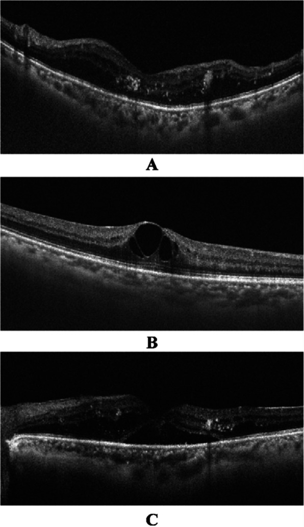
Purpose: This study aimed to observe the morphological changes and visual outcomes in diabetic macular edema (DME) subtypes based on optical coherence tomography classification following intravitreal injection.
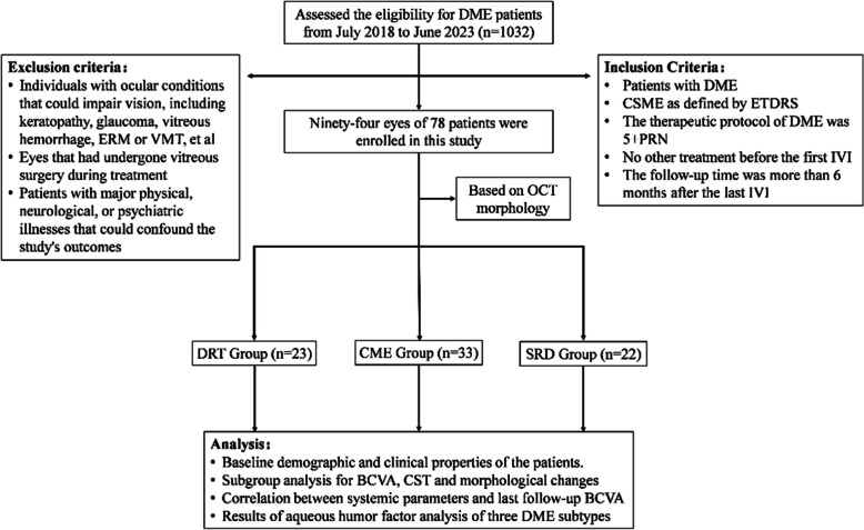
Methods: Ninety-four eyes from seventy-eight patients were enrolled between July 1, 2018, and June 30, 2023. Best-corrected visual acuity (BCVA), central subfield thickness, and morphological changes were examined at each follow-up visit. Aqueous humor samples were obtained before intravitreal injection of anti-vascular endothelial growth factor or cataract surgery. The inflammatory cytokine profile in the aqueous humor of each DME subtype was analyzed to explore potential contributions.
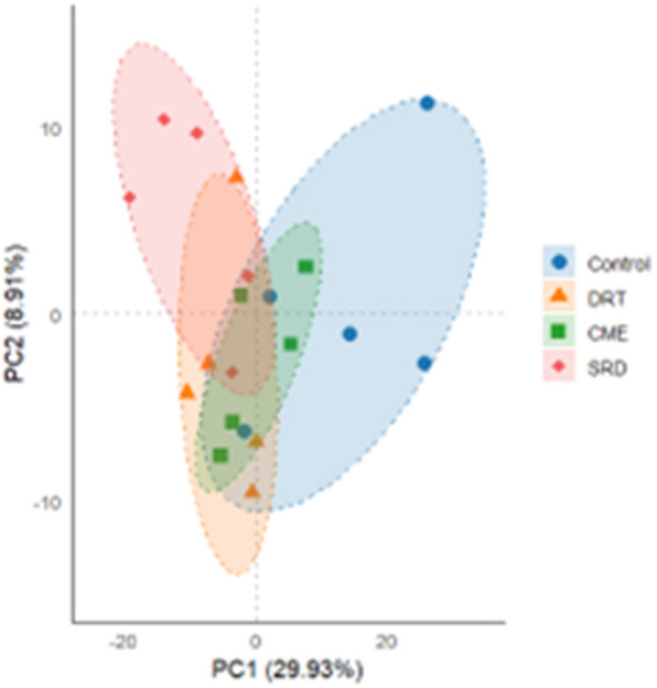
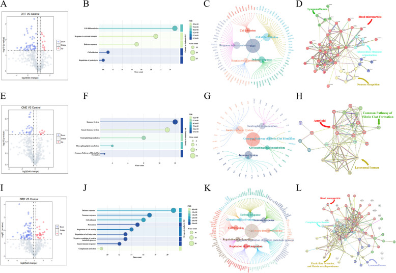
Results: The BCVA in the diffuse retinal thickening group was significantly improved at the last follow-up (p = 0.006), but no significant change in central subfield thickness was observed. Conversely, the BCVA in the cystoid macular edema and serous retinal detachment groups did not significantly improve, although a significant reduction in central subfield thickness was observed in both groups at the final follow-up (both p < 0.05). Principal component analysis of proteomic data revealed clear segregation between samples from patients with different DME subtypes and those from the control group. 59 proteins were found to be significantly regulated in the diffuse retinal thickening group compared to those in the control group. Of these, 10 proteins were upregulated, whereas 49 were downregulated. In the cystoid macular edema group, 30 proteins were significantly regulated, with 8 proteins upregulated and 22 downregulated. In the serous retinal detachment group, 87 proteins showed significant changes: 24 exhibited increased expression and 63 decreased expression.
Conclusions: The diffuse retinal thickening subtype showed a better response and cystoid macular edema was the most common morphological subtype of DME after intravitreal injection treatment in real-world clinical observations. The variations in protein expression of aqueous humor suggested distinct pathological mechanisms for each DME subtype, laying the groundwork for more targeted treatment strategies.
Keywords: Cystoid macular edema; Diabetic macular edema; Diffuse retinal thickening; Intravitreal injection; Optical coherence tomography; Serous retinal detachment.
© 2025. The Author(s).
Conflict of interest statement
Declarations. Ethics approval and consent to participate: All participants provided signed informed consent before enrollment. Ethical approval was granted by the Ethics Committee of Tianjin Medical University Eye Hospital (approval number 2023KY-24). We adhered to all institutional regulations regarding the ethical use of human volunteer information and samples. Consent for publication: Not Applicable. Competing interests: The authors declare no competing interests.
Figures
Similar articles
-
Vitrectomy as an Adjunct to Treat-and-Extend Anti-VEGF Injections for Diabetic Macular Edema: The Vitrectomy in Diabetic Macular Oedema (VIDEO) Randomized Clinical Trial.JAMA Ophthalmol. 2024 Sep 1;142(9):837-844. doi: 10.1001/jamaophthalmol.2024.2777. JAMA Ophthalmol. 2024. PMID: 39115867 Free PMC article. Clinical Trial.
-
Assessing the Role of Statins as an Adjunctive Anti-VEGF Therapy for Clinically Significant Macular Edema (CSME) in Type 2 Diabetes Mellitus.Rom J Ophthalmol. 2025 Apr-Jun;69(2):219-227. doi: 10.22336/rjo.2025.35. Rom J Ophthalmol. 2025. PMID: 40698099 Free PMC article. Clinical Trial.
-
Anti-vascular endothelial growth factor combined with intravitreal steroids for diabetic macular oedema.Cochrane Database Syst Rev. 2018 Apr 18;4(4):CD011599. doi: 10.1002/14651858.CD011599.pub2. Cochrane Database Syst Rev. 2018. PMID: 29669176 Free PMC article.
-
Simultaneous inhibition of fibroblast growth factor-2 and vascular endothelial growth factor-a with RC28-E in diabetic macular edema: a phase 2 randomised trial.Br J Ophthalmol. 2025 Jun 23;109(7):784-790. doi: 10.1136/bjo-2024-326006. Br J Ophthalmol. 2025. PMID: 40122579 Clinical Trial.
-
Anti-vascular endothelial growth factor for diabetic macular oedema: a network meta-analysis.Cochrane Database Syst Rev. 2017 Jun 22;6(6):CD007419. doi: 10.1002/14651858.CD007419.pub5. Cochrane Database Syst Rev. 2017. Update in: Cochrane Database Syst Rev. 2018 Oct 16;10:CD007419. doi: 10.1002/14651858.CD007419.pub6. PMID: 28639415 Free PMC article. Updated.
References
-
- Wang FH, Liang YB, Zhang F, et al. Prevalence of Diabetic Retinopathy in Rural China: The Handan Eye Study. Ophthalmology. 2009;116:461–7. - PubMed
-
- Li JQ, Welchowski T, Schmid M, et al. Prevalence, incidence and future projection of diabetic eye disease in Europe: a systematic review and metaanalysis. Eur J Epidemiol. 2020;35:11–23. - PubMed
-
- Teo ZL, Tham YC, Yu M, et al. Global prevalence of diabetic retinopathy and projection of burden through 2045: systematic review and meta-analysis. Ophthalmology. 2021;128:1580–91. - PubMed
MeSH terms
Substances
Grants and funding
LinkOut - more resources
Full Text Sources
Medical
