Review
. 2025 Jul 1;23(1):477.
doi: 10.1186/s12951-025-03541-5.

Stimuli-responsive smart materials enabled high-performance biosensors for liquid biopsies

Affiliations
Review

Stimuli-responsive smart materials enabled high-performance biosensors for liquid biopsies

Xiaoqi Gao et al. J Nanobiotechnology. .

Abstract

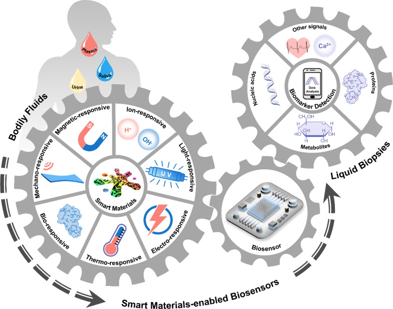
Liquid biopsies have emerged as a key tool that enables personalized medicine, enabling precise detection of biochemical parameters to tailor treatments to individual needs. Modern biosensors enable real-time detection, precise diagnosis, and dynamic monitoring by rapidly analyzing biomarkers such as nucleic acids, proteins, and metabolites in bodily fluids like blood, saliva, and urine. Despite their potential, many biosensors are still constrained by mono-functionality, sub-optimal sensitivity, bulky designs, and complex operation requirements. Recent advances in stimuli-responsive smart materials present a promising pathway to overcome these limitations. These materials enhance biomarker signal transduction, release, or amplification, leading to improved sensitivity, simplified workflows, and multi-target detection capabilities. Further exploration of the integration of these smart materials into biosensing is therefore essential. To this end, this review critically examines and compares recent progress in the development and application of physical, chemical, and biochemical stimuli-responsive smart materials in biosensing. Emphasis is placed on their responsiveness mechanisms, operational principles, and their role in advancing biosensor performance for biomarker detection in bodily fluids. Additionally, future perspectives and challenges in developing versatile, accurate, and user-friendly biosensors for point-of-care and clinical applications using these smart materials are discussed.

Keywords: Biosensors; Liquid biopsy; Smart materials; Stimuli-responsive materials.


Conflict of interest statement

Declarations. Ethics approval and consent to participate: Not applicable. Competing interests: The authors declare no competing interests.

Figures

Fig. 1
Fig. 1
Smart materials enabled biosensors for liquid biopsy and their applications
Fig. 2
Fig. 2
Responsive mechanisms of micro- and nanobubbles under ultrasound stimuli and their working principles when applied in biosensors. A Schematic illustration of the generation of micro- and nanobubbles through i phase-changeable nanodroplets, ii gas vesicles and iii engineered microbubbles under an ultrasound field. The generated nanobubbles undergo cavitation and final implosion as acoustic pressure increases. B The working principle underlying nanobubbles’ applications in detecting biomarkers in bodily fluids. Their barrier-breaking effect improves detecting sensitivity by facilitating extratumoral biomarker release. PFC: perfluorocarbon; GV: gas vesicles; ctDNA: circulating tumor DNA; miRNA: microRNA; cfDNA: cell-free DNA; qPCR: quantitative polymerase chain reaction
Fig. 3
Fig. 3
Applications of microbubble-assisted focused ultrasound (FUS)-induced BBB opening in biosensing. A Sonobiopsy for minimally invasive detection of glioblastoma-derived ctDNA. i The system set up. iiv Concentration of ctDNA and cfDNA in plasma after ultrasound treatment. Reproduced with permission [43]. Copyright 2022, Ivyspring International Publisher. B Ultrasound-mediated BBB opening for increasing cfDNA plasma level. i Representative fluorescent images of cfDNA plasma level at various time points post sonication on mouse head. iiiv Optimal acoustic power and optimal blood collection time post sonication. Reproduced with permission [44]. Copyright 2021, Oxford University Press. BBB: blood–brain barrier; cfDNA: cell-free DNA; ctDNA: circulating tumor DNA; US: ultrasound only; MB: microbubble only; SS: single sonication; DS: double sonication
Fig. 4
Fig. 4
Representative classifications of acoustic robots and their working principles when applied in biosensors. A Schematic illustration of the structures of different types of acoustic robots. B The working principle underlying the applications of acoustic robots in detecting biomarkers in bodily fluids. i Ultrasound propulsion makes functionalized nanoparticles intracellular sensors, and ii the acoustic assemble effect enhances detecting sensitivity by amplifying fluorescent signals. CEA: carcinoembryonic antigen
Fig. 5
Fig. 5
Applications of acoustic robots in biosensing. A Acoustic nanomotors for intracellular detection of human papillomavirus-associated head and neck cancer. i Working principle. ii, iii Fluorescent intensity for each condition listed. iv Fluorescent images of modified nanomotors after 15-min incubation with a HPV-negative or b, c HPV-positive cells under b static conditions or a, c ultrasound field. Reproduced with permission [61]. Copyright 2019, SAGE Publications Inc. B Acoustic aggregation of functionalized nanoparticles to assess the carcinoembryonic antigen (CEA) level in saliva. i Working principle of the ratiometric fluorescent platform based on modified Eu-MOFs. ii Fluorescent images of modified Eu-MOFs at different concentrations of CEA under acoustic aggregation. iii The linear relationship between CEA concentration and the fluorescence ratio of green and red of modified Eu-MOFs. iv Comparison of the proposed ratiometric platform and commercial enzyme-linked immunosorbent assay kit for CEA detection of salivary samples. Reproduced with permission [62]. Copyright 2023, American Chemical Society. Eu-MOFs: europium metal–organic frameworks
Fig. 6
Fig. 6
Representative examples of piezoelectric materials and their application in biosensing. A-i, ii Schematic illustrations of two types of piezoelectric materials and iii the piezoelectric property. B The working principle underlying piezoelectric materials’ application in detecting blood pressure. C A thin, soft, miniaturized system (TSMS) using piezoelectric material PZT 5H for continuous wireless monitoring of artery blood pressure. i, ii Schematic illustration of the blood propagation and generated piezo response. iii, iv The piezo response and the converted pulse waveform. v The BP measurement accuracy of the TSMS compared with commercial CNAP. Reproduced with permission [67]. Copyright 2023, Springer Nature. PVDF: polyvinylidene fluoride; PZT: lead zirconate titanate; BP: blood pressure; CNAP: continuous noninvasive artery pressure
Fig. 7
Fig. 7
Classifications, responsive mechanisms and typical applications of liquid metal. A Liquid metal’s three typical embodiments. B Schematic illustrations of the resistance changes of LM composites in response to external stress. C The working principle underlying LM’s application in detecting blood pressure. D A wearable 3D-printed rigid microbump-integrated LM-based pressure sensor (3D-BLiPS). i Schematic view of the proposed 3D-BLiPS. ii, iii Effect of the microbump on pressure sensitivity. iv Dynamic response of the sensor to the application of varying pressure levels. v, vi Continuous epidermal pulse and ECG signals for PTT calculation before and after exercise, respectively (PTT0 = 278 ms, PTT1 = 238 ms). The SBP and DBP after exercise were estimated to be 138.4 ± 4.2 and 66.8 ± 1.4 mmHg, respectively. Reproduced with permission [79]. Copyright 2019, Wiley. LM: liquid metal; ECG: electrocardiogram; PTT: pulse transit time; SBP: systolic blood pressure; DBP: diastolic blood pressure
Fig. 8
Fig. 8
Representative classifications of artificial enzyme mimics and their working principle when applied in biosensors. A Schematic illustration of i conjugated microporous polymer (CMP) and ii nanozymes. B The working principle underlying artificial enzyme mimics’ applications in detecting biomarkers in bodily fluids. GO: graphene oxide; MOF: metal–organic framework; ROS: reactive oxygen species; LSPR: localized surface plasmon resonance
Fig. 9
Fig. 9
Applications of artificial enzyme mimics in biosensing. A Light-responsive oxidase mimic of CMP for urease sensing. i The working principle and iiiv feasibility of the CMP-PQx-based fluorescent sensor. v The linear relationship between urease concentration and the fluorescence intensity ratio (F/F0). Reproduced with permission [86]. Copyright 2020, Elsevier. B A colorimetric sensor for exosomalmiR-21 detection based on the visible light-triggered oxidase mimic of MAA. i The working principle and ii feasibility of MAA-based fluorescent sensor. iii The linear relationship between exosomal miR-21 concentration and the UV–vis absorbance at 652 nm. Reproduced with permission [102]. Copyright 2021, Elsevier. C A photosensitized metal–organic framework (PSMOF)-enabled colorimetric biosensor for cellular GSH detection. i The working principle and ii feasibility of PSMOF-based colorimetric biosensor. iii The linear relationship between GSH concentration and the UV–vis absorbance at 652 nm. Reproduced with permission [93]. Copyright 2019, American Chemical Society. CMP-PQx: CMP containing pyrazino[2,3-g] quinoxaline; MAA: 10-methyl-2-amino-acridone; miR-21: microRNA-21; UV–vis absorbance: UV–visible absorbance; GSH: glutathione
Fig. 10
Fig. 10
Representative classifications of quantum dots (QDs) and the working principle when applied in biosensors. A Schematic illustrations of metallic QDs and cadmium QDs. B The working principle underlying QDs’ applications in detecting biomarkers in bodily fluids. QDs: quantum dots; C atom: carbon atom; GQDs: graphene quantum dots; CQDs: carbon quantum dots; CPDs: carbon polymeric dots
Fig. 11
Fig. 11
Applications of QDs in biosensing. A A photoelectrochemical biosensor using CdSe QDs-decorated ZnIn2S4 nanosheets for ATP detection. i The working principle and iiiv feasibility of the CdSe/ZnIn2S4-based PEC sensor. v The linear relationship between the logarithm of the ATP concentration and the photocurrent change. Reproduced with permission [129]. Copyright 2021, Elsevier. B QD-based molecular beacons for quantitative detection of nucleic acids. i Schematic illustration of the designed fluorescent probe, QD525/DHP-Cy3 complex and ii the working principle of the fluorescent biosensor. The concentration of iii, iv ssDNA and v, vi lcrVRNA was determined by detecting the PL ratio of Cy3 to QD525. Reproduced with permission [130]. Copyright 2022, American Chemical Society. C A single QD-based biosensor for detection of METTL3/14 complex activity in breast cancer tissues. i The working principle and ii feasibility. iii The linear relationship between the logarithm of the METTL3/14 complex concentration and the Cy5 fluorescence intensity. Reproduced with permission [131]. Copyright 2023, Elsevier. PEC: photoelectrochemical; CdSe QDs: lead selenide quantum dots; ZnIn2S4: zinc indium sulfide; ATP: adenosine triphosphate; DHP: DNA hairpin; QD525: CdSe/CdS/ZnS core/shell/shell quantum dot with an emission peak at 528 nm; PL: photoluminescence
Fig. 12
Fig. 12
Representative classifications of light-responsive MOF and their working principles when applied in biosensors. A Schematic illustrations of luminescent MOFs for fluorescent biosensors and non-luminescent MOFs for PEC biosensors. B The working principle underlying MOFs’ applications in detecting biomarkers in bodily fluids. MOF: metal–organic framework
Fig. 13
Fig. 13
Applications of light-responsive MOFs in biosensing. A A dual-emissive MOF-biosensor for ratiometric detection of GSH. i The working principle and ii feasibility of the MOF-based fluorescent biosensor. iii The linear relationship between the logarithm of the GSH concentration and the quenching efficiency, defined as [(F565/F440)0/(F565/F440)]. Reproduced with permission [140]. Copyright 2019, Elsevier. B A Eu-MOFs enabled PEC biosensor for AFP detection. i Schematic illustration of the working principle. ii The photocurrent responses for the PEC immunosensing interface assembling: a a bare GCE, b an Eu-MOF@AuNPs/GCE, c an anti-AFP/Eu-MOF@AuNPs/GCE, d an anti-AFP(BSA)/Eu-MOF@AuNPs/GCE and e an AFP/anti-AFP(BSA)/Eu-MOF@AuNPs/GCE. PEC responses of the immunosensor. iii The concentration of AFP from a to i: 0.002, 0.02, 0.05, 0.1, 0.2, 1.0, 2.0, 8.0, 15.0 ng mL−1. iv The linear relationship between the logarithm of the AFP concentration and the photocurrent decrement ΔI. Reproduced with permission [141]. Copyright 2022, Elsevier. GSH: glutathione; CQDs: carbon quantum dots; AFP: alpha-fetoprotein; Eu-MOFs: Europium-based metal organic frameworks; GCE: glassy carbon electrode; BSA: bovine serum albumin; AuNPs: gold nanoparticles
Fig. 14
Fig. 14
Representative classifications of plasmonic nanoparticles and their working principle when applied in biosensors. A Schematic illustrations of three optical phenomena when plasmonic nanoparticles are encountered with biomolecules. B The working principle underlying plasmonic nanoparticles’ applications in detecting biomarkers in bodily fluids. LSPR: localized surface plasmon resonance; MEF: metal-enhanced fluorescence; SERS: surface-enhanced Raman scattering; PSA: prostate-specific antigen
Fig. 15
Fig. 15
Applications of plasmonic nanoparticles in biosensing. A A portable colorimetric biosensor based on the LSPR mechanism for colorectal cancer-associated miRNAs assessment. i Schematic illustration of the working principle. The linear relationship between the miRNAs concentration and the imaging intensity in the green channel in ii, iii urine samples and iv, v serum samples, respectively. Reproduced with permission [150]. Copyright 2023, American Chemical Society. B A MEF-based biosensor for detecting the Parkinson’s disease biomarker, AIMP-2. i Schematic illustration of the working principle. ii The fluorescent images at different AIMP-2 concentrations. iii The nonlinear relationship between the logarithmic concentration of AIMP-2 and the relative fluorescent intensity. Reproduced with permission [148]. Copyright 2024, Elsevier. C A SERS-based biosensor using Ag NPs immunocolloidal probes for quantitively detecting f-PSA. i Schematic illustration of the working principle and ii feasibility of the SERS-based biosensor. iii The linear relationship between the f-PSA concentration and the SERS intensity ratio (I1330/I1074). Reproduced with permission [149]. Copyright 2023, Elsevier. AIMP-2: aminoacyl-tRNA synthetase complex interacting multi-functional protein 2; f-PSA: free prostate-specific antigen
Fig. 16
Fig. 16
The working principle underlying electro-responsive piezoelectric materials’ applications in biosensing. A Schematic illustration of the decrease in the quartz oscillation frequency of piezoelectric materials when biomolecules are absorbed on their rigid surface. B The working principle underlying QCM in detecting biomarkers in bodily fluids. QCM: quartz crystal microbalance
Fig. 17
Fig. 17
The working principle underlying conductive polymers’ applications in detecting biomarkers in bodily fluids
Fig. 18
Fig. 18
Representative type of MNPs and the working principles when applied in biosensors. A-i Schematic illustration of functionalized MNPs. MNPs-enabled biosensors based on the ii magnetoresistive property, iii Néel relaxation and iv Brownian relaxation. B The working principle underlying MNPs’ applications in detecting biomarkers in bodily fluids. MNPs: magnetic nanoparticles; PSA: prostate-specific antigen; CEA: carcinoembryonic antigen
Fig. 19
Fig. 19
Applications of MNPs in biosensing. A A magnetic immunoassay analyzer based on the magnetoresistive mechanism for simultaneously detecting twelve tumor markers. i Schematic illustration of the setup and working principle of the GMR immunoassay biosensor. ii The linear relationship between the logarithm of CEA concentration and the resistance change using two different capture antibodies. iii The linear relationship between the logarithm of PSA concentration and the resistance change using two different capture antibodies. Reproduced with permission [198]. Copyright 2019, Elsevier. B A wash-free volumetric-MPS biosensor for quantifying SARS-CoV-2 spike and nucleocapsid proteins. Schematic illustration of i the setup and ii working principle of the GMR immunoassay biosensor. iii The 3rd harmonics monotonically increase as the concentration of nucleocapsid protein decreases (highlighted green areas). Reproduced with permission [199]. Copyright 2021, American Chemical Society. C A Critical Offset Magnetic PArticle SpectroScopy (COMPASS) for sensitive point-of-care diagnostics. Schematic illustration of i the working principle and ii feasibility of the COMPASS. iii Results for three different blood sera. Reproduced with permission [200]. Copyright 2022, Springer Nature. D A Brownian relaxation-based MPS biosensor for detecting streptavidin. Schematic illustration of i the working principle; and ii feasibility of the proposed MPS biosensor. iii The relationship between streptavidin concentration and the ratio of the 3rd to the 5th harmonics (R35) at different driven frequencies. Reproduced with permission [195]. Copyright 2019, American Chemical Society. MNPs: magnetic nanoparticles; GMR: giant magnetoresistance; CEA: carcinoembryonic antigen; PSA: prostate-specific antigen; MPS: MNPs-based magnetic particle spectroscopy
Fig. 20
Fig. 20
Classifications, responsive mechanisms and typical applications of thermochromic materials. A Schematic illustration of thermochromic mechanisms of three thermo-responsive materials including cholesteric liquid crystals (CLCs), leuco dye systems and phase-change materials (PCMs). B The working principle underlying thermochromic materials’ applications in detecting biomarkers in bodily fluids. C A thermochromic paper-based photothermal biosensor for rapid screening of acute myocardial infarction. Schematic illustration of i the working principle and ii feasibility of the photothermal biosensor. iii The linear relationship between the concentration of cTnI protein and the maximum temperature. cTnI protein: cardiac troponin I protein. Reproduced with permission [211]. Copyright 2022, American Chemical Society
Fig. 21
Fig. 21
Representative type of pH-responsive chromophores and fluorescent materials and their working principles when applied in biosensors. A Schematic illustration of pH-responsive i chromophores and ii fluorophores. B The working principle underlying MNPs’ applications in detecting biomarkers in bodily fluids. PL: photoluminescence
Fig. 22
Fig. 22
Applications of pH-responsive chromophores and fluorescent materials in biosensing. A An optical fibre biosensor for acetylcholine detection based on pH sensitive fluorescent CQDs. Schematic illustration of i the working principle and ii feasibility. iii The linear relationship between the AchCl concentration and the I0/I value, defined as the fluorescence intensity of the fiber biosensor absence and presence of AchCl in test solution. Reproduced with permission [221]. Copyright 2022, Elsevier. B A pH-responsive fluorometric biosensor based on SiQDs and 4-NP for urease activity detection. i Schematic illustration of the working principle. ii, iii The linear relationship between the pH and the fluorescence intensity. iv Specificity of the proposed fluorometric biosensor. v, vi The linear relationship between the urease concentration and the fluorescence intensity. Reproduced with permission [222]. Copyright 2022, Elsevier. C A pH-responsive ratiometric fluorescence system based on AIZS QDs and Aza for urea detection. i Schematic illustration of the working principle. ii The linear relationship between the pH and the ratio of the fluorescence intensity F455/F566. iii The linear relationship between the urea concentration and ratio of the fluorescence intensity F455/F566. Reproduced with permission [223]. Copyright 2022, Elsevier. CQDs: carbon quantum dots; Ach: acetylcholine; AchE: acetylcholinesterase; AchCl: acetylcholine chloride; SiQDs: silicon quantum dots; 4-NP: 4-nitrophenol; AIZS QDs: Zn doped AgInS2 quantum dots; Aza: azamonardine
Fig. 23
Fig. 23
Responsive mechanism of ion-responsive AIEgens and their working principle when applied in biosensors. A Schematic illustration of two ion-responsive mechanisms including i metal-bridged crosslinking (MBC) and ii coordination-induced complexation (CIC). B The working principle underlying ion-responsive AIEgens’ applications in detecting biomarkers in bodily fluids. C A fluorescent assay of H2O2 and glucose based on a sensitive CuNCs-Ce3+ fluoroprobe. Schematic illustration of i the working principle and ii feasibility. iii Fluorescence spectra of CuNCs-Ce3+ with 160 μL of the different glucose concentrations. am 0 mM; 0.05 mM; 0.07 mM; 0.1 mM; 0.2 mM; 0.3 mM; 0.5 mM; 0.7 mM; 1 mM; 5 mM; 10 mM; 15 mM; 20 mM). iv The linear relationship between the glucose concentrations and the values of (F0F)/F0. v The specificity of the proposed fluorescent assay. Reproduced with permission [235]. Copyright 2021, Springer Heidelberg. AIEgens: aggregation-induced emission luminogens; CuNCs-Ce3+: copper nanoclusters-Ce(III)
Fig. 24
Fig. 24
Responsive mechanisms of biomolecule-responsive AIEgens and their working principle when applied in biosensors. A Schematic illustrations of i direct detection and ii indirect detection using biomolecule-responsive AIEgens in biosensing. B The working principle underlying biomolecule-responsive AIEgens’ applications in detecting biomarkers in bodily fluids. C An AIE-based fluorescent probe for detecting HSA. Schematic illustration of i the working principle and ii feasibility. iii The linear relationship between albumin concentration and the fluorescent intensity (II0) at 490 nm. Reproduced with permission [248]. Copyright 2019, American Chemical Society. D A turn-off strategy to analyze ALP activity. Schematic illustration of i the working principle and ii feasibility. iii The linear relationship between ALP concentration and the fluorescent intensity. Reproduced with permission [249]. Copyright 2023, Elsevier. AIEgens: aggregation-induced emission luminogens; RIM: restriction of intramolecular motion; HSA: human serum albumin; ALP: alkaline phosphatase; PPi: pyrophosphate ion; Pi: phosphate ions; TPE-Py: tetraphenylethene-substituted pyridinium salt
Fig. 25
Fig. 25
Responsive mechanisms of DNA hydrogels and the working principle when applied in biosensors. A Schematic illustrations of biomolecule-responsive mechanisms for constructing colorimetric, fluorescent and PEC biosensors. B The working principle underlying DNA hydrogels’ applications in detecting biomarkers in bodily fluids. C A colorimetric biosensor for microRNA detection based on DNA-AuNP hybrid hydrogel. i Schematic illustration of the working principle. ii The value in the green channel of solutions with different miRNA-21 concentrations. ai 0 nM; 0.05 nM; 0.5 nM; 2.5 nM; 5 nM; 25 nM;50 nM; 100 nM; 200 nM). iii The linear relationship between miRNA-21 concentration and the logarithm of the relative green value (Log(G0/G)). Reproduced with permission [260]. Copyright 2023, Elsevier. D A PEC biosensor for miRNA analysis using TiO2NP-embedded DNA hydrogels. i Schematic illustration of the working principle. ii) The PEC signal of the target at various concentrations ai 0 fM; 1 fM; 5.0 fM; 10 fM; 50 fM; 100 fM; 1.0 pM; 10 pM; 100 pM). iii The linear relationship between the logarithmic concentration of the miRNA-155 and the photocurrent value. Reproduced with permission [261]. Copyright 2021, Springer Vienna. AuNP: gold nanoparticle; PEC: photoelectrochemical

Similar articles

References

    1. Brlek P, Bulić L, Bračić M, Projić P, Škaro V, Shah N, Shah P, Primorac D. Implementing whole genome sequencing (WGS) in clinical practice: advantages, challenges, and future perspectives. Cells. 2024;13(6):504. - PMC - PubMed
    1. Martínez-Rodríguez F, Limones-González JE, Mendoza-Almanza B, Esparza-Ibarra EL, Gallegos-Flores PI, Ayala-Luján JL, Godina-González S, Salinas E, Mendoza-Almanza G. Understanding cervical cancer through proteomics. Cells. 2021;10(8):1854. - PMC - PubMed
    1. Song J, Wang H, Meng X, Li W, Qi J. A hypoxia-activated and microenvironment-remodeling nanoplatform for multifunctional imaging and potentiated immunotherapy of cancer. Nat Commun. 2024;15(1):10395. - PMC - PubMed
    1. Song P, Wu LR, Yan YH, Zhang JX, Chu T, Kwong LN, Patel AA, Zhang DY. Limitations and opportunities of technologies for the analysis of cell-free DNA in cancer diagnostics. Nat Biomed Eng. 2022;6(3):232–45. - PMC - PubMed
    1. Lo YMD, Han DSC, Jiang P, Chiu RWK. Epigenetics, fragmentomics, and topology of cell-free DNA in liquid biopsies. Science. 2021;372(6538):eaaw3616. - PubMed

LinkOut - more resources