. 2025 Jul 1;56(1):128.
doi: 10.1186/s13567-025-01559-z.

A bacteriophage-based virus-like particle vaccine induces cross-reactive neutralising antibodies against porcine epidemic diarrhoea viruses (PEDV)

Affiliations

A bacteriophage-based virus-like particle vaccine induces cross-reactive neutralising antibodies against porcine epidemic diarrhoea viruses (PEDV)

Jixiang Gu et al. Vet Res. .

Abstract

Although vaccines against porcine epidemic diarrhoea viruses (PEDV) are available, PED outbreaks continue to occur in many countries due to the emergence of new variants. Therefore, further endeavours are necessary to develop efficient and broadly protective vaccines. In this context, we present a nanoparticle vaccine candidate, referred to as AP205-S1, which successfully elicited antibody responses in mice and pigs. The vaccine was created by coupling the S1 protein of PEDV-KB2013, a G-II strain, to a bacterially expressed AP205-VLP using the SpyCatcher/SpyTag system. The AP205-S1 vaccine demonstrated an intact and homogenous viral particle structure, incorporating E. coli-derived ssRNA. Upon administration in mice, AP205-S1 induced high levels of S1-specific IgG antibodies in both serum and the gastrointestinal tract, particularly following a booster dose. Importantly, these antibodies were capable of neutralising PEDV in vitro, suggesting that the vaccine can generate protective antibodies against PEDV infection. Notably, the antibodies elicited by AP205-S1 exhibited cross-neutralising potential against a G-I strain, PEDV-AH2018-HF1, which was preserved in our lab. Additionally, S1-specific IgG antibodies were stimulated in piglets following immunisation with AP205-S1, and these antibodies could neutralise PEDV in vitro. Interestingly, piglets immunised with AP205-S1 exhibited lower viral loads compared to control piglets following a viral challenge. In conclusion, we developed a VLP-based vaccine candidate against PEDV, which demonstrated excellent immunogenicity in both mice and piglets, potentially providing protection against viral infection. Our work offers an effective option for preventing future PEDV epidemics.

Keywords: AP205-VLP; PEDV; neutralizing antibody; vaccine.


Conflict of interest statement

Declarations. Ethics approval and consent to participate: All animal experiments followed the Animal Ethics Procedures and Guidelines of the People’s Republic of China. The mouse experiment protocols were approved by the Animal Care and Use Committee of Anhui Agricultural University (Approval No. SYXK (Anhui) 2016-007). The pig experiment protocols were approved by the Animal Welfare Committee of Northwest A&F University (Approval No. ICAUC-2024-CVM032). Competing interests: Lisha Zha is involved in a pet vaccine company and owns shares. All the other authors declare that they have no competing interests.

Figures
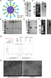
Figure 1
Figure 1
Generation and characterisation of the AP205-S1 vaccine. A Diagram of the way displaying S1 protein on surface of AP205-VLP via SpyTag and SpyCatcher specific bonds; B SDS-PAGE image of purified AP205-SpyCatcher (lane 1); C Agarose gel images of purified AP205-SpyCatcher (lane 1), left: UV light, right: Coomassie blue staining; D SDS-PAGE image of different molar ratios of AP205-SpyCatcher to S1-SpyTag, lane 1: AP205-SpyCatcher alone, lane 2: S1-SpyTag alone; lane 3: 1:2, lane 4: 1:1, lane 5: 2:1, lane 6: 3:1; E Western blot image of the generated AP205-S1 using monoclonal anti-AP205 antibody, lane 1: AP205-SpyCatcher alone, lane 2: S1-SpyTag alone; lane 3: AP205-S1 (1:1 reaction); F Agarose gel images of the formed AP205-S1 (lane 1), left: UV light, right: Coomassie blue staining; G Dynamic light scattering reports of AP205-SpyCatcher and formed AP205-S1; H TEM images of AP205-SpyCatcher and AP205-S1.
Figure 2
Figure 2
S1-specific IgG antibody responses in mouse sera after immunisation. A Mice immunization regimen. Serum and feces samples were collected weekly. B ELISA results of AP205-SpyCatcher, S1 or AP205-S1 immunised mice sera against S1 protein at different time points. Each point represented 5 mice, and the error bar was shown as ± SEM. C S1-specific IgG antibody titres of immunised sera at different time points. Each point represented one mouse, and the error bar was shown as ± SEM. D Plasma cell counts that secrete S1-specific IgG antibodies in immunised spleens at day 49. One well of spots was displayed in each group. P ≤ 0.05 was shown *, P ≤ 0.01 as **.
Figure 3
Figure 3
IFA images of immunised mice sera to recognise PEDV KB2013 viruses in vitro. A Day 49, mouse serum immunised with AP205-SpyCatcher. B, C mice sera immunised with S1 protein at days 28 (B) and 49 (C). D: Mouse serum immunised with AP205-S1 (day 28). DAPI-stained nucleus and FITC reflected mouse IgG antibodies bound to viruses. Serum samples at day 49 and day 28 were diluted 1:1000 or 1:100, respectively.
Figure 4
Figure 4
Avidity ELISA results of immunised mice sera against S1 protein. A ELISA curves of OD450nm values with diluted sera washed with(red) or without (black) 7 M urea. B Area under the curve values of avidity ELISA curves. P ≤ 0.05 was shown *, P ≤ 0.001 as ***.
Figure 5
Figure 5
S1-specific IgA antibody levels in immunised mice sera. A ELISA curves of day 28 and 49 sera. Each point represented 5 mice, and the error bar was shown as ± SEM. B S1-specific IgA titres of each group at days 28 and 49. P ≤ 0.05 was shown *, P ≤ 0.01 as **.
Figure 6
Figure 6
Immunised mice sera to neutralise PEDV (KB2013) viruses in vitro. A IFA images of day 49 sera immunised with AP205-SpyCatcher, S1 protein or AP205-S1 to block viruses from infecting Vero cells. DAPI-stained nucleus and FITC reflected viral N protein-specific antibodies derived from rabbit, i.e. the infected viruses. AP205-SpyCatcher immunised sera were diluted 1:10, and S1 protein and AP205-S1 immunised sera were diluted 1:100. B Neutralisation titres of immunised sera from day 21 to day 49. The dilution folds of sera to completely (100%) inhibit CPE in Vero cells were determined as titres. P ≤ 0.05 was shown *, P ≤ 0.01 as **.
Figure 7
Figure 7
Antibody responses in the intestinal mucosa of mice at day 42 after prime immunisation. A ELISA curves of S1-specific IgG antibodies in faecal supernatants. B IFA images of mouse faecal supernatants to bind to PEDV (KB2013) viruses in vitro. DAPI-stained nucleus and FITC reflected mouse IgG antibodies bound to viruses. C IFA images of mouse faecal supernatants to neutralise PEDV (KB2013) viruses from infecting Vero cells. DAPI-stained nucleus and FITC reflected viral N protein-specific antibodies derived from rabbit, i.e. the infected viruses. Faecal supernatants were not diluted. P ≤ 0.05 was shown *.
Figure 8
Figure 8
Recognition and neutralisation of immunised mice sera to PEDV AH2018-HF1 (G-I strain) virus. A IFA images of mouse sera at day 49 to bind to AH2018-HF1 viruses in vitro. DAPI-stained nucleus and FITC reflected mouse IgG antibodies bound to viruses. Serum samples were diluted 1:1000. B IFA images of mouse sera at day 49 to neutralise AH2018-HF1 viruses from infecting Vero cells. DAPI-stained nucleus and FITC reflected viral N protein-specific antibodies derived from rabbit, i.e. the infected viruses. Serum samples were diluted 1:10. C Neutralisation titres of immunised sera at day 49. The dilution folds of sera to completely (100%) inhibit CPE in cells caused by AH2018-HF1 infection were determined as titres. P ≤ 0.05 was shown *.
Figure 9
Figure 9
Mucosal antibodies from immunised mice to recognise and neutralise the PEDV AH2018-HF1 (G-I strain) virus. A IFA images of mouse faecal supernatants on day 42 to bind to AH2018-HF1 viruses in vitro. DAPI-stained nucleus and FITC reflected mouse IgG antibodies bound to viruses. B IFA images of mouse faecal supernatants on day 42 to neutralise AH2018-HF1 viruses from infecting Vero cells. DAPI-stained nucleus and FITC reflected viral N protein-specific antibodies derived from rabbit, i.e. the infected viruses. Faecal supernatants were not diluted.
Figure 10
Figure 10
Protection efficacy of AP205-S1 in piglets against PEDV infection after AP205-S1 immunisation. A Immunisation and challenge regimen of pig experiments. B S1-specific IgG titres in pig sera after challenge. C Neutralisation titres of pig sera after challenge. D IFA results of day 0 pig sera to bind to PEDV in vitro. Sera were diluted 1:1000. DAPI-stained nuclei and FITC reflected pig IgG antibodies bound to viruses. E Diarrhoea scores of piglets after challenge. F PEDV viral loads in pig rectal swabs after challenge.

Similar articles

References

    1. Jung K, Saif LJ, Wang QH (2020) Porcine epidemic diarrhea virus (PEDV): an update on etiology, transmission, pathogenesis. Virus Res 286:198045 - PMC - PubMed
    1. Li M, Pan YY, Xi Y, Wang M, Zeng QY (2023) Insights and progress on epidemic characteristics, genotyping, and preventive measures of PEDV in China: a review. Microb Pathog 181:106185 - PubMed
    1. Wang QH, Vlasova AN, Kenney SP, Saif LJ (2019) Emerging and re-emerging coronaviruses in pigs. Curr Opin Virol 34:39–49 - PMC - PubMed
    1. Turlewicz-Podbielska H, Pomorska-Mól M (2021) Porcine coronaviruses: overview of the state of the art. Virol Sin 36:833–851 - PMC - PubMed
    1. Lei JL, Miao YQ, Bi WR, Xiang CH, Li W, Zhang RT, Li Q, Yang ZQ (2024) Porcine epidemic diarrhea virus: etiology, epidemiology, antigenicity, and control strategies in China. Animals (Basel) 14:294 - PMC - PubMed

MeSH terms