

The effects of prone position on optic nerve sheath diameter and intraocular pressure in elective lumbar disc surgery
- PMID: 40597635
- PMCID: PMC12210980
- DOI: 10.1186/s12871-025-03204-w
The effects of prone position on optic nerve sheath diameter and intraocular pressure in elective lumbar disc surgery
Abstract
Background: The objective of this study was to determine the efficacy of intraocular pressure (IOP) measurements in diagnosing elevated increased intracranial pressure (ICP) by examining the correlation between optic nerve sheath diameter (ONSD) and IOP in the prone position.
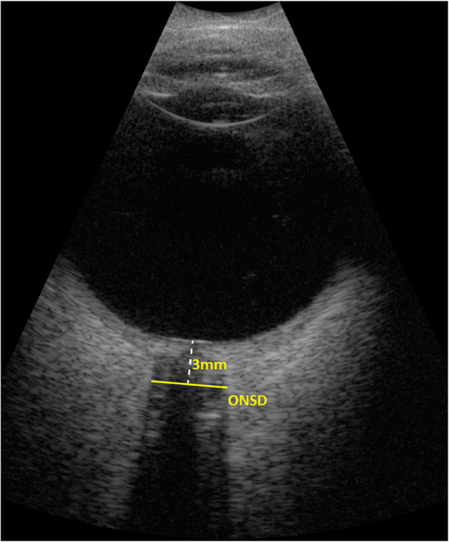
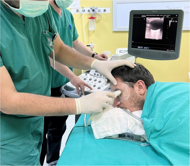
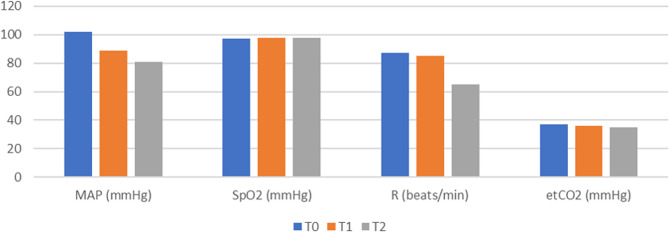
Methods: This prospective observational study included patients in the American Society of Anesthesiologists (ASA) 1–2 risk group, aged 18–65 years, who were scheduled for elective surgery. ONSD and IOP measurements were performed with ultrasound and Tonopen XL.
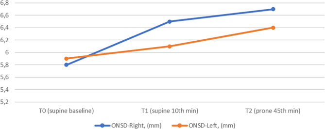
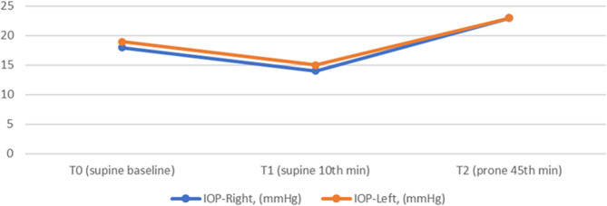
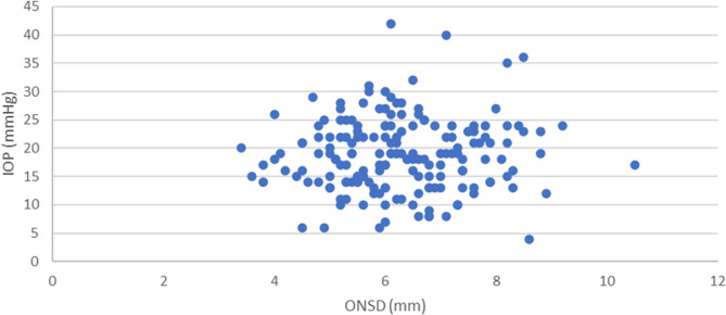
Results: Data from 59 patients were analysed. Spearman’s correlation analysis was used to determine the relationship between quantitative data. Significance was considered at p < 0.05. ONSD and IOP measurements were significantly higher in the prone position (p = 0.001). However, no correlation was found between ONSD and IOP measurements.
Conclusion: We found that ONSD and IOP measurements increased in the prone position, but these two measurements were not correlated. ONSD can be used in the diagnosis and follow-up of elevated ICP. Since there are many factors affecting IOP values, we concluded that IOP would not be effective in the diagnosis and follow-up of ICP.
Keywords: Intracranial pressure; Intraocular pressure; Optic nerve; Prone position.
Conflict of interest statement
Declarations. Ethics approval and consent to participate: Ethical approval was obtained from the Recep Tayyip Erdogan University Non-invasive Clinical Research Ethics Committee (01.09.2021-2021/155). This prospective observational study was conducted in the operating theatre of Recep Tayyip Erdogan University Training and Research Hospital between September 2021 and September 2022. The study was conducted within the guidelines of the World Medical Association’s Declaration of Helsinki. The study was conducted in accordance with the guidelines and regulations regarding human experimentation and/or the use of human tissue samples. Informed consent for participation was obtained from all participants in the study. Consent for publication: Not applicable. Competing interests: The authors declare no competing interests.
Figures
References
-
- Dubourg J, Javouhey E, Geeraerts T, Messerer M, Kassai B. Ultrasonography of optic nerve sheath diameter for detection of Raised intracranial pressure: a systematic review and meta-analysis. Intensive Care Med. 2011;37:1059–68. 10.1007/s00134-011-2224-2. - PubMed
-
- Sallam A, Abdelaal Ahmed Mahmoud M, Alkhatip A, Kamel MG, Hamza MK, Yassin HM, Hosny H, Younis MI, Ramadan E, Algameel HZ, Abdelhaq M, Abdelkader M, Mills KE, Mohamed H. The diagnostic accuracy of noninvasive methods to measure the intracranial pressure: A systematic review and Meta-analysis. Anesth Analg. 2021;132:686–95. 10.1213/ANE.0000000000005189. - PubMed
-
- Kristiansson H, Nissborg E, Bartek J, Andresen M, Reinstrup P, Romner B. Measuring elevated intracranial pressure through noninvasive methods: a review of the literature. J Neurosurg Anesthesiol. 2013;25:372–85. 10.1097/ANA.0B013E31829795CE. - PubMed
-
- Kristiansson H, Nissborg E, Bartek J, Andresen M, Reinstrup P, Romner B, Kristiansson H, Nissborg E, Bartek J Jr, Andresen M, Reinstrup P, Romner B. Reply to comment by Albin on measuring elevated intracranial pressure through noninvasive methods: a review of the literature. J Neurosurg Anesthesiol. 2014;26:407. 10.1097/ANA.0000000000000044. - PubMed
LinkOut - more resources
Full Text Sources
