. 2025 Jul 1;26(1):325.
doi: 10.1186/s12882-025-04222-z.

Inhibition of Axl attenuates acute kidney injury by alleviating inflammation via SOCS3 downregulation in tubular epithelial cells

Affiliations

Inhibition of Axl attenuates acute kidney injury by alleviating inflammation via SOCS3 downregulation in tubular epithelial cells

Xin Kang et al. BMC Nephrol. .

Abstract

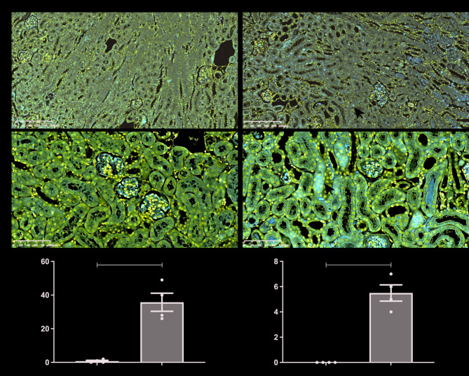
Background: In acute kidney injury (AKI), inflammatory crosstalk between tubular epithelial cells (TECs) and immune cells drives disease progression. Although the Axl-SOCS3 axis in myeloid cells typically suppresses inflammation, TEC-specific SOCS3 deletion paradoxically protects against AKI, suggesting a cell type-specific pro-inflammatory role.

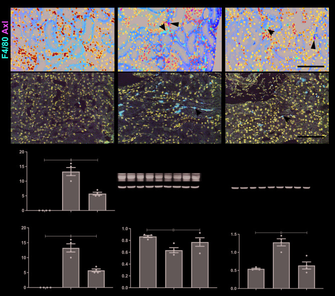
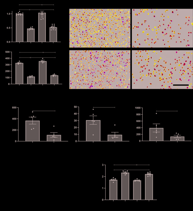
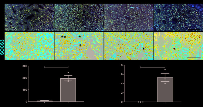
Methods: We induced AKI via bilateral ischemia/reperfusion (IRI) in mice. The Axl-specific pharmacological inhibitor R428 was administered via subcutaneous injection immediately post-IRI, with plasma and kidney samples collected 24 h later. To assess the effects of SOCS3 in TECs, small interfering RNA was used to silence SOCS3 in cisplatin injured HK2 cells. Axl/SOCS3 expression levels were assessed in human AKI biopsies.

Results: In AKI patients and IRI mice, Axl was upregulated in interstitial immune cells, while SOCS3 increased in TECs. Axl inhibition by R428 attenuated renal injury, reducing inflammatory infiltration, NF-κB p65 phosphorylation, and TEC SOCS3 expression. Notably, SOCS3 knockdown in TECs suppressed NF-κB activation and IL-1β/IL-6 production, implicating Axl-SOCS3 as a pro-inflammatory amplifier in AKI.

Conclusion: The Axl-SOCS3 axis exacerbates AKI by reinforcing NF-κB-driven inflammation in TECs, creating a vicious cycle between immune cells and TECs. Targeting this cross-cellular pro-inflammatory pathway offers a promising therapeutic strategy for AKI.

Keywords: Acute kidney injury; Axl; Inflammation; Suppressor of cytokine signaling 3; Tubular epithelial cells.


Conflict of interest statement

Declarations. Ethics approval and consent to participate: All animal experiments were approved by the Institutional Animal Care and Use Committee of Tianjin Medical University General Hospital (Approval Number: IRB2022-ZWFL-070). All patients provided written informed consent. The clinical protocols were performed in accordance with the principles of the Declaration of Helsinki and approved by the Ethics Committee of Tianjin Medical University General Hospital (Approval Number: IRB2020-YX-114-01). Consent for publication: Not applicable. Competing interests: The authors declare no competing interests.

Figures

Fig. 1
Fig. 1
Expression patterns of Axl and SOCS3 in kidneys following IRI-induced AKI. (A-B) Representative immunohistochemical staining of Axl (A) and SOCS3 (B) in renal tissues. Arrows indicate positively stained cells/tubules. Scale bars: 200 μm (A), 100 μm (B). (C) Quantitative analysis of Axl-positive cells/100× (n = 4 mice/group). (D) Quantitative analysis of SOCS3-positive tubules/200× (n = 4). Data are shown as mean ± SEM. ** P < 0.01
Fig. 2
Fig. 2
Effects of Axl inhibitor on mice with IRI-induced AKI. (A) PAS-stained kidney sections showing tubular injury. Scale bar, 200 μm. (B) Immunofluorescence co-staining of Axl (green) and E-cadherin (red). The arrows indicate the positive staining of Axl. Scale bar, 100 μm. (C) Semiquantitative renal pathological scores (n = 5). (D-E) Plasma Cr (D) and BUN (E) levels (n = 4). (F) Quantification of Axl-positive cells/200× (n = 4). (G) Representative Western blot of Axl expression. (H) Densitometric analysis of Axl/β-actin ratio (n = 6). Data are shown as mean ± SEM or median ± interquartile range (IQR). *P < 0.05, **p < 0.01, and P****Р<0.0001
Fig. 3
Fig. 3
Effects of Axl inhibitor on renal inflammation in mice with IRI-induced AKI. (A) Immunofluorescence co-staining of F4/80 (red) and Axl (green). Arrows indicate F4/80-positive macrophages. (B) Ly6G immunohistochemistry showing neutrophil infiltration. (C-D) Quantification of F4/80- (C) and Ly6G- (D) positive cells/400× (n = 4). (E-F) Western blot analysis (E) and quantification (F) of Arg-1 expression. (G-H) Western blot analysis (G) and quantification (G) of p-p65 expression (n = 4). Scale bar, 100 μm. Data are shown as mean ± SEM. **P < 0.01
Fig. 4
Fig. 4
Reduction of SOCS3 expression in TECs of IRI-induced AKI mice by Axl inhibition. (A) Immunofluorescence co-staining of SOCS3 (green) and Ecadherin (red). The arrows indicate the positive staining of SOCS3. Scale bar, 100 μm. (B) Quantification of SOCS3-positive cells/400× (n = 4). (C-D) Western blot analysis (C) and quantification (D) of SOCS3 expression (n = 4). Data are shown as mean ± SEM or median ± interquartile range (IQR). *P < 0.05
Fig. 5
Fig. 5
Impact of SOCS3 knockdown in cisplatin-treated HK2 cells. (A) Cell viability assessed by CCK-8 assay. (B) Representative Ki67 immunofluorescence (green). (C) Quantification of Ki67-positive cells/400× (n = 4). Scale bar, 100 μm. (D) The relative mRNA expression of SOCS3, IL-1β, and IL-6 determined by qRT-PCR (n = 5). (E-F) Western blot analysis (E) and quantification (F) of p-p65 levels (n = 6). Data are shown as mean ± SEM. *P < 0.05, **P < 0.01
Fig. 6
Fig. 6
Expressions of Axl and SOCS3 in kidneys of patients with AKI. (A) Representative Axl immunohistochemistry (400×). (B) SOCS3 immunofluorescence (400×). The arrows indicate the positive staining of Axl and SOCS3. Scar bar, 100 μm. (C-D) Quantification of Axl-positive cells (C) and SOCS3-positive tubules/400× (n = 1–3 patients). Data are shown as mean ± SEM. *P < 0.05

Similar articles

References

    1. Ronco C, Bellomo R, Kellum JA. Acute kidney injury. Lancet. 2019;394(10212):1949–64. - PubMed
    1. Bonventre JV, Yang L. Cellular pathophysiology of ischemic acute kidney injury. J Clin Invest. 2011;121(11):4210–21. - PMC - PubMed
    1. Markó L, Vigolo E, Hinze C, Park JK, Roël G, Balogh A, Choi M, Wübken A, Cording J, Blasig IE, et al. Tubular epithelial NF-κB activity regulates ischemic AKI. J Am Soc Nephrol. 2016;27(9):2658–69. - PMC - PubMed
    1. Zhu C, Wei Y, Wei X. AXL receptor tyrosine kinase as a promising anti-cancer approach: functions, molecular mechanisms and clinical applications. Mol Cancer. 2019;18(1):153. - PMC - PubMed
    1. Bian Q, Anderson JC, Zhang XW, Huang ZQ, Ebefors K, Nyström J, Hall S, Novak L, Julian BA, Willey CD, et al. Mesangioproliferative kidney diseases and Platelet-Derived growth Factor-Mediated AXL phosphorylation. Kidney Med. 2021;3(6):1003–e10131001. - PMC - PubMed

MeSH terms

LinkOut - more resources