Review
. 2025 Jul 1;26(1):316.
doi: 10.1186/s12882-025-04260-7.

The role and mechanism of TSC in kidney diseases: a literature review

Affiliations
Review

The role and mechanism of TSC in kidney diseases: a literature review

Qiulin Luo et al. BMC Nephrol. .

Abstract

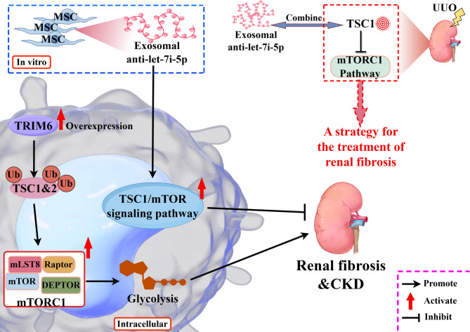
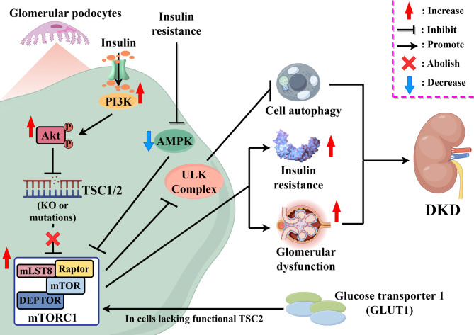
Background: Tuberous sclerosis complex (TSC) is an autosomal dominant genetic disorder characterized by multisystem involvement, primarily caused by loss-of-function mutations in the TSC1 or TSC2 genes. TSC is a key integrator of metabolic signaling and cellular stress and has become an important regulator in several kidney diseases. TSC1 and TSC2 can be used not only as genetic markers for disease diagnosis, but also as potential immunotherapeutic targets for kidney disease. Recent studies on the pathogenesis of TSC may provide guidance for developing new treatment strategies for kidney diseases.

Key messages: Therefore, we systematically reviewed the molecular biology of TSC and their signaling pathway, regulation of cell metabolism, and immune response in acute renal injury, chronic kidney disease, diabetic kidney disease, renal cysts, benign and malignant intrarenal tumors, and renal angiomyolipomas. We also summarize the efficacy and adverse effects of mTOR inhibitors in the treatment of TSC-related kidney diseases.

Keywords: Immunotherapeutic; Kidney diseases; Tuberous sclerosis complex; mTOR inhibitors.


Conflict of interest statement

Declarations. Ethical approval: None required for reviews. Informed consent to participate: It does not apply to reviews. Competing interests: The authors declare no competing interests.

Figures

Fig. 1
Fig. 1
Patterns of TSC1/2 role in kidney disease (by Figdraw). The deletion of TSC1/2 in the kidney results in a range of renal disease responses. TSC1/2 deletion results in the activation of the mTORC1 signaling pathway, which subsequently induces insulin resistance, increases the secretion of growth-promoting factors, and enhances the activity of TFE3 and TFEB. Concurrently, increased MITF expression promotes cell growth, invasion, and migration, consequently accelerating the progression of DKD, PKD, RCC, and RAML. In the context of AKI and CKD, the early stages of IRI are characterized by TSC1 deletion-induced macrophage polarization toward the M1 phenotype, which in turn leads to increased renal dysfunction. Conversely, in the repair stage of IRI, TSC1 deletion reduced macrophage polarization toward M2, thereby attenuating renal fibrosis. Additionally, the mTORC1-JNK signaling pathway, mitochondrial homeostasis, and glycolysis play a role in the occurrence and development of AKI and CKD. DKD: Diabetic kidney disease; PKD: Polycystic kidney disease; RCC: Renal cell carcinoma; RAML: Renal angiomyolipomas; AKI: Acute kidney injury; CKD: Chronic kidney disease; MITF: Microphthalmia transcription factor; TSC1/TSC2-|-: TSC1/TSC2 deletion; Fnip1/PKD1-|-; Fnip1/PKD1 deletion; Foxi1/TSC1 dKO: Foxi1/TSC1 double knockout; CAII/TSC1 dKO: CAII/TSC1 double knockout
Fig. 2
Fig. 2
Schematic diagram of the TSC1/2 signaling pathway (by Figdraw). TSC genes is localized in the cytoplasm and serves as a critical molecular hub that integrates the PI3K-Akt and mTOR signaling pathways. During cellular regulation, growth factors, such as insulin, activate PI3K, PI3K then phosphorylates PIP2 to PIP3. PIP3 phosphorylates and activates Akt through the intermediary action of the kinase PDK1. PI3K activity is negatively modulated by PTEN, a phosphatase that dephosphorylates PIP3 to PIP2. Akt regulates the function of TSC by phosphorylating multiple sites on the TSC2 subunit, thereby inhibiting its suppressive effect on mTORC1. Additionally, TSC negatively regulates mTORC1 by inhibiting Rheb, an activator of mTORC1. AMPK activates the TSC, thereby enhancing its inhibitory effect on mTOR. Upon binding to GTP, Rheb triggers mTORC1 activation, which initiates a series of phosphorylation reactions. This leads to p70S6K activation and 4EBP1 phosphorylation, promoting protein synthesis and cell proliferation. PI3K: Phosphoinositide 3-kinase; PIP2: Phosphatidylinositol (4,5)-bisphosphate; PIP3: Phosphatidylinositol (3,4)-bisphosphate; PTEN: Phosphatase and tensin homolog; PDK1: Pyruvate dehydrogenase kinase 1; Akt: Protein kinase B; Rheb: Ras homolog enriched in brain; GDP: Guanosine diphosphate; GTP: Guanosine triphosphate; AMPK: AMP-activated Protein Kinase; TSC1/TSC2: Tuberous sclerosis complex 1/2; mTORC1/2: Mechanistic target of rapamycin complex 1/2; 4EBP1: Eukaryotic initiation factor 4E-binding protein 1; S6K: Ribosomal S6 kinase; 70S6K: 70 kDa ribosomal S6 kinase; P: Phosphorylation
Fig. 3
Fig. 3
Schematic illustration of the molecular interactions and signaling pathways involved in the association between TSC and acute kidney injury development (by Figdraw). In mouse kidneys undergoing IRI or LPS-induced injury, deletion of TSC1 in fibroblasts caused activation of Rheb, which in turn activated the mTORC1 signaling pathway, causing a significant increase in the downstream abundance of p-S6, and consequently protecting renal tubular cells from acute injury. Meanwhile, EDN1 was upregulated in renal fibroblasts after TSC1 knockdown and was dependent on the mTORC1-JNK pathway, which improved renal function in LPS-induced AKI mice. In a cisplatin-induced mouse model of AKI, deletion of Rheb1 signaling in renal tubular cells resulted in tubular cell death, mitochondrial defects, and worsening of AKI. In the Lyz-TSC1 CKO mouse model, deletion of TSC1 promoted macrophage polarization toward M1 early in the disease, leading to increased renal impairment after IRI. In contrast, during the repair phase of IRI, TSC1 deficiency led to a reduction in macrophage polarization toward the M2 type, which attenuated renal fibrosis. IRI: Ischemia-reperfusion injury; LPS: Lipopolysaccharides; TSC1: Tuberous sclerosis complex 1; Rheb: Ras homolog enriched in brain; p-S6: Phospho-S6; mTORC1: Mechanistic target of rapamycin complex 1; EDN1: Endothelin−1; mTORC1-JNK: Mechanistic Target of Rapamycin Complex 1-c-Jun N-terminal kinase; AKI: Acute kidney injury; Lyz-TSC1 CKO: Myeloid cell-specific knockout of TSC1; Rheb1: Ras homolog enriched in brain 1
Fig. 4
Fig. 4
Schematic illustration of the molecular interactions and signaling pathways involved in the association between TSC and renal fibrosis and CKD development (by Figdraw). Overexpression of TRIM6 leads to increased ubiquitination levels of TSC1 and TSC2, thereby promoting activation of the mTORC1 pathway and exacerbating renal fibrosis. Additionally, TSC1-related mTORC1 signaling mediates the progression of renal interstitial fibrosis by regulating glycolysis in RTE cells. Notably, in an in vitro model of TGF-β-induced renal fibrosis, exosomes derived from mesenchymal stem cells (MSC) carrying anti-let−7i−5p activate the TSC1/mTOR signaling pathway, thereby alleviating renal fibrosis. Furthermore, in a UUO-induced renal fibrosis model, TSC1 serves as the binding target for the anti-let−7i−5p exosomes. TSC1 significantly impacts the progression of renal fibrosis by regulating the mTORC1 pathway, and targeting this pathway may offer a novel therapeutic strategy for renal fibrosis. TRIM6: Tripartite Motif Containing 6; TSC1/TSC2: Tuberous sclerosis complex 1/2; mTORC1: Mechanistic target of rapamycin complex 1; RET: Proximal tubular epithelial; TGF-β: Transforming Growth Factor β; UUO: unilateral ureteral obstruction; CKD: Chronic kidney disease; TSC1&2 Ub: TSC1&2 Ubiquitination
Fig. 5
Fig. 5
Schematic illustration of the molecular interactions and signaling pathways involved in the association between TSC and diabetic kidney disease development (by Figdraw). TSC is a key inhibitor of mTORC1. Abnormal activation of mTORC1 in glomerular podocytes leads to glomerular dysfunction, as well as induces insulin resistance and may promote the progression of diabetic kidney disease. In vitro, insulin-induced activation of PI3K-Akt signaling inhibits TSC1 or TSC2 signaling, which promotes activation of the mTORC1 pathway. Furthermore, in cells with knocked-out TSC1 or TSC2, the mTORC1 pathway was significantly activated, leading to the progression of diabetic kidney disease. When the AMPK pathway is inactivated, it activates the TSC-mTOR signaling pathway, which inhibits the ULK kinase complex and prevents autophagy initiation, thereby exacerbating diabetic kidney disease. TSC: Tuberous sclerosis complex; mTORC1: Mechanistic target of rapamycin complex 1; PI3K-Akt: Phosphoinositide 3-kinase-Protein kinase B; TSC1/TSC2: Tuberous sclerosis complex 1/2; AMPK: AMP-activated Protein Kinase; ULK: Unc−51 Like Autophagy Activating Kinase; DKD: Diabetic kidney disease
Fig. 6
Fig. 6
Schematic illustration of the molecular interactions and signaling pathways involved in the association between TSC and renal cysts and polycystic kidney disease development (by Figdraw). TSC1pt-KO mice exhibited proliferation and hypertrophy of renal proximal tubule cells with cystic degeneration. In vivo, metformin treatment resulted in increased AMPK phosphorylation; activation of pro-apoptotic factors BAD and cleaved caspase−3; and concomitant blockade of the pro-survival factor Akt. This inhibited the aberrant renal proliferation induced by TSC1 deletion, which was reversed by the AMPK inhibitor dosomorphine. In mouse RTE cells, knockdown of Fnip1 resulted in decreased AMPK activity and increased mTORC1 pathway activity. Notably, when Fnip1 and TSC1 were co-deleted, the mTORC1 signaling pathway was massively activated, leading to a significant acceleration in the development of PKD compared to deletion of Fnip1 or TSC1 alone. When PKD1 was knocked down in renal mesangial cells, the kidney was more likely to progress toward cystic degeneration.In addition, mutations in TSC1 and PKD1 in glomerular mesangial cells may exacerbate the development of renal cystic degeneration by inducing the secretion of growth-promoting factors, which promotes the proliferation of renal tubular cells. It is noteworthy that TSC1/Foxil-dKO and CAII/TSC1-dKO mice have a lower cystic load than TSC1-KO mice. TSC1pt-KO: Renal proximal tubule TSC1-specific knockout; AMPK: AMP-activated Protein Kinase; BAD: Bcl−2 Associated Agonist of Cell Death; Akt: Protein kinase B; TSC1: Tuberous sclerosis complex 1; RTE: Proximal renal tubular epithelial; Fnip1: Folliculin Interacting Protein 1; mTORC1: Mechanistic target of rapamycin complex 1; PKD: polycystic kidney disease; PKD1: Polycystic Kidney Disease 1; Foxil: Forkhead transcription factor 1; TSC1/Foxil-dKO: TSC1/Foxil double knockout; CAII: Carbonic anhydrase 2; CAII/TSC1-dKO: CAII/TSC1 double knockout; TSC1-KO: TSC1-knockout
Fig. 7
Fig. 7
Schematic illustration of the molecular interactions and signaling pathways involved in the association between TSC and renal cell carcinoma development (by Figdraw). In a mouse model with heterozygous deletion of TSC2 and spontaneous inactivation of the second allele, some cystadenomas developed into eosinophilic high-grade RCC as the mice aged. TSC2 gene deletion is a potential risk factor for the development of renal cystic lesions and renal cell carcinomas, especially in elderly patients. Activation of mTORC1 triggered by TSC2 deletion increases the activity of TFE3 and TFEB. At the molecular level, mTOR and TFE3/TFEB may be located in a mutually driven feed-forward signaling loop. In addition, GPNMB was highly expressed in TSC2-deficient mouse renal tumors and human renal cell lines, and GPNMB expression was regulated by mTOR kinase and TFEB/TFE3 in vitro. These findings provide new ideas for the diagnosis, typing and treatment of TSC1/2/mTOR-associated renal tumors. TSC1/2: Tuberous sclerosis complex 1/2; RCC: renal cell carcinoma; mTORC1: Mechanistic target of rapamycin complex 1; TFE3: Transcription Factor E3; TFEB: Transcription Factor EB; GPNMB: Glycoprotein nonmetastatic B
Fig. 8
Fig. 8
Schematic illustration of the molecular interactions and signaling pathways involved in the association between TSC and renal angiomyolipomas development (by Figdraw). In patients with TSC1 mutations, RAML usually occurs at a later age with relatively small lesion size and growth potential, whereas patients with TSC2 mutations are predisposed to develop RAML at an earlier age and have a higher probability of bleeding and hematuria complications. Studies suggest that human proximal renal tubular epithelial cells may be the origin of RAML. Interestingly, reduced expression of MITF negatively regulates RAML development by inhibiting cell growth, invasion, and migration. It can act by regulating CyR61, which suggests that CyR61 is a novel target for MITF to regulate RAML progression, and both MITF and CyR61 are expected to be potential targets for future clinical treatment of RAML. TSC1/2: Tuberous sclerosis complex 1/2; RAML: Renal angiomyolipomas; MITF: Microphthalmia transcription factors; CyR61: Cysteine-Rich Angiogenic Protein 61

References

    1. Eckardt K-U, Coresh J, Devuyst O, Johnson RJ, Köttgen A, Levey AS, et al. Evolving importance of kidney disease: from subspecialty to global health burden. Lancet. 2013;382:158–69. 10.1016/s0140-6736(13)60439-0. - PubMed
    1. Dare AJ, Fu SH, Patra J, Rodriguez PS, Thakur JS, Jha P. Renal failure deaths and their risk factors in India 2001–13: nationally representative estimates from the million death study. Lancet Global Health. 2017;5:e89–95. 10.1016/s2214-109x(16)30308-4. - PubMed
    1. Kurts C, Meyer-Schwesinger C. Protecting the kidney against autoimmunity and inflammation. Nat Rev Nephrol. 2019;15:66–8. 10.1038/s41581-018-0095-2. - PubMed
    1. Tang PM-K, Nikolic-Paterson DJ, Lan H-Y. Macrophages: versatile players in renal inflammation and fibrosis. Nat Rev Nephrol. 2019;15:144–58. 10.1038/s41581-019-0110-2. - PubMed
    1. Henske EP, Jóźwiak S, Kingswood JC, Sampson JR, Thiele EA. Tuberous sclerosis complex. Nat Reviews Disease Primers. 2016;2:16035. 10.1038/nrdp.2016.35. - PubMed

MeSH terms

Substances

LinkOut - more resources