

Comparison of cardiac magnetic resonance and speckle tracking echocardiography in cardiac evaluation of children with acute myocarditis with preserved left ventricular function
- PMID: 40597834
- PMCID: PMC12219550
- DOI: 10.1186/s12880-025-01772-4
Comparison of cardiac magnetic resonance and speckle tracking echocardiography in cardiac evaluation of children with acute myocarditis with preserved left ventricular function
Abstract
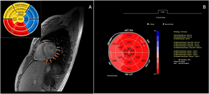
This study aimed to evaluate the reliability and efficacy of speckle tracking echocardiography (STE) compared to cardiac magnetic resonance (CMR) in assessing left ventricular function and segmental involvement in patients with acute myocarditis and preserved left ventricular systolic function. We analyzed conventional echocardiography, two-dimensional STE, and CMR findings in 33 pediatric patients (aged 0-18 years) diagnosed with acute myocarditis. The STE results were compared with CMR findings. The mean patient age was 14.67 years (± 2.88), with 13 (39.4%) females and 20 (60.6%) males. The mean ejection fraction (EF) was 68.54% (± 6.54), and the mean fractional shortening (FS) was 38.20% (± 5.34). All patients had an EF greater than 55%, with no detected wall motion abnormalities. Mild pleural effusion was observed in 4 (12.1%) patients. A significantly reduced left ventricular global longitudinal strain (LV-GLS) pattern was detected in 45.4% (n = 15) of patients, with an average LV-GLS value of -18.12% (± 3.5). The LV-GLS reduction was uniformly distributed across all left ventricular segments (LV-GLSAP2: -18.1 ± 3.85, LV-GLSAP3: -17.33 ± 4.34, LV-GLSAP4: -18.88 ± 4.20). STE measurements showed a mean left ventricular end-diastolic volume of 71.54 ± 24.41 and an end-systolic volume of 37.62 ± 16.42, with a mean EF of 48.52 ± 9.39%. CMR identified widespread myocardial contrast enhancement in 25 (75.7%) patients. When comparing STE to CMR, using an LV-GLS cut-off value of -18%, the sensitivity and specificity for diagnosing myocarditis were 52% and 63%, respectively. Lowering the cut-off to -16% reduced sensitivity to 40% but increased specificity to 75%. No significant association was found between decreased LV-GLS values (<-18%) and late gadolinium enhancement on CMR or regional edema (p > 0.05). Our findings suggest that STE is a valuable diagnostic tool for detecting cardiac involvement, particularly in focal cases of pediatric acute myocarditis with normal EF. While CMR remains the gold standard, STE provides a practical, accessible alternative for monitoring disease progression in suspected myocarditis cases.
Keywords: Cardiac magnetic resonance imaging; Myocarditis; Speckle tracking echocardiography.
© 2025. The Author(s).
Conflict of interest statement
Declarations. Ethics approval and consent to participate: The study was conducted in accordance with the principles of the Declaration of Helsinki and was approved by the Ethics in Research Committee of the School of Medicine of the Celal Bayar University of Manisa (11.11.2020/ 20.478.486). All protocols were carried out by following the guidelines of Declaration of Helsinki. All participants or their guardians were informed of the specific details of the study and signed the informed consents before enrollment. Consent for publication: Not Applicable. Competing interests: The authors declare no competing interests.
Figures
Similar articles
-
Reference Ranges of Left Ventricular Strain Measures by Two-Dimensional Speckle-Tracking Echocardiography in Children: A Systematic Review and Meta-Analysis.J Am Soc Echocardiogr. 2016 Mar;29(3):209-225.e6. doi: 10.1016/j.echo.2015.11.016. Epub 2015 Dec 30. J Am Soc Echocardiogr. 2016. PMID: 26747685 Free PMC article.
-
Artificial Intelligence Performance in Cardiac Magnetic Resonance Strain Analysis for Aortic Stenosis: Validation with Echocardiography and Healthy Controls.Medicina (Kaunas). 2025 May 22;61(6):950. doi: 10.3390/medicina61060950. Medicina (Kaunas). 2025. PMID: 40572638 Free PMC article.
-
Validating real-time three-dimensional echocardiography against cardiac magnetic resonance, for the determination of ventricular mass, volume and ejection fraction: a meta-analysis.Clin Res Cardiol. 2024 Mar;113(3):367-392. doi: 10.1007/s00392-023-02204-5. Epub 2023 Apr 20. Clin Res Cardiol. 2024. PMID: 37079054 Free PMC article.
-
Assessing Left Ventricular Pathology in Patients with Ebstein Anomaly Using Cardiovascular Magnetic Resonance: Looking Past the Right Heart.Pediatr Cardiol. 2025 Aug;46(6):1676-1683. doi: 10.1007/s00246-024-03585-8. Epub 2024 Jul 20. Pediatr Cardiol. 2025. PMID: 39033244
-
Global Longitudinal Strain and Cardiac Events in Patients With Immune Checkpoint Inhibitor-Related Myocarditis.J Am Coll Cardiol. 2020 Feb 11;75(5):467-478. doi: 10.1016/j.jacc.2019.11.049. J Am Coll Cardiol. 2020. PMID: 32029128 Free PMC article.
References
-
- Biesbroek PS, Beek AM, Germans T, Niessen HW, van Rossum AC. Diagnosis of myocarditis: current state and future perspectives. Int J Cardiol. 2015;191:211–9. - PubMed
-
- Caforio AL, Pankuweit S, Arbustini E, Basso C, Gimeno-Blanes J, Felix SB, et al. Current state of knowledge on aetiology, diagnosis, management, and therapy of myocarditis: a position statement of the European society of cardiology working group on myocardial and pericardial diseases. Eur Heart J. 2013;34:2636–48. (48a-48d). - PubMed
-
- Sengupta PP, Korinek J, Belohlavek M, Narula J, Vannan MA, Jahangir A, Khandheria BK. Left ventricular structure and function: basic science for cardiac imaging. J Am Coll Cardiol. 2006;48:1988–2001. - PubMed
Publication types
MeSH terms
LinkOut - more resources
Full Text Sources
Medical
Research Materials
Miscellaneous
