

OCTA-based assessment of macular and peripapillary vessel changes during the menstrual cycle
- PMID: 40597908
- PMCID: PMC12220553
- DOI: 10.1186/s12886-025-04204-7
OCTA-based assessment of macular and peripapillary vessel changes during the menstrual cycle
Abstract
Background: To investigate the effect of menstrual cycle-related hormonal fluctuations on retinal and choroidal microvasculature using swept-source optical coherence tomography angiography (SS-OCTA).
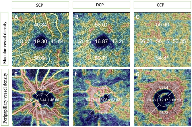
Methods: In this prospective study, a cohort of 31 healthy women with regular menstrual cycles was enrolled. SS-OCTA imaging was performed during three standardized menstrual phases: early follicular (day 3), ovulatory (day 14), and mid-luteal (day 21). Macular and peripapillary regions were evaluated using 3 × 3 mm and 4.5 × 4.5 mm scan protocols, respectively. Vessel density (VD) in the superficial capillary plexus (SCP), deep capillary plexus (DCP), and choriocapillaris (CC) was automatically quantified using the device's software. Choroidal thickness (CT) was manually measured in all quadrants, and the foveal avascular zone (FAZ) area was manually delineated by two independent graders. To control for diurnal variation, all measurements were conducted between 11:00 a.m. and 12:00 p.m.
Results: A statistically significant decrease in macular CT was observed across the menstrual phases (p = 0.002), along with significant variations in peripapillary CT in the temporal and inferior quadrants (p = 0.004 for both). VD in the peripapillary region showed significant differences in the superior quadrant of the SCP (p = 0.040), the inferior quadrant of the DCP (p = 0.008), and in the temporal and inferior quadrants of the CC (p = 0.011 and p = 0.007, respectively). In the macula, CC-VD in the inferior quadrant differed significantly (p = 0.030). FAZ area remained stable throughout the cycle. Post-hoc analysis revealed significant differences between the early follicular, ovulatory, and mid-luteal phases.
Conclusion: Hormonal fluctuations throughout the menstrual cycle appear to influence both retinal and choroidal microvasculature, particularly in the choriocapillaris and peripapillary regions. These findings underscore the importance of considering menstrual phase when interpreting OCTA measurements in women of reproductive age, to improve diagnostic accuracy and consistency in clinical and research settings.
Keywords: Menstrual cycle; Optical coherence tomography angiography; Retinal microvascular structure.
© 2025. The Author(s).
Conflict of interest statement
Declarations. Ethics approval and consent to participate: The study protocol received ethical approval from the Hamidiye Ethics Committee at the University of Health Sciences and adhered to the tenets of the Declaration of Helsinki. Written informed consent was provided from all participants enrolled in the study. Competing interests: The authors declare no competing interests.
Figures
Similar articles
-
Comparison of microvasculature between neovascular, non-neovascular chronic central serous chorioretinopathy cases and asymptomatic fellow eyes using swept-source optical coherence tomography angiography.Int Ophthalmol. 2025 Jul 18;45(1):297. doi: 10.1007/s10792-025-03673-5. Int Ophthalmol. 2025. PMID: 40679632
-
Longitudinal Changes in Macular Vessel Density in High Myopia on Optical Coherence Tomography Angiography.Ophthalmic Res. 2025;68(1):342-351. doi: 10.1159/000543975. Epub 2025 May 19. Ophthalmic Res. 2025. PMID: 40388900
-
Analyzing the Impact of a New β3 Adrenergic Agonist on Chorioretinal and Peripapillary Vessel Density.Neurourol Urodyn. 2025 Sep;44(7):1503-1511. doi: 10.1002/nau.70108. Epub 2025 Jun 25. Neurourol Urodyn. 2025. PMID: 40566846
-
Macular Optical Coherence Tomography Angiographic Study in Children with a History of Prematurity: A Systematic Review and Meta-Analysis.Curr Eye Res. 2025 Feb;50(2):111-123. doi: 10.1080/02713683.2024.2397034. Epub 2024 Aug 29. Curr Eye Res. 2025. PMID: 39210511
-
Optical coherence tomography angiography measurements in multiple sclerosis: a systematic review and meta-analysis.J Neuroinflammation. 2023 Mar 27;20(1):85. doi: 10.1186/s12974-023-02763-4. J Neuroinflammation. 2023. PMID: 36973708 Free PMC article.
References
-
- Pašalić E, Tambuwala MM, Hromić-Jahjefendić A, Endometriosis. Classification, pathophysiology, and treatment options. Pathol Res Pract. 2023;251:154847. 10.1016/j.prp.2023.154847. - PubMed
-
- Constantini NW, Dubnov G, Lebrun CM. The menstrual cycle and sport performance. Clin Sports Med. 2005;24(2):e51–xiv. 10.1016/j.csm.2005.01.003. - PubMed
-
- Toker E, Yenice O, Akpinar I, et al. The influence of sex hormones on ocular blood flow in women. Acta Ophthalmol Scand. 2003;81(6):617–24. 10.1111/j.1395-3907.2003.00160.x. - PubMed
-
- Cavdar E, Ozkaya A, Alkin Z, et al. Changes in tear film, corneal topography, and refractive status in premenopausal women during menstrual cycle. Cont Lens Anterior Eye. 2014;37(3):209–12. 10.1016/j.clae.2013.11.005. - PubMed
-
- Akar ME, Taskin O, Yucel I, et al. The effect of the menstrual cycle on optic nerve head analysis in healthy women. Acta Ophthalmol Scand. 2004;82(6):741–5. 10.1111/j.1600-0420.2004.00351.x. - PubMed
MeSH terms
LinkOut - more resources
Full Text Sources
Miscellaneous
