

Early dynamics of Toxoplasma gondii infection in sheep inoculated at mid-gestation with archetypal type II oocysts
- PMID: 40598310
- PMCID: PMC12218951
- DOI: 10.1186/s13567-025-01557-1
Early dynamics of Toxoplasma gondii infection in sheep inoculated at mid-gestation with archetypal type II oocysts
Abstract
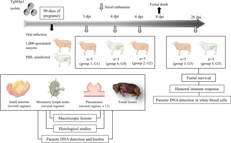
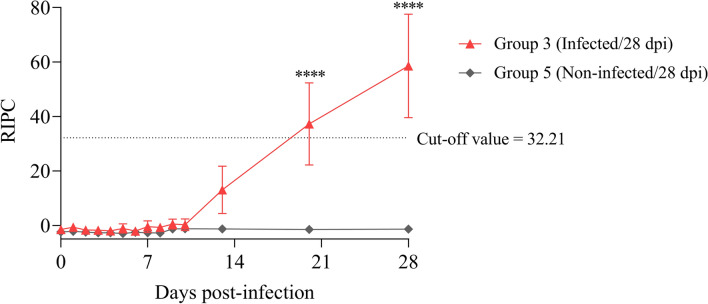
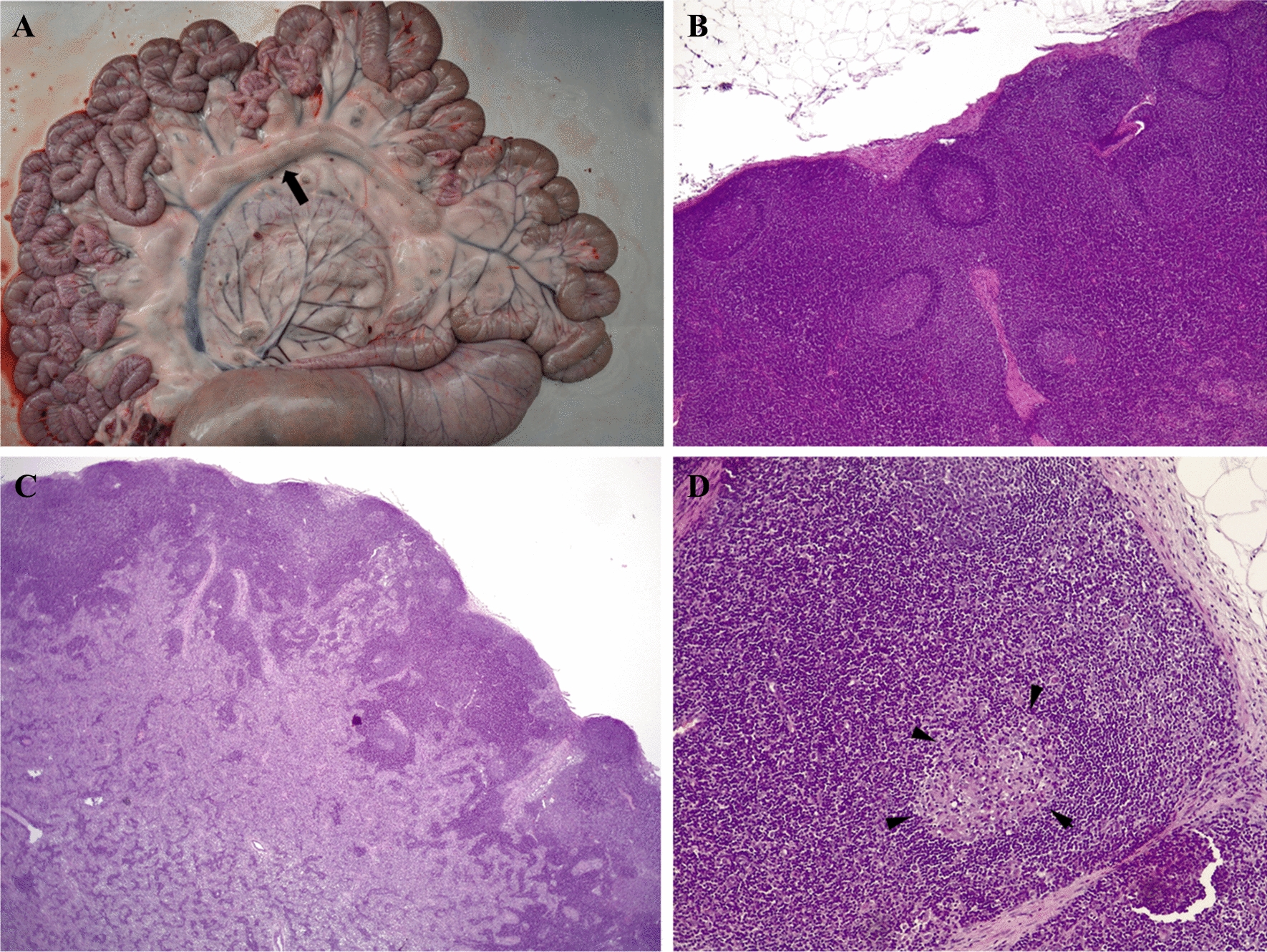
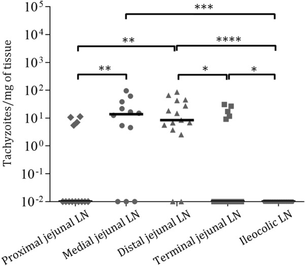
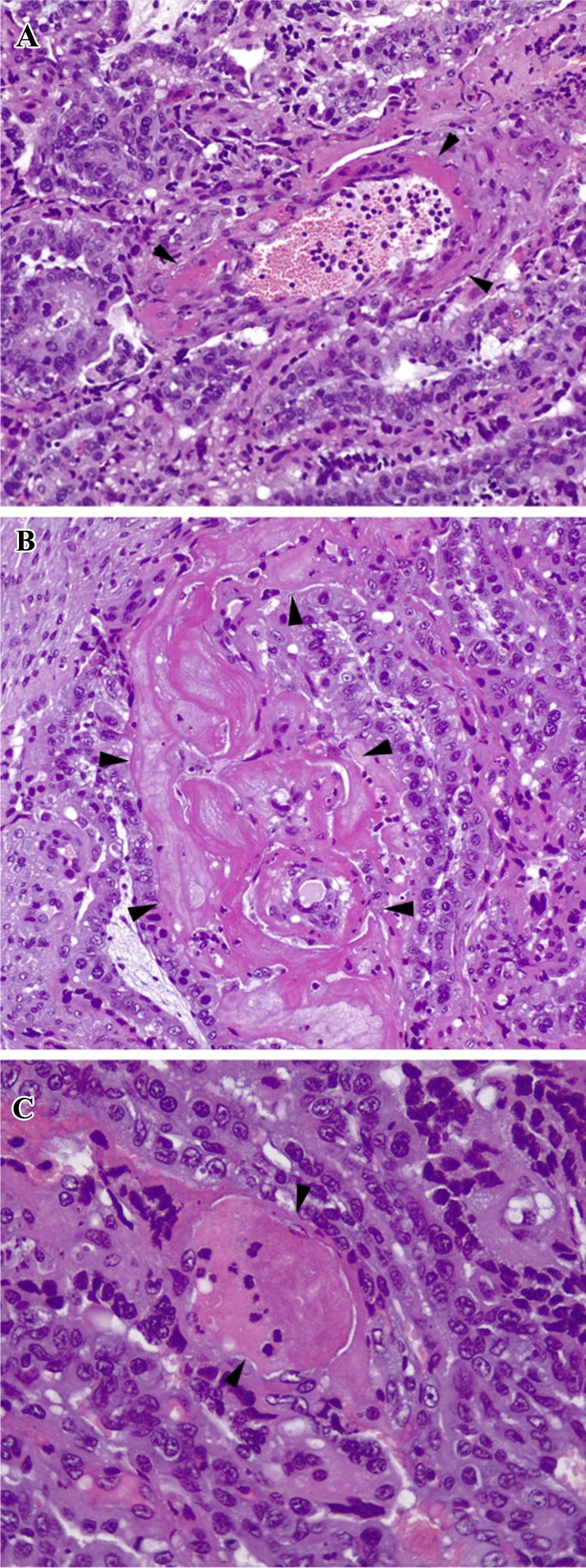
Early abortion is a clinical presentation of ovine toxoplasmosis that occurs in the second week post-infection (pi), which is characterised by placental infarcts, foetal leukomalacia and absence of parasites in the placenta and foetal tissues. The pathogenic mechanism of early abortion is unknown, and descriptions of the early dynamics of T. gondii infection in pregnant sheep are scarce. The aim of this study was to investigate the presence of lesions and parasite DNA in the small intestine, mesenteric lymph nodes and placenta/foetus, that could be key during the first week after oral infection in sheep at mid-pregnancy. In the small intestine, lesions were rare and parasite DNA detection rates were low (3-8%), with the highest parasite DNA detection and burden found on day 6 pi in the Peyer's patches of the medial jejunum. In the mesenteric lymph nodes, adenomegaly and microscopic lesions were mainly observed on day 6 pi. Parasite DNA was detected in 11% and 61.2% of the samples from mesenteric lymph nodes on days 3 and 6 pi, respectively, with higher parasite DNA detection rates and burdens in the medial and distal jejunal lymph nodes on day 6 pi. In the placentomes, on day 6 pi, gross lesions were not observed, although significant histological changes, such as endothelial activation and vascular thrombosis, were found in 18.6% and 8.3% of the placentomes, respectively. These findings lay the groundwork for future research aimed at elucidating the precise mechanisms underlying early abortions following T. gondii infection in pregnant sheep.
Keywords: Toxoplasma gondii; early abortion; early infection dynamics; mesenteric lymph nodes; mid-gestation; placental thrombosis; sheep; small intestine.
© 2025. The Author(s).
Conflict of interest statement
Declarations. Competing interests: The authors declare that they have no competing interests.
Figures

Similar articles
-
Placental thrombosis in acute phase abortions during experimental Toxoplasma gondii infection in sheep.Vet Res. 2014 Jan 29;45(1):9. doi: 10.1186/1297-9716-45-9. Vet Res. 2014. PMID: 24475786 Free PMC article.
-
Toxoplasma gondii parasites induce a localized myeloid cell immune response surrounding parasites in the brain during acute infection.mBio. 2025 Jul 9;16(7):e0081025. doi: 10.1128/mbio.00810-25. Epub 2025 Jun 10. mBio. 2025. PMID: 40492741 Free PMC article.
-
Preliminary seroprevalence study of zoonotic abortigenic agents in the abortion inexperienced sheep population in the Northern Cyprus.Vet Res Commun. 2025 Jun 10;49(4):219. doi: 10.1007/s11259-025-10790-0. Vet Res Commun. 2025. PMID: 40493315 Free PMC article.
-
Antiretrovirals for reducing the risk of mother-to-child transmission of HIV infection.Cochrane Database Syst Rev. 2011 Jul 6;(7):CD003510. doi: 10.1002/14651858.CD003510.pub3. Cochrane Database Syst Rev. 2011. PMID: 21735394
-
Antiretrovirals for reducing the risk of mother-to-child transmission of HIV infection.Cochrane Database Syst Rev. 2007 Jan 24;(1):CD003510. doi: 10.1002/14651858.CD003510.pub2. Cochrane Database Syst Rev. 2007. Update in: Cochrane Database Syst Rev. 2011 Jul 06;(7):CD003510. doi: 10.1002/14651858.CD003510.pub3. PMID: 17253490 Updated.
References
-
- Dubey JP (2021) Toxoplasmosis of Animals and Humans. CRC Press, Boca Raton, Florida, USA
-
- Gutiérrez-Expósito D, Tejerina F, Gutiérrez J, Fernández-Escobar M, Ortega-Mora LM, Mantecón AR, Dagleish MP, Pérez V, Benavides J (2021) Direct economic losses of Toxoplasma gondii abortion outbreaks in two Spanish sheep flocks. Vet Parasitol Reg Stud Reports 26:100623 - PubMed
-
- Vallejo R, Benavides J, Arteche-Villasol N, Sánchez-Sánchez R, Calero-Bernal R, Ferreras MC, Criado M, Pérez V, Ortega-Mora LM, Gutiérrez-Expósito D (2023) Experimental infection of sheep at mid-pregnancy with archetypal type II and type III Toxoplasma gondii isolates exhibited different phenotypic traits. Vet Parasitol 315:109889 - PubMed
MeSH terms
Substances
Grants and funding
LinkOut - more resources
Full Text Sources
Research Materials
Miscellaneous
