. 2025 Jul 1;18(1):69.
doi: 10.1186/s13045-025-01715-0.

CAR-T cells targeting CCR9 and CD1a for the treatment of T cell acute lymphoblastic leukemia

Affiliations

CAR-T cells targeting CCR9 and CD1a for the treatment of T cell acute lymphoblastic leukemia

Néstor Tirado et al. J Hematol Oncol. .

Abstract

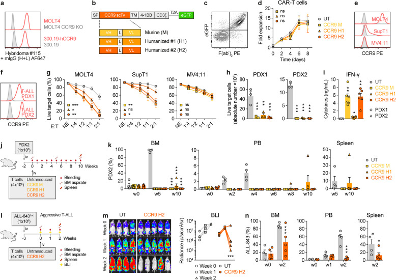
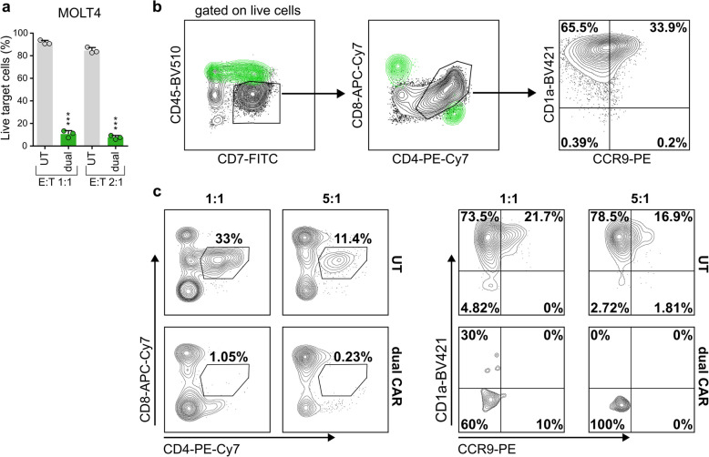
T cell acute lymphoblastic leukemia (T-ALL) is an aggressive malignancy characterized by high rates of induction failure and relapse, and effective targeted immunotherapies are lacking. Despite promising clinical progress with genome-edited CD7-directed CAR-T cells, which present significant logistical and regulatory issues, CAR-T cell therapy in T-ALL remains challenging due to the shared antigen expression between malignant and healthy T cells. This can result in CAR-T cell fratricide, T cell aplasia, and the potential for blast contamination during CAR-T cell manufacturing. Recently described CAR-T cells target non-pan-T antigens, absent on healthy T cells but expressed on specific T-ALL subsets. These antigens include CD1a (NCT05679895), which is expressed in cortical T-ALL, and CCR9. We show that CCR9 is expressed on >70% of T-ALL patients (132/180) and is maintained at relapse, with a safe expression profile in healthy hematopoietic and non-hematopoietic tissues. Further analyses showed that dual targeting of CCR9 and CD1a could benefit T-ALL patients with a greater blast coverage than single CAR-T cell treatments. We therefore developed, characterized, and preclinically validated a novel humanized CCR9-specific CAR with robust and specific antileukemic activity as a monotherapy in vitro and in vivo against cell lines, primary T-ALL samples, and patient-derived xenografts. Importantly, CCR9/CD1a dual-targeting CAR-T cells showed higher efficacy than single-targeting CAR-T cells, particularly in T-ALL cases with phenotypically heterogeneous leukemic populations. Dual CD1a/CCR9 CAR-T therapy may prevent T cell aplasia and obviate the need for allogeneic transplantation and regulatory-challenging genome engineering approaches in T-ALL.


Conflict of interest statement

Declarations. Competing interests: PM is a cofounder of OneChain Immunotherapeutics, a spin-off company from the Josep Carreras Leukemia Research Institute which has licensed the CCR9 binder (PCT/EP2024/053734). The remaining authors report no conflicts of interest in this work.

Figures

Fig. 1
Fig. 1
CCR9 is a safe and specific target for T-ALL. a Flow cytometry analysis of CCR9 in patient-matched leukemic blasts and normal (n) CD4+ and CD8+ T cells in 180 T-ALL patients. Left panels, representative flow cytometry plot. b, c Expression of CCR9 in the cohort, with T-ALL samples classified by developmental stage (b) or disease stage, at diagnosis (Dx) or relapse (Rel) (c). Three cases with Dx-Rel matched samples are color-coded. d UMAP representation showing organ/tissue annotation and CCR9 expression levels in 483,152 cells from human healthy tissues (Tabula Sapiens scRNAseq dataset). e Violin plots for CCR9 expression levels across tissues identified in (d). f-h CCR9 expression in the indicated leukocyte populations in relevant hematological tissues: thymus (f, n=4), PB (g, n =18), and BM (h, n=13)
Fig. 2
Fig. 2
CCR9 CAR-T cells are highly effective against T-ALL. a Anti-CCR9 hybridoma specifically stains CCR9-expressing cells. b Cartoon of second-generation CAR constructs. Three scFv versions were generated: scFv derived from the murine (M) hybridoma, and two humanized candidates (H1 and H2). SP, signal peptide; VH and VL, heavy and light chains; L, linker; TM, CD8 transmembrane domain. c Representative flow cytometry plot showing successful detection of transduced CAR-T cells as measured by co-expression of surface F(ab’)2 (scFv) and eGFP reporter. d Proliferation curves for UT and the indicated CCR9 CAR-transduced T cells (n=3). e CCR9 expression in the target T-ALL cell lines MOLT4 (CCR9high) and SupT1 (CCR9dim) and the CCR9neg AML cell line MV4;11. Isotype control stainings in gray. f CCR9 expression in two T-ALL PDXs. g 24 h cytotoxicity mediated by the different murine and humanized CCR9 CAR-T cells against the indicated cell lines at different effector:target (E:T) ratios (n=5). NE, no effector T cells. h Absolute numbers of live target PDX cells after co-culture with UT or the indicated CCR9 CAR-T cells for 24 h at an E:T ratio of 1:1 (n=3). i IFN-γ production by the indicated CCR9 CAR-T cells upon 24 h co-culture with PDXs (E:T 1:1, n=3 per PDX). j In vivo experimental design for the assessment of the efficacy of the three indicated CCR9 CAR-T cells against a T-ALL PDX (PDX2, n=4–6 mice/group). k Flow cytometry follow-up of tumor burden in BM, PB, and spleen in the different treatment groups indicated in (j). l In vivo experimental design for the assessment of the efficacy of the selected CCR9 H2 CAR-T cells against a highly aggressive Luciferase-bearing T-ALL PDX (ALL-843, n=4–5 mice/group). m Weekly bioluminescence imaging of mice. Left panel, bioluminescence images. Right panel, bioluminescence quantification. n Flow cytometry follow-up of tumor burden in BM, PB, and spleen after treatment with UT or CCR9 H2 CAR-T cells. Plots show mean ± SEM
Fig. 3
Fig. 3
Co-expression of CCR9 and CD1a in T-ALL and molecular strategies for dual targeting with CAR-T cells. a Flow cytometry analysis of CCR9 and CD1a expression in blasts from 180 T-ALL primary samples, 20% expression cut-off was set to define positivity for each marker. b CCR9 and CD1a immunophenotypes in 24 representative T-ALL patient samples. Cut-off thresholds for antigen positivity were determined using isotype controls. c Cartoons of all CAR constructs tested. Top panels, eight different configurations of tandem CAR constructs with different arrangements of the humanized scFvs (CCR9-CD1a vs. CD1a-CCR9 and VH-VL vs. VL-VH). Bottom panels, four distinct configurations of bicistronic CAR constructs (CCR9-CD1a vs. CD1a-CCR9). Different signal peptides derived from CD8α (SP1), human IgG1 (SP2), and murine IgG1 (SP3) were used for bicistronic CARs. The CCR9 H2 scFv was used in all versions. d Transduction efficiencies of single, co-transduced, tandem, and bicistronic CAR-T cells. e Cytotoxicity assays comparing the specificity and efficiency of the different single, co-transduced, tandem, and bicistronic CAR-T cells against combinatorial phenotypes of MOLT4 cells at a 1:1 E:T ratio after 24 h of co-culture (n=3–6). UT T cells were used as controls. Plots show mean ± SEM
Fig. 4
Fig. 4
Efficacy of CAR-T cells co-transduced with single CAR vectors. a Flow cytometry analysis of MOLT4 cells CRISPR/Cas9-engineered to express combinatorial CCR9/CD1a phenotypes (+/+, –/+, +/–, –/–).b In vitro cytotoxicity assays of the different phenotypes of MOLT4 cells with single CAR (CD1a H or CCR9 H2) T cells or CCR9/CD1a dual-targeting CAR-T cells at different E:T ratios after 24 h of co-culture (n=3). c In vivo experimental design for the assessment of the efficacy of CCR9- and CD1a-targeting CAR-T cells against a CCR9+CD1a+ T-ALL PDX (PDX2) (n=8–14 mice/group). d Flow cytometry follow-up of tumor burden in PB, spleen, and BM after treatment with the indicated CAR treatments. Frequencies of relapsing mice (>1% blasts) for each tissue are indicated. e Expression of CCR9 and CD1a in CAR-T-resistant blasts. Plots show mean ± SEM
Fig. 5
Fig. 5
Superior efficacy of dual CCR9 and CD1a CAR-T cells for the treatment of T-ALL with phenotypically heterogeneous leukemic populations. a Combinatorial phenotypes (CCR9+CD1a+, CCR9+CD1a, CCR9CD1a+) of T-ALL cells were mixed at a ratio of 1:1:1 to reproduce phenotypically heterogenous leukemic samples. b Relative (left) and absolute (right) numbers of live mixed target cells after a time-course cytotoxicity with UT, single CAR (CCR9 H2 or CD1a H) or CCR9/CD1a dual-targeting CAR-T cells at a 1:1 E:T ratio (n=3). c Cytokine production by the indicated CAR-T cells upon 24 h co-culture with target cells (n=3). d In vivo experimental design for the assessment of CCR9- and CD1a-targeting dual CAR-T cells against phenotypically heterogeneous Luc-bearing T-ALL target cells (n=6 mice/group). e Weekly bioluminescence imaging of mice (n=6 mice/group). Left panel, bioluminescence (BLI) images. Right panel, BLI quantification. f Flow cytometry analysis of BM tumor burden at the endpoint. Plots show mean + SEM
Fig. 6
Fig. 6
Patient-derived dual CCR9/CD1a CAR-T cells effectively eliminate leukemic cells in vitro. a Cytotoxicity of patient-derived CCR9/CD1a dual CAR-T cells against MOLT4 cells was assessed by flow cytometry. Target cells were pre-stained with eFluor 670 dye and exposed to either UT or dual CAR-T for 48 h at the indicated E:T ratio. The bars show the percentage of eFluor670+7−AAD cells (n=3). Plots show mean + SD. P values were calculated using a paired t-test (***p<0.001). b FACS showing the phenotype of the T-ALL sample selected for autologous CAR-T cells generation. The black population are blasts (CD45dimCD7+CD4/CD8+CD1a++CCR9low/+). The cells depicted in green are normal single CD4+ or single CD8+ T cells that were used for transduction (following CD1a+ cells depletion using the autoMACS separator). (c) FACS analysis of the cytotoxic assay. The total bone marrow-derived T-ALL sample was exposed to autologous UT or dual CAR-T cells for 24 h at a given E:T ratio. Contour plots show the percentages of CD4/CD8-double-positive blasts (gated on CD3 live cells) (left panel) and the CCR9/CD1a levels in both treatment groups (right panel)

References

    1. Litzow MR, Ferrando AA. How I treat T-cell acute lymphoblastic leukemia in adults. Blood. 2015;126:833–41. - PubMed
    1. Vora A, et al. Treatment reduction for children and young adults with low-risk acute lymphoblastic leukaemia defined by minimal residual disease (UKALL 2003): a randomised controlled trial. Lancet Oncol. 2013;14:199–209. - PubMed
    1. Schrappe M, et al. Late MRD response determines relapse risk overall and in subsets of childhood T-cell ALL: results of the AIEOP-BFM-ALL 2000 study. Blood. 2011;118:2077–84. - PubMed
    1. Marks DI, et al. T-cell acute lymphoblastic leukemia in adults: clinical features, immunophenotype, cytogenetics, and outcome from the large randomized prospective trial (UKALL XII/ECOG 2993). Blood. 2009;114:5136–45. - PMC - PubMed
    1. Fielding AK, et al. Outcome of 609 adults after relapse of acute lymphoblastic leukemia (ALL); an MRC UKALL12/ECOG 2993 study. Blood. 2007;109:944–50. - PubMed

LinkOut - more resources