. 2025 Jul 1;16(1):331.
doi: 10.1186/s13287-025-04439-7.

Tensile stress promotes the chondrogenic ability of condylar cartilage stem/progenitor cells in the temporomandibular joint via the Piezo1-Ca2+-Prkca pathway

Affiliations

Tensile stress promotes the chondrogenic ability of condylar cartilage stem/progenitor cells in the temporomandibular joint via the Piezo1-Ca2+-Prkca pathway

Wuyi Gong et al. Stem Cell Res Ther. .

Abstract

Background: Tensile force is a key regulator for condylar cartilage remodeling in the temporomandibular joint (TMJ), and this biomechanical characteristic underlies the mechanisms of mandibular growth modification in orthodontic practice. Cartilage stem/progenitor cells (CSPCs) in the superficial layer of condylar cartilage play an essential part in the development and remodeling of condylar cartilage. However, the regulatory role of tensile force on condylar CSPCs remains unclear. This study aimed to investigate the impact of tensile loading on condylar CSPCs and explore the molecular mechanisms within.

Methods: The mandibular advancement (MA) model was constructed to apply tensile force on the condylar cartilage in vivo. Flow cytometry and transcriptome sequencing were utilized to assess the percentage of CSPCs and gene expression in the superficial layer of rat condylar cartilage. Lineage tracing with cathepsin K (Ctsk) in mice was employed to trace the differentiation of CSPCs. 10% equibiaxial dynamic strain was loaded on rat CSPCs for cell stretching in vitro. GsMTx4 was used to inhibit the Piezo1 channel, and the calcium chelating agent BAPTA was used to block the Ca2+ influx of rat CSPCs. siRNA was applied to knock down the protein kinase C alpha (Prkca) of rat CSPCs in vitro and in vivo.

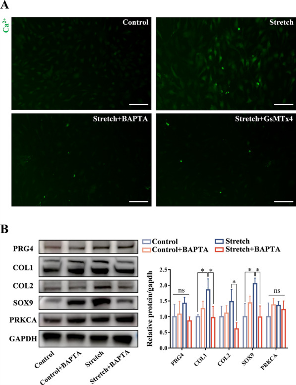
Results: Cartilage thickening and a transient reduction of the CSPCs proportion in the superficial layer of the condylar cartilage were observed after 1 week of MA. The ratio of Ctsk and type II collagen double-positive cells climbed in the first week after MA, and 2 weeks later, the ratio of Ctsk and EdU double-positive cells rose. The expression level of chondrogenic-related genes, Piezo1, and Prkca was elevated in CSPCs after tensile loading. GsMTx4 and BAPTA could block the Ca2+ influx into CSPCs caused by tensile stress. Furthermore, BAPTA and siPrkca could inhibit the stretch-induced chondrogenesis of CSPCs.

Conclusions: We uncovered that tensile stress could cause a transient shrinkage of the CSPCs pool in condylar cartilage, resulting from the accelerated chondrogenesis of CSPCs. Tensile force could promote the chondrogenic ability of CSPCs via the Piezo1-Ca2+-Prkca pathway. This study suggested a new regulatory route for mandibular growth modification in orthodontic practice.

Keywords: Cartilage; Cartilage stem/progenitor cells; Mandibular advancement; Mandibular condyle; Mechanical stress; Temporomandibular joint.


Conflict of interest statement

Declarations. Ethics approval and consent to participate: All the procedures of animal experiments were approved by the Animal Welfare Committee of the School and Hospital of Stomatology, Tongji University, under no. 2024-DW-31. The title of the approved project was “Research on the identification of mechanical force-responsive subpopulation in mouse fibrocartilage stem cells and the underlying mechanisms”. The date of approval was February 22, 2024. Consent for publication: Not applicable. Competing interests: The authors declare that they have no competing interests.

Figures

Fig. 1
Fig. 1
Mandibular advancement (MA) in rats leads to condylar tissue remodeling and cartilage stem/progenitor cells (CSPCs) pool alteration. A The schematic illustration shows the workflow of the MA model establishment in rats. B The rat's lower incisors were trimmed by 3 mm every 3 days for 2 weeks for MA achievement, and lateral X-ray images were captured at the end of trimming. C Representative safranin O/fast green staining results of rat condyles after 1 and 2 weeks of Sham or MA operation. Dashed lines mark the superficial layer (SL) and the whole condylar cartilage (CC). Scale bar: 100 μm. D The average SL and CC thickness of rat condyles in the Sham and MA groups, n = 5. E The superficial layer of rat condylar cartilage was separated by microsurgical forceps, and the representative HE staining results are shown. Scale bar: 50 μm. F Representative flow cytometry gating strategy of CD90+, CD29+, CD45, CD11b/c CSPCs digested from the superficial layer of rat condylar cartilage after 1 and 2 weeks of Sham or MA operation. G The percentage of CD90+, CD29+, CD45, CD11b/c CSPCs in the superficial layer of rat condylar cartilage after 1 and 2 weeks of Sham or MA operation, n = 5. Data are presented as mean ± SD. *p < 0.05, ***p < 0.001; ns, not significant
Fig. 2
Fig. 2
The chondrogenic differentiation and proliferation of cathepsin K (Ctsk)+ CSPCs are activated in the MA mouse model. A Representative type II collagen (Col2) immunofluorescence staining images of condyles from tdTomato; Ctsk-Cre mice at 3 and 14 days old. Arrows indicate Ctsk and Col2 double-positive cells in the condylar cartilage. Scale bar: 50 μm. B The schematic diagram displays the workflow of the MA model establishment in tdTomato; Ctsk-Cre mice. C The lower incisors of tdTomato; Ctsk-Cre mice were trimmed by 1 mm every 3 days for 2 weeks to achieve MA. D Representative Col2 immunofluorescence staining images of condyles from tdTomato; Ctsk-Cre mice after 1 and 2 weeks of Sham or MA operation. Scale bar: 100 μm. E Representative EdU immunofluorescence staining images of condyles from tdTomato; Ctsk-Cre mice after 1 and 2 weeks of Sham or MA operation. Scale bar: 100 μm. F Quantitative analysis of the percentage of Ctsk, Col2, and Ctsk, EdU double-positive cells in the condylar cartilage from tdTomato; Ctsk-Cre mice in the Sham and MA groups, n = 5. Data are presented as mean ± SD. ***p < 0.001; ns, not significant
Fig. 3
Fig. 3
MA enhances the expression of chondrogenic-related genes, Piezo1, and protein kinase C alpha (Prkca) in the superficial layer of rat condylar cartilage. A The principal component analysis of transcriptome sequencing samples (the superficial layers of rat condylar cartilage after 1 week of Sham or MA operation), n = 5. B Transcriptome sequencing reveals the 15 most differentially expressed genes in the superficial layer of rat condylar cartilage between the Sham and MA groups in a heatmap, n = 5. Arrows indicate the genes of interest. C The volcano plot of the differentially expressed genes between the Sham and MA groups. D qPCR detection of chondrogenic markers, Mmp13, Piezo1, and Prkca in the superficial layer of rat condylar cartilage after 1 week of Sham or MA operation, n = 5. Data are presented as mean ± SD. **p < 0.01, ***p < 0.001
Fig. 4
Fig. 4
Tensile loading enhances the expression of chondrogenic-related genes, Piezo1, and Prkca in rat condylar CSPCs. A The flow cytometric analysis of surface markers CD90, CD29, CD45, and CD11b/c in isolated rat condylar CSPCs at the second passage. B Representative pictures of oil red, alizarin red, and alcian blue staining after trilineage differentiation induction of rat condylar CSPCs in vitro. Scale bar: 100 μm. C, D qPCR detection (n = 6 biological replicates) and representative western blots of chondrogenic markers and Prkca in rat CSPCs after 10% tensile loading for 24 or 48 h in vitro. Full-length blots are presented in Figure S1. E, F Representative Piezo1 immunofluorescence staining images of rat condylar CSPCs after 10% tensile loading for 24 or 48 h in vitro, and the corresponding quantitative analysis, n = 3 biological replicates. Scale bar: 100 μm. Data are presented as mean ± SD. **p < 0.01, ***p < 0.001; ns, not significant
Fig. 5
Fig. 5
Blocking Piezo1 and Ca2+ influx reduces the chondrogenic effect of tensile loading on rat condylar CSPCs. A Representative calcium immunofluorescence staining images of rat condylar CSPCs after 10% tensile loading for 48 h with or without the pretreatment of 10 μM calcium chelator BAPTA or 5 μM Piezo1 inhibitor GsMTx4. Scale bar: 100 μm. B Western blot detection and the corresponding quantitative analysis of chondrogenic markers and Prkca in rat condylar CSPCs after 10% tensile loading for 48 h with or without the pretreatment of 10 μM calcium chelator BAPTA, n = 3 biological replicates. Full-length blots are presented in Figure S2. Data are presented as mean ± SD. *p < 0.05; ns, not significant
Fig. 6
Fig. 6
Prkca knockdown diminishes the chondrogenic effect of tensile stress on rat condylar CSPCs. A Western blot detection and the corresponding quantitative analysis of chondrogenic markers and Prkca in rat condylar CSPCs after 10% tensile loading for 48 h with siNC or siPrkca pretreatment in vitro, n = 3 biological replicates. Full-length blots are presented in Figure S3. B The schematic diagram shows the workflow of in vivo Prkca silencing in condylar CSPCs by locally injecting in vivo siPrkca into the joint capsules of rats. C qPCR detection and representative western blots of Prkca in the superficial layer of rat condylar cartilage with local in vivo siPrkca injection for 1 and 2 weeks, n = 6. Full-length blots are presented in Figure S4. DF Representative safranin O/fast green and immunohistochemistry staining images of rat condyles after 1 and 2 weeks of MA with siNC or siPrkca injection. Dashed lines mark the superficial layer (SL) and the whole condylar cartilage (CC). Scale bar: 100 μm. G Quantitative analysis of the average SL and CC thickness, ratio of Col2-positive areas, and percentage of Sox9-positive cells in rat condylar cartilage after 1 and 2 weeks of MA with siNC or siPrkca injection, n = 5. Data are presented as mean ± SD. *p < 0.05, **p < 0.01, ***p < 0.001; ns, not significant
Fig. 7
Fig. 7
The proposed regulatory mechanism in condylar CSPCs under tensile stress

Similar articles

References

    1. Qiao Y, Yi D, Reed DA, Mercuri LG, Chen D, Oh C-D. A novel approach to establishing a temporomandibular joint fibrocartilage cell line. J Dent Sci. 2022;17(3):1378–86. - PMC - PubMed
    1. Fan Y, Cui C, Li P, Bi R, Lyu P, Li Y, et al. Fibrocartilage stem cells in the temporomandibular joint: insights from animal and human studies. Front Cell Dev Biol. 2021;9:665995. - PMC - PubMed
    1. Rogers-DeCotes AW, Porto SC, Dupuis LE, Kern CB. ADAMTS5 is required for normal trabeculated bone development in the mandibular condyle. Osteoarthr Cartil. 2021;29(4):547–57. - PMC - PubMed
    1. Purcell P, Jheon A, Vivero MP, Rahimi H, Joo A, Klein OD. Spry1 and spry2 are essential for development of the temporomandibular joint. J Dent Res. 2012;91(4):387–93. - PMC - PubMed
    1. Nickel JC, Iwasaki LR, Gonzalez YM, Gallo LM, Yao H. Mechanobehavior and ontogenesis of the temporomandibular joint. J Dent Res. 2018;97(11):1185–92. - PMC - PubMed

MeSH terms

LinkOut - more resources