. 2025 Jul 1;18(1):246.
doi: 10.1186/s13071-025-06808-w.

New findings on the male reproductive system and spermatozoa of Aedes aegypti (Diptera: Culicidae)

Affiliations

New findings on the male reproductive system and spermatozoa of Aedes aegypti (Diptera: Culicidae)

Henrique Barbosa da Silva et al. Parasit Vectors. .

Abstract

Background: Aedes aegypti is one of the most important arbovirus vectors, characterized by its widespread distribution and exceptional reproductive capacity. This study reexamines the male reproductive system (MRS) of this species, focusing on its morphology throughout post-embryonic development and the structure of its spermatozoa.

Methods: We analyzed the MRS of A. aegypti in the larval L4, pupal, and adult stages using bright-field light microscopy, fluorescent microscopy, and transmission electron microscopy techniques. Spermatozoa measurements were made using the ImageJ software.

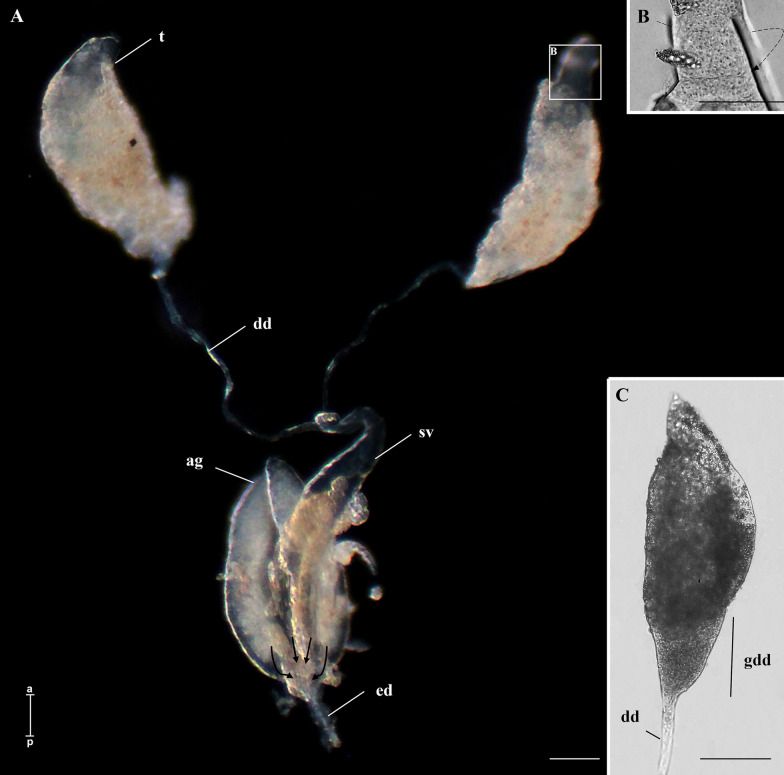
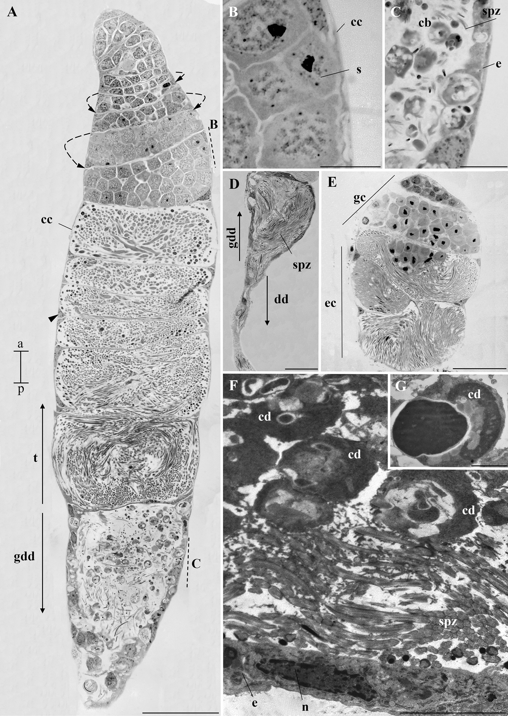
Results: In L4 larvae, the MRS was composed of the two testes, a thin deferent duct, and a pair of seminal vesicles. The MRS is fully developed in pupae and adults, with two testes, deferent ducts, seminal vesicles, accessory glands, and an ejaculatory duct. Histological sections revealed that each testis is formed by a single follicle, which appeared to spiral at all stages. In pupae and adults, the testes showed germ cells at different stages of development, while the goblet portion of the deferent duct contained cytoplasmic bodies and spermatozoa. In adults, the seminal vesicles were filled with spermatozoa soon after emergence. Secretions from accessory glands were of the apocrine type. The spermatozoa were thin and long, measuring around 335 µm in length. Ultrastructural analysis revealed a very short acrosome covering the apical nucleus, in the flagellar region, an axoneme with the 9 + 9 + '1' microtubule pattern typical of mosquitoes, and two mitochondrial derivatives along the flagellum, narrowing at the terminal portion.

Conclusions: Our analysis revealed a clear link between testicular development and spermatogenesis. In addition, we identified seminal vesicles at all life stages and accessory glands visible only in pupae and adults. The characterization of sperm structure and ultrastructure indicated similarities with other mosquito species. Finally, our study provided valuable information that may support research in comparative biology and reproduction.

Keywords: Microscopy; Mosquito reproduction; Sexual development; Spiraled testicular follicle.


Conflict of interest statement

Declarations. Ethics approval and consent to participate: Not applicable. Consent for publication: All authors read and approved the final manuscript. Competing interests: The authors declare no competing interests.

Figures

Fig. 1
Fig. 1
Whole-mount of the MRS of immature A. aegypti. A Testis (t) of L4 larva with a spiral shape (dotted arrow) continuous with the deferent duct (dd). B MRS of a pupa. The spiral shape (dotted arrow) of the testis (t) is more evident than in the L4. Each deferent duct (dd) has a dilated portion forming the seminal vesicles (sv). Note the presence of a pair of accessory glands (ag) and the ejaculatory duct (ed). Inset I shows that the ag and sv converge into the ed (arrows). In both instars, fatty bodies (fb) are noted. Scale bars: 100 µm. a, anterior region; p, posterior region
Fig. 2
Fig. 2
Whole-mount of MRS of A. aegypti adults. A Testis (t), deferent duct (dd), seminal vesicles (sv), accessory glands (ag), and ejaculatory duct (ed). The arrows indicate that the accessory glands and the seminal vesicles empty into the ejaculatory duct. B The spiral shape of the testis (dotted arrow). C Detail of the testis showing the goblet of the deferent duct (gdd). Scale bars: 100 µm. a, anterior region; p, posterior region
Fig. 3
Fig. 3
Histology of the testis of immature A. aegypti. A Testis of larva with evident spiraling of the testicular follicle (dotted arrow). Note the cytoplasm of cystic cells (cc) and their nucleus (arrowhead). The cysts contain young cells of the spermatogenic lineage. Note the presence of globular cells throughout the organ. B Testis of pupa. The spiraling of the testicular follicle (dotted arrow) is more pronounced. The cytoplasm of cystic cells (cc) and their nucleus (arrowhead) are also visible. Globular cells (newer in the spermatogenic lineage) are seen in the upper region of the testis while more elongated cells (older in the spermatogenic lineage) are seen in the lower regions of the testis. C Goblet of the deferent duct in the testis of a pupa. See the presence of an epithelium (e) in this region. Spermatozoa (spz) and cytoplasmic bodies can be seen in the lumen of the goblet. Staining: toluidine blue. Scale bar: 50 µm. a, anterior region; p, posterior region
Fig. 4
Fig. 4
Histology and ultrastructure of the testis of adult A. aegypti. A Longitudinal histological section of the testis (t) and goblet of the deferent duct (gdd), revealing the spiral aspect of the testis (dotted arrow) and the cytoplasm of cystic cells (cc) covering it, detailed in B. Cystic cell nuclei (arrowhead) are present throughout the testis. The gdd is located posterior to the t and is further detailed in C. B Cytoplasm of cystic cells (cc), developing sperm cell (s). C Details of the gdd showing epithelium (e), numerous cytoplasmic bodies (cb), and spermatozoa (spz). D Histological section of the goblet of the deferent duct (gdd) and the deferent duct (dd). Note the presence of numerous spermatozoa (spz) in the lumen. E Cross section of the testis of A. aegypti at 21 days of age. The dotted arrows indicate the spiral shape of the organ. Note the presence of globular cells (gc, newer in the spermatogenic lineage) and elongated cells (ec, older in the spermatogenic lineage), indicating that spermatogenesis is still occurring at this age. F Ultrastructure obtained by transmission electron microscopy of the goblet of the deferent duct, identified by the presence of the epithelium (e) and a lumen filled with spermatozoa (spz). n, nucleus. Note the presence of cellular debris (cd) of various shapes in F and G. Staining AE: toluidine blue. Scale bars: A, D, E: 50 µm; B, C: 10 µm; F: 5 µm; G: 1 µm
Fig. 5
Fig. 5
Histology of seminal vesicles and accessory glands of immature A. aegypti. A Seminal vesicle (sv) of a larva, showing a wide lumen (L). B Anterior portion of the pupal seminal vesicles (sv), covered by a musculature (arrowhead) surrounding each vesicle completely. C Posterior portion of the same sv as in B, with a narrower lumen (L) and no individualized muscular covering for each gland (arrowhead). D Longitudinal section of a pupal accessory gland (ag) surrounded by musculature (arrowhead). E Cross-section of two pupal accessory glands, showing the lumen (L) and the surrounding musculature (arrowhead). F Detail of secretory cells in the ag with apocrine secretions (as). Staining: toluidine blue. Scale bars: A, B, D, and E: 50 µm; C: 25 µm; F: 10 µm. a, anterior region; p, posterior region
Fig. 6
Fig. 6
Histological sections of the seminal vesicle and accessory gland of adult A. aegypti. A Longitudinal section of two seminal vesicles (sv) filled with spermatozoa (spz). B Details of a seminal vesicle, showing a simple epithelium (e) and a muscular layer (m) surrounding the vesicle. Spermatozoa (spz) are visible in the lumen. C Junction point of two accessory glands (ag) and the ejaculatory duct (ed). D Longitudinal section of an accessory gland (ag) filled with apocrine secretions produced by the glandular epithelium. The ag has two distinct regions; the anterior region is stained more lightly than the posterior region. Musculature (arrowhead). EF Longitudinal sections showing details of the secretions (*) in the anterior (E) and posterior (F) regions of the gland. G Junction point (arrow) of an ag with a seminal vesicle (sv), also highlighting the ejaculatory duct (ed). H Details of the cuticle (c) in the ejaculatory duct. Staining: toluidine blue. Scale bars: AG: 25 µm; H: 10 µm. a, anterior region; p, posterior region
Fig. 7
Fig. 7
A. aegypti spermatozoa. A Overview of the spermatozoon showing the head (h) and flagellum (f). B Detail of the head (h) of a spermatozoon. C Nucleus (n) of a spermatozoon stained with DAPI. D DAPI and light microscopy overlay, demonstrating that the nucleus (n) occupies nearly the entire head region. f, flagellum. E Terminal portion (*) of the flagellum (f). F Spermatozoon with the centriolar adjunct (ca) highlighted. G Spermatozoa from a newly emerged adult. Staining: A, B, E, G: Giemsa. F crystal violet and eosin. Scale bars: A and G: 50 µm; BF: 10 µm. a, anterior region; p, posterior region
Fig. 8
Fig. 8
Electron micrograph of A. aegypti spermatozoa. A Anterior region of the head, showing the nucleus (n) surrounded by the acrosome (ac). B Several spermatozoa where the nucleus (n) is visible in I: the insertion of the axoneme (ax) into the nucleus (n); II: the centriole adjunct (ca) region containing two mitochondrial derivatives (md) and the axoneme (ax); III: the median portion of the flagellum also with two mitochondrial derivatives (md) and the axoneme (ax), and the final portion of the flagellum (fpf). C Median portion of the flagellum, highlighting the 9 + 9 + ‘1’ arrangement of the axoneme (ax) and the two mitochondrial derivatives (m). D Final portions of the sperm flagella (arrowhead), in one of them the disintegrating axoneme (dax) can be observed. Scale bar: 200 nm
Fig. 9
Fig. 9
Diagram of the spermatozoa of A. aegypti, indicating their structures and the locations where they are found: nucleus (n), acrosome (ac), flagellum (f), axoneme (ax), centriolar adjunct (ca), mitochondrial derivatives (m), and disintegrated axoneme (dax)

References

    1. Carvalho FD, Moreira LA. Why is Aedes aegypti Linnaeus so successful as a species? Neotrop Entomol. 2017;46:243–55. - PubMed
    1. Consoli RA, Oliveira RL. Principais mosquitos de importância sanitária no Brasil. 1st ed. Fiocruz; 1994.
    1. Lima-Camara TN. Emerging arboviruses and public health challenges in Brazil. Rev Saúde Pública. 2016;50:36–42. - PMC - PubMed
    1. Franklinos LHV, Jones KE, Redding DW, Abubakar-Ibraim. The effect of global change on mosquito-borne disease. Lancet Infect Dis. 2019;19:302–19. - PubMed
    1. Word Health Organization: Dengue—Global situation; 2025. https://www.who.int/emergencies/disease-outbreak-news/item/2024-DON518. Accessed 12 Jan 2025.

MeSH terms

LinkOut - more resources