. 2025 Jul 1;23(1):309.
doi: 10.1186/s12964-025-02292-y.

Aurantio-obtusin modulates Wilms Tumour 1 within the breast tumour microenvironment reducing immunosuppression and tumour growth

Affiliations

Aurantio-obtusin modulates Wilms Tumour 1 within the breast tumour microenvironment reducing immunosuppression and tumour growth

Rui Li et al. Cell Commun Signal. .

Abstract

Introduction: Cancer associated fibroblasts (CAFs) contribute to tumourigenesis and immune tolerance within the tumour microenvironment (TME). Therefore, inhibiting the pro-tumourigenic function of CAFs can be a viable therapeutic approach. However, targeting CAFs is challenging due to the lack of specific markers. The objective of this study is to identify CAF specific therapeutic targets that have the potential to enhance tumour immunity and reduce tumour growth.

Methods: RNA sequencing was performed on CAFs and normal fibroblasts (NFs) from the same breast cancer patient. Wilms tumour-1 (WT1) was identified as a gene upregulated in CAFs. WT1 levels in CAFs were manipulated using plasmid overexpression of-or siRNA downregulation of WT1. Co-culture assays were performed to evaluate the role of CAF-derived WT1 in T cell proliferation and differentiation using flow cytometry. Western blot and ELISA were performed to interrogate the mechanism of action of WT1 within CAFs. Three-dimensional patient-derived organoids (PDOs) that encompassed the tumour immune-microenvironment were established to determine the therapeutic potential of targeting CAF-derived WT1.

Results: WT1, a transcription factor, regulates signal transducer and activator of transcription (STAT) 1/3 levels, promotes programmed death ligand 1 (PD-L1) expression and indoleamine 2,3-dioxygenase (IDO) expression in CAFs. CAF-derived WT1 reduces the proliferation of CD4+ and CD8+ T cells and enhances the differentiation of naïve T cells into regulatory T cells (Tregs), thus producing an immunosuppressive TME. Reducing CAF WT1 levels results in less immunosuppressive CAFs, smaller PDOs and increased levels of cytotoxic granzyme B+ (GZMB+) T cells within the TME. Standard chemotherapeutic agents, paclitaxel (PTX) and doxorubicin (DOX), increase WT1 levels in CAFs enhancing their ability to suppress T cell proliferation. However, Aurantio-obtusin (AO, a DOX analogue) decreases WT1 expression in CAFs reducing their ability to suppress T cell proliferation. AO causes decreased PDO size which correlates with increased levels of T cells within the TME.

Conclusions: Therapeutic targeting of the WT1/STAT1/3/PD-L1/IDO axis in CAFs with AO has the potential to enhance T cell activity and reduce Treg percentage within the TME, thereby enhancing tumour immunity and reducing tumourigenesis.

Keywords: Breast cancer; Cancer associated fibroblast; Immunosuppression; Tumour microenvironment; Wilms’ tumour 1.


Conflict of interest statement

Declarations. Ethics approval and consent to participate: Ethical approval was obtained from the research ethics committee at the University of Galway (Ref: 45/05). After written informed consent, fresh specimens of human breast tumours were harvested from patients undergoing surgery at University College Hospital Galway. All patient material and clinical information were obtained from the Cancer Biobank at the University of Galway. Consent for publication: Not applicable. Competing interests: SJE and PGL are employees and shareholders of Orbsen Therapeutics Ltd. RL, DOC, BD, POB, XH, NG, MK and LRB have no conflicts of interest to declare.

Figures
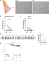
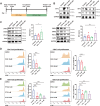
Fig. 1
Fig. 1
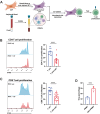
WT1 is upregulated in breast CAFs. A Schematic demonstrating CAF and NF sampling from breast. Morphology of cancer associated fibroblasts (CAFs) and normal fibroblasts (NFs) isolated from the same BC patient. Scale bar, 100 μm. B RNA-seq data indicated WT1 was upregulated in CAFs compared to paired NFs. n = 12, P < 0.01). C qPCR demonstrated higher expression of WT1 in a larger CAF cohort compared to paired NFs (n = 22, P < 0.0001). D WT1 protein levels were determined in CAFs and paired NFs. Actin was used as a loading control. E Overall survival curve of WT1 in breast cancer patients from Kaplan–Meier database
Fig. 2
Fig. 2
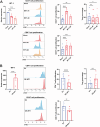
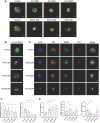
CAFs regulate T cell proliferation and differentiation into Tregs. A Schematic representation of CAF and PBMC co-culture and T cell proliferation assay. B Flow cytometry analysis of CFSE-labelled CD4+ (B) and CD8+ (C) T cell proliferation and CD4+CD25+FoxP3.+ Treg quantitation (D). Representative experiment is shown of n = 3–5 biological replicates. Data represents mean ± SEM; * P < 0.05, ** P < 0.01, *** P < 0.001, and **** P < 0.0001
Fig. 3
Fig. 3
WT1 regulates the ability of CAFs to suppress T cell proliferation and induce Treg differentiation. A RNA levels of WT1 were determined in WT1 knockdown CAFs. TBP was used as a loading control. CAFs with WT1 knockeddown were co-cultured with PBMCs and CFSE-labelled CD4+ and CD8+ T cell proliferation and CD4+CD25+FoxP3+ Treg quantitation were determined by flow cytometry. B RNA levels of WT1 were determined in WT1 overexpressed CAFs. TBP was used as a loading control. CAFs with WT1 overexpressed were co-cultured with PBMCs and CFSE-labelled CD4+ and CD8+ T cell proliferation and CD4+CD25+FoxP3.+ Treg quantitation were determined by flow cytometry. Representative experiment is shown of n = 3–5 biological replicates. Data represents mean ± SEM; * P < 0.05, ** P < 0.01, *** P < 0.001, and **** P < 0.0001
Fig. 4
Fig. 4
WT1 regulates the STAT1/3 pathway in CAFs. A Protein levels of WT1, STAT1, STAT3, PD-L1 and the TGFβ receptor were determined in WT1 knockdown (A) and WT1 overexpressed (B) CAFs by Western blotting. Actin was used as a loading control. IDO release was determined by ELISA. RNA levels of PD-L1 was determined by qRT-PCR. TBP was used as a loading control. Representative experiment is shown of n = 3–5 biological replicates. Data represents mean ± SEM; * P < 0.05, ** P < 0.01, *** P < 0.001, and **** P < 0.0001
Fig. 5
Fig. 5
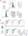
CAF-derived WT1 effects cancer cells indirectly via tumour infiltrating T-lymphocyte (TIL) activity. A CAFs were co-cultured with tumour cell line T47D. CFSE-labelled tumour cell (EpCAM+) proliferation was determined by flow cytometry. CAFs with WT1 knockdown were co-cultured with T47Ds and tumour cell proliferation and tumour cell number were determined by flow cytometry. B CAFs were co-cultured with tumour cells and TILs from the same patient. Flow cytometry median fluorescence intensity (MFI) was used to measure EpCAM.+ cells. Representative experiment is shown of n = 3 biological replicates. Data represents mean ± SEM; * P < 0.05, ** P < 0.01
Fig. 6
Fig. 6
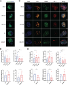
CAF-derived WT1 effects breast PDOs growth, TIL recruitment and TIL activity. A 3D PDOs were established using CAFs, tumour cells and TILs from same patient. PDOs were stained with tumour epithelial marker EpCAM (green) and the nuclear counterstain DAPI (blue). WT1 knockdown and WT1 overexpressed CAFs were used to generate PDOs to determine the effect of CAF-derived WT1 on organoid growth and tumour cell content. Scale bar, 100 μm. B Graphical representation showing the effect of manipulating CAF WT1 on organoid size and epithelial content. The organoid size and tumour cells were measured by DAPI+ area and EpCAM+ area by Fiji and normalized by the smallest sicon or EV samples. C WT1 knockdown and WT1 overexpressed CAFs were used to generate PDOs to determine the effect of CAF-derived WT1 on T cell recruitment CD3 (yellow), and T cell activity GZMB (green) and PD1 (red) within PDOs. DAPI was used as nuclear counterstain (blue). Scale bar, 100 μm. Representative experiment is shown. D Graphical representation showing the effect of manipulating CAF WT1 on T cell recruitment and activity. T cell infiltration and activation were quantified as a percentage of CD3/GZMB/PD1-positive cells among DAPI+ cells. n = 4 biological replicates. Data represents mean ± SEM; * P < 0.05, ** P < 0.01
Fig. 7
Fig. 7
Chemotherapeutic drugs DOX and PTX increase WT1 levels in CAFs and enhance their ability to inhibit T cell proliferation. A Schematic overview of drug treatment, co-culture of CAFs and PBMCs followed by T cell proliferation assay. B Protein expression of WT1 and p53 in CAFs following DOX and PTX treatment was determined by Western blotting. GAPDH was used as a loading control. C Protein levels of STAT1, STAT3, PD-L1 levels. IDO release from DOX and PTX treated CAFs was determined by ELISA. D DOX treated CAFs were co-cultured with PBMCs and CFSE-labelled CD4+ and CD8+ T cell proliferation was determined by flow cytometry (E) PTX treated CAFs were co-cultured with PBMCs and CFSE-labelled CD4+ and CD8.+ T cell proliferation was determined by flow cytometry. Representative experiment is shown of n = 3 biological replicates. Data are mean ± SEM; * P < 0.05, ** P < 0.01, *** P < 0.001, and **** P < 0.0001
Fig. 8
Fig. 8
AO reduces WT1 levels in CAFs and reduces the ability of CAFs to suppress T cell proliferation. A Comparison of chemical structures of DOX and AO. B Protein levels of WT1, STAT1, STAT3, and PD-L1 after AO treatment of CAFs were determined by Western blotting. Actin was used as a loading control. C Dose–response curves of DOX, PTX and AO treatment of CAFs. P1 and P3 (Luminal A), P2 (TNBC). Curves were generated by non-linear regression analysis by GraphPad. D AO treated CAFs were co-cultured with PBMCs and CFSE-labelled CD4+ and CD8.+ T cell proliferation was determined by flow cytometry. Representative experiment is shown of n = 3 biological replicates. Data represent mean ± SEM; * P < 0.05, and ** P < 0.01
Fig. 9
Fig. 9
AO effects tumour cell viability, TIL recruitment and TIL activity within PDOs. A PDOs were established and treated with increasing concentrations of AO or DOX. PDOs were stained with Calcein AM (green) and Ethidium homodimer-1 (red) to determine the amount of live and dead cells respectively. Scale bar, 100 μm. B AO-treated PDOs were stained with EpCAM (green) and counterstained with DAPI (blue) to determine the effect of AO on organoid growth and tumour cell content. Scale bar, 100 μm. C Graphical representation showing the effect of AO on organoid size and epithelial content. The organoid size and tumour cells were measured by DAPI+ area and EpCAM+ area by Fiji and normalized to the smallest control sample. D AO-treated PDOs were stained with CD3 (yellow), GZMB (green) and PD1 (red) to determine the effect of AO on T cell recruitment and T cell activity within PDOs. DAPI was used as nuclear counterstain (blue). Scale bar, 100 μm. E Graphical representation showing the effect of AO on T cell recruitment and activity. T cell infiltration and activation were quantified of percentage of CD3/GZMB/PD1-positive cells among DAPI.+ cells. Representative experiment is shown of n = 3 biological replicates. Data are mean ± SEM; * P < 0.05, ** P < 0.01
Fig. 10
Fig. 10
Proposed model highlighting how WT1 in CAFs modulates immune evasion and tumour growth in the breast TME. WT1 is upregulated in breast patient-derived CAFs. WT1 is a transcription factor, regulating expression of STAT1/STAT3 in CAFs. STAT1/STAT3 regulate PD-L1 expression and IDO release in CAFs. Both PD-L1 and IDO inhibit T cell activity and cytotoxic GZMB levels within the TME, contributing to increased tumour growth. Targeting WT1 in CAFs reduces the release of immunosuppressive factors into the TME. T cells are more active and are able to limit tumour growth

Similar articles

References

    1. Wang R, Zeng H, Xiao X, Zheng J, Ke N, Xie W, Lin Q, Zhang H. Identification of prognostic biomarkers of breast cancer based on the immune-related gene module. Autoimmunity. 2023;56(1):2244695. - PubMed
    1. Cho S, Joo B, Park M, Ahn SJ, Suh SH, Park YW, Ahn SS, Lee SK. A radiomics-based model for potentially more accurate identification of subtypes of breast cancer brain metastases. Yonsei Med J. 2023;64(9):573–80. - PMC - PubMed
    1. Serageldin MA, Kassem AB, El-Kerm Y, Helmy MW, El-Mas MM, El-Bassiouny NA. The effect of metformin on chemotherapy-induced toxicities in non-diabetic breast cancer patients: a randomised controlled study. Drug Saf. 2023;46(6):587–99. - PMC - PubMed
    1. Luo J, Li X, Wei KL, Chen G, Xiong DD. Advances in the application of computational pathology in diagnosis, immunomicroenvironment recognition, and immunotherapy evaluation of breast cancer: a narrative review. J Cancer Res Clin Oncol. 2023;149(13):12535–42. - PMC - PubMed
    1. Chang X, Ma Z, Zhu G, Lu Y, Yang J. New perspective into mesenchymal stem cells: molecular mechanisms regulating osteosarcoma. J Bone Oncol. 2021;29:100372. - PMC - PubMed

MeSH terms