

Utilizing MRI and CT to identify risk factors associated with cage subsidence
- PMID: 40598416
- PMCID: PMC12210715
- DOI: 10.1186/s40001-025-02797-9
Utilizing MRI and CT to identify risk factors associated with cage subsidence
Abstract
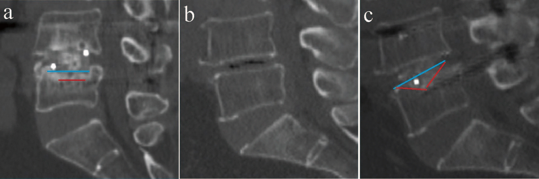
Objectives: To identify risk factors associated with cage subsidence (CS) following single segment transforaminal lumbar interbody fusion (TLIF) and unilateral biportal endoscopic lumbar interbody fusion (ULIF) and to compare the predictive performance of various bone quality assessment methods using MRI and CT images.
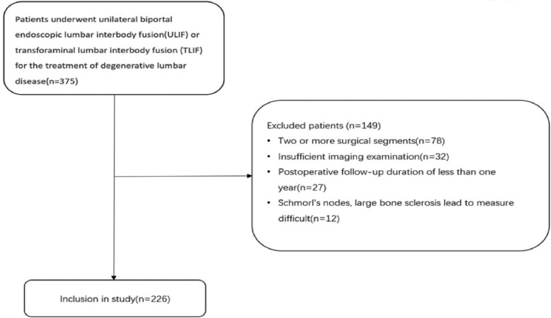
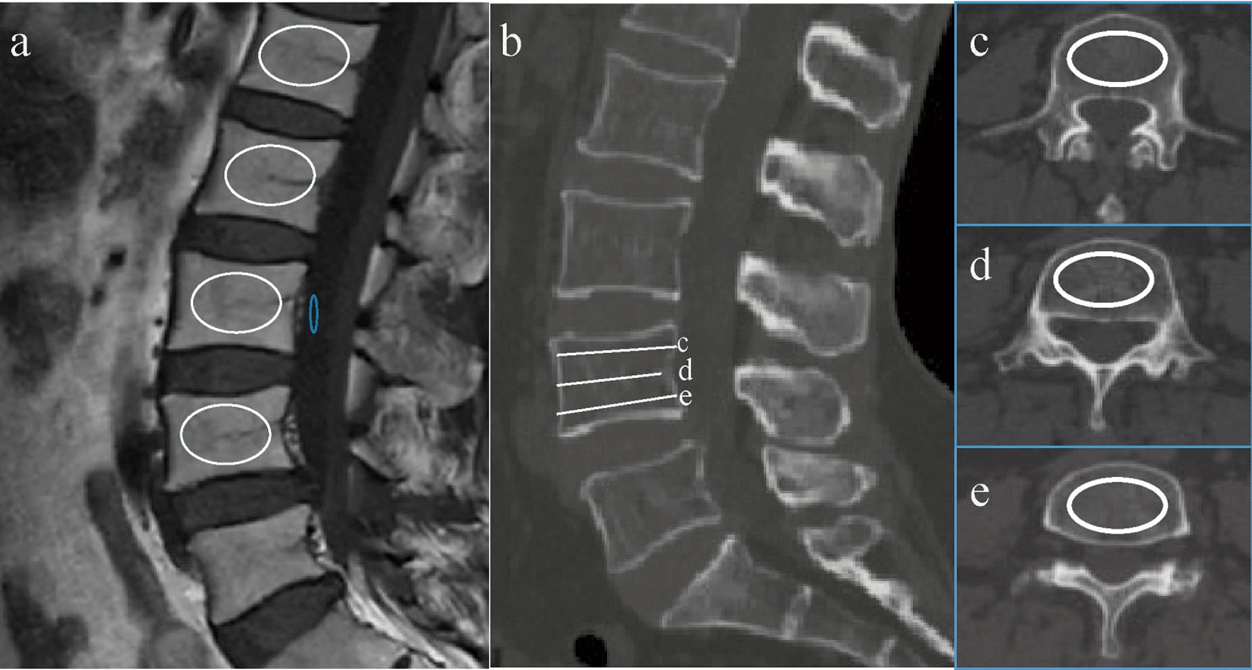
Methods: A total of 226 patients from 2021 to 2023 who underwent ULIF/TLIF because of lumbar disc herniation and lumbar spinal stenosis were enrolled. The subsidence of the cage into the vertebral body exceeding 2 mm was defined as CS and diagnosed using CT scans. Immediate endplate destruction (IED) was defined by CT and VBQ was measured through T1-weighted lumbar MRI. The independent sample t-test was employed to examine the risk factors associated with CS. Additionally, risk factors associated with CS were identified using logistic regression analysis. Lastly, the comparative predictive values were assessed through ROC curve analysis.
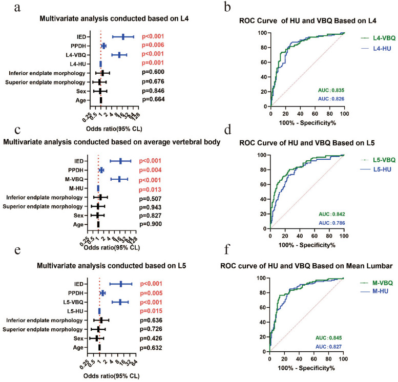
Results: Logistic regression analysis revealed that increased postoperative posterior disc height (PPDH), higher segmental VBQ scores, higher mean VBQ (M-VBQ) scores, decreased segmental HU values, decreased mean HU (M-HU) values and immediate endplate destruction (IED) were associated with the occurrence of CS. The area under the curve (AUC) of the VBQ score was higher than that of the HU value, both in segment and in average.
Conclusions: The incidence of CS was lower in ULIF compared to TLIF. High VBQ scores, low HU values, high PPDH and the presence of IED were associated with an increased risk of CS. Notably, the predictive value of both VBQ scores and HU values were high for CS, with the former potentially outperforming the latter.
Keywords: Cage subsidence; Hounsfield units; Unilateral biportal endoscopic lumbar interbody fusion immediate endplate destruction; Vertebral bone quality.
© 2025. The Author(s).
Conflict of interest statement
Declarations. Ethics approval and consent to participate: This study was supported by the Review of Ethics Committee in Clinical Research (ECCR) of the First Affiliated Hospital of Wenzhou Medical University (KY2024-R074).According to the Regulations and Rules of"Ethical Reviews for Biomedical Research Involving Human Subjects"(2023) the National Health Commission of PRC,"Declaration of Helsinki"of WMA, and"International Ethical Guidelines for Human Biomedical Research"of CIOMS, the project was approved by ECCR. Competing interests: The authors declare no competing interests.
Figures
Similar articles
-
MRI based paraspinal muscle mass predicts early cage subsidence after posterior lumbar interbody fusion.Sci Rep. 2025 Jul 29;15(1):27712. doi: 10.1038/s41598-025-13217-7. Sci Rep. 2025. PMID: 40731052 Free PMC article.
-
Predictive value of vertebral specificity of bone mineral density for cage subsidence among patients undergoing anterior cervical diskectomy and fusion: a retrospective study.Eur Spine J. 2025 Jun;34(6):2207-2218. doi: 10.1007/s00586-025-08859-0. Epub 2025 Apr 15. Eur Spine J. 2025. PMID: 40232368
-
A survival analysis for predictors of implant subsidence following 1- or 2-level transforaminal lumbar interbody fusion.J Neurosurg Spine. 2025 May 9;43(1):42-51. doi: 10.3171/2025.1.SPINE24923. Print 2025 Jul 1. J Neurosurg Spine. 2025. PMID: 40344766
-
Evaluating the prognostic role of computed tomography Hounsfield units in anticipating spinal outcomes post-instrumentation: a systematic review and meta-analysis.Eur Spine J. 2025 Apr;34(4):1420-1432. doi: 10.1007/s00586-025-08737-9. Epub 2025 Mar 8. Eur Spine J. 2025. PMID: 40055220
-
Influence of the geometric and material properties of lumbar endplate on lumbar interbody fusion failure: a systematic review.J Orthop Surg Res. 2022 Apr 10;17(1):224. doi: 10.1186/s13018-022-03091-8. J Orthop Surg Res. 2022. PMID: 35399075 Free PMC article.
References
-
- Heemskerk JL, et al. Long-term clinical outcome of minimally invasive versus open single-level transforaminal lumbar interbody fusion for degenerative lumbar diseases: a meta-analysis. Spine J. 2021;21(12):2049–65. - PubMed
-
- Vazan M, et al. Minimally invasive transforaminal lumbar interbody fusion versus open transforaminal lumbar interbody fusion: a technical description and review of the literature. Acta Neurochir (Wien). 2017;159(6):1137–46. - PubMed
-
- Liu G, et al. Clinical outcomes of unilateral biportal endoscopic lumbar interbody fusion (ULIF) compared with conventional posterior lumbar interbody fusion (PLIF). Spine J. 2023;23(2):271–80. - PubMed
MeSH terms
LinkOut - more resources
Full Text Sources
Medical
