. 2025 Jul 1;23(1):308.
doi: 10.1186/s12964-025-02273-1.

Peritoneal cavity-derived small extracellular vesicles from aged tumor-naïve hosts promote ovarian cancer adhesion and invasion

Affiliations

Peritoneal cavity-derived small extracellular vesicles from aged tumor-naïve hosts promote ovarian cancer adhesion and invasion

Reihaneh Safavi-Sohi et al. Cell Commun Signal. .

Abstract

Background: Epithelial ovarian cancer (OvCa) remains a leading cause of mortality among gynecological cancers. Metastasis to the peritoneum, characterized by tumor cell adhesion to and invasion of the mesothelial lining of the abdominal cavity, represents a critical early event in OvCa metastatic progression. The median age of diagnosis is 63 and there exists a strong correlation between advanced age, OvCa incidence and disease stage. Moreover, the aged peritoneal cavity represents a permissive niche for metastatic dissemination.

Methods: To investigate age-related factors that influence host-tumor communication in metastatic progression, the current study isolated small extracellular vesicles (sEVs) from the peritoneal lavage of healthy tumor-naïve young (3-6 month) and aged (20-22 month) mice. sEVs were analyzed using LC-MS/MS to identify sEV protein cargoes and incubated with murine and human OvCa cells to evaluate effect on pro-metastatic behaviors.

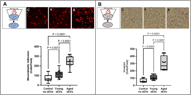
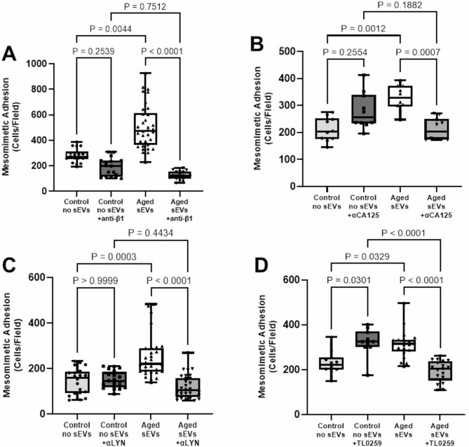
Results: Treatment of human or murine OvCa cells with sEVs from healthy aged hosts significantly enhanced adhesion to peritoneal mesothelial cells in a three-dimensional in vitro meso-mimetic culture assay and to the intact omentum in a short-term in vivo adhesion assay relative to OvCa cells treated with sEVs from young hosts. OvCa cell invasion of collagen gels was also enhanced by aged host-derived sEVs. Proteomic analysis of sEV protein cargos identified differentially expressed proteins in sEVs obtained from aged vs. young hosts that may play a significant role in regulation of adhesion. This was confirmed using meso-mimetic adhesion assays with function blocking antibodies or small molecule inhibitors, supporting a potential role for several proteins in promoting intra-peritoneal dissemination in the aged host.

Conclusions: Results suggest that sEVs derived from the aged peritoneal microenvironment can contribute significantly to disease progression, highlighting sEV-mediated host: tumor communication as a potential therapeutic target for intervention in OvCa progression or recurrence in the aged host.

Keywords: Adhesion; Aging; Extracellular vesicle; Invasion; Mesothelium; Metastasis; Ovarian cancer; Proteomics.


Conflict of interest statement

Declarations. Ethics approval and consent to participate: Studies involving mice were carried out with the approval of the University of Notre Dame Animal Care and Use Committee. Consent for publication: Not applicable. Competing interests: The authors declare no competing interests.

Figures

Fig. 1
Fig. 1
sEV isolation and characterization. (A) Young (3–6 month) and aged (20–22 month) healthy mice (n = 3/group) were subjected to peritoneal lavage once a week for 3 weeks. Lavage fluids from each mouse were pooled prior to sEV isolation. (B) sEV isolation was accomplished using a NanoEX™ system, a bionanoparticle isolation technology microfluidic cassette housing an asymmetric nanopore membrane, according to the manufacturer’s specifications. (C) sEVs derived from young and aged peritoneal lavage (20 ug) were electrophoresed on a 9% SDS polyacrylamide gel and western-blotted to PVDF membrane. Blots were probed with antibodies to CD63 and TSG101 at a 1:100 dilution, and with a peroxidase-conjugated rabbit secondary antibody at a 1:4000 dilution. (D) NanoSight NS300 nanoparticle tracking analysis was performed immediately following sEV isolation. sEVs typically had a mean size slightly larger than 100 nm with little large particle contaminants. Plots show nanoparticle size and concentration. (E) Transmission electron microscopy of sEVs from young and aged mouse lavages. Plot shows nanoparticle size
Fig. 2
Fig. 2
sEVs from aged host tumor-naïve lavage promote in vitro meso-mimetic adhesion and invasion of human OvCa cells. sEVs (5 × 107) purified from young or aged host peritoneal lavage were added to OVCAR5 cells (5 × 105) and incubated for 24 h. (A) Mesomimetic adhesion assay. Upper panel: assay schematic and representative images of adherent cells treated with control (C), young (Y) and aged (A) sEVs. OvCa cells were dyed with Mito-Tracker Red CMXRos dye and cells (5 × 104) were added to a monolayer of LP9 human peritoneal mesothelial cells on type I collagen coated wells 20 min. Non adherent cells were gently rinsed away with PBS and adherent cells were enumerated by counting cells in five 4X fields. (B) 3D collagen invasion assay. Upper panel: assay schematic and representative images of invaded cells adherent to the underside of the filter after treatment with control (C), young (Y) and aged (A) sEVs. OvCa cells (5 × 105) were added to a Boyden chamber containing 0.8 μm filter and coated with type I collagen (20 ug). After incubating 48 h, the filter was removed and invaded cells on the bottom of the filter were enumerated by counting cells in five 4X fields. Experiments were performed a minimum of three times and evaluated using student’s t-test
Fig. 3
Fig. 3
sEVs from aged host tumor-naïve lavage promote in vitro meso-mimetic and in vivo omental adhesion of murine OvCa cells. sEVs (5 × 107) purified from young or aged host peritoneal lavage were added to RFP-tagged ID8-Trp53-/- (5 × 105) and incubated for 24 h. (A) OvCa cells (5 × 104) were then added to a monolayer of LP9 peritoneal mesothelial cells on type I collagen coated wells20 min. Non adherent cells were gently rinsed away with PBS and adherent cells were enumerated by counting cells in five 4X fields. Experiments were performed in triplicate. (B-D) To evaluate initial adhesive events in vivo, sEV-treated cells (6 × 106) were injected i.p. into female C57Bl/6 mice. (B) After 24 h, a midline dissection was made and the peritoneal cavity imaged in situ. (C, D) Dissected omenta were imaged ex vivo and RFP fluorescence quantified using ImageJ. Data obtained from n = 3 mice/group
Fig. 4
Fig. 4
Proteomic analysis of sEVs isolated from young vs. aged peritoneal lavage. (A) Statistical summary of protein identification; the number of MS run, MS1, MS2, PSM, identified peptides, sequences, proteins, and protein groups in aged (A) and young (Y) EVs. (B) Venn diagrams display the unique and overlapping protein and protein group identifications across the A and Y groups. (C) Volcano plot showing differentially expressed proteins. Black symbols– upregulated proteins; red symbols– downregulated proteins. Selection criteria: up-regulated (FC ≥ 2 and significance ≥ 13) and down-regulated (FC < 0.5 and significance ≥ 13)
Fig. 5
Fig. 5
Comparison of ‘molecular function’ categories and ‘protein classes’ in protein datasets obtained from sEVs isolated from young vs aged peritoneal lavage. (A) Pie chart of gene ontology ‘molecular function’ categories in protein datasets from aged and young lavage sEVs. The number of proteins classified as ‘binding’ is highlighted in each group. (B) Pie chart of gene ontology ‘protein classes’ identified in protein datasets from aged and young lavage sEVs
Fig. 6
Fig. 6
Blocking sEV-associated proteins abrogates the enhanced meso-mimetic adhesion observed with aged sEVs. sEVs (5 × 107) purified from peritoneal lavage obtained from aged hosts or control (PBS) were incubated with function-blocking antibodies directed against (A) β1 integrin (2 µg), (B) CA125 (MUC16, 1 µg) or (C) LYN kinase (1 µg) in a total volume of 200 ul for 3 h prior to adding to OvCa cells for 24 h. The meso-mimetic adhesion assay was then performed as described in Fig. 3. (D) sEVs (5 × 107) purified from peritoneal lavage obtained from aged hosts or control (PBS) were incubated with the Lyn kinase inhibitor TL0259 (0.1 nM) for 3 h prior to adding to OvCa cells for 24 h. The meso-mimetic adhesion assay was then performed as described in Fig. 3. Assays were performed in triplicate. Data were analyzed using Kruskal-Wallis test and Dunn’s multi-comparison test

References

    1. Huang J, Chang WC, Ngai CH, Lok V, Zhang L, Lucero-Prisno DE, et al. Worldwide burden, risk factors, and temporal trends of ovarian cancer: a global study. Cancers. 2022;14. 10.3390/cancers14092230 - PMC - PubMed
    1. Howlader N et al., SEER Cancer Statistics Review, 1975–2014. Natl Cancer Inst. vol. Bethesda, MD, Apr. 2017. https://seer.cancer.gov/csr/1975_2014/
    1. Lengyel E. Ovarian cancer development and metastasis. Am J Path. 2010;177:1053–64. - PMC - PubMed
    1. Lengyel, et al. Epithelial ovarian cancer experimental models. Oncogene. 2014;33:3619–33. - PMC - PubMed
    1. Yancik R. Ovarian cancer: age contrasts in incidence, histology, disease stage at diagnosis, and mortality. Cancer. 1993;71:517–23. - PubMed

LinkOut - more resources