. 2025 Jul 1;23(1):464.
doi: 10.1186/s12951-025-03540-6.

Brain targeted lipid nanoparticles with Hv1 inhibitors alleviate neuroinflammation post-ischemic stroke

Affiliations

Brain targeted lipid nanoparticles with Hv1 inhibitors alleviate neuroinflammation post-ischemic stroke

Zeyu Yang et al. J Nanobiotechnology. .

Abstract

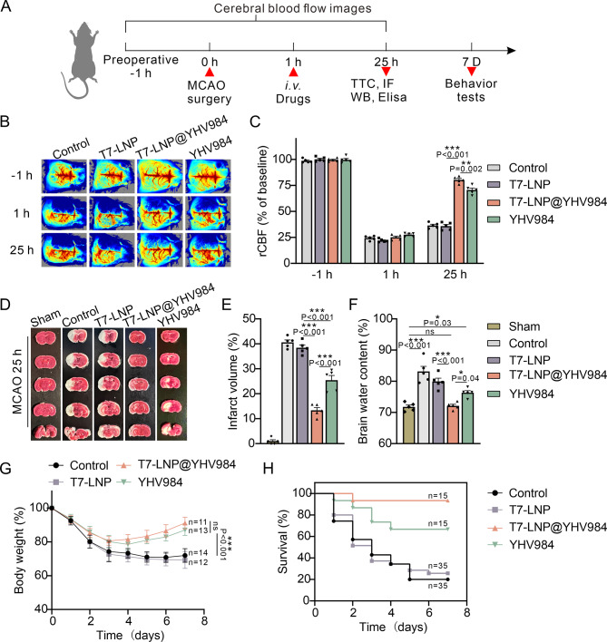
Background: Ischemic stroke (IS) represents a significant global health burden. Current therapeutic options face problems such as window narrowing and reperfusion injury risk. Moreover, with increasing aging and risk factors, novel treatment strategies are urgently needed. NADPH oxidase (NOX)-mediated oxidative stress in microglia is a critical mechanism driving neuroinflammation and cell death. Hv1, a voltage-gated proton channel highly expressed in microglia, synergizes with NOX to generate reactive oxygen species (ROS), exacerbating brain damage. YHV984, a potent Hv1 inhibitor, alleviates post-IS neuroinflammation but faces clinical limitations due to potential toxic side effects and solubility issues. To improve the physicochemical and pharmacokinetic properties of YHV984 for specific Hv1 inhibition in the brain, the multifunctional nanoparticles consisting of a T7-targeting peptide and lipid nanoparticles (LNP) were developed to deliver YHV984 (T7-LNP@YHV984).

Results: The results demonstrated that T7-LNP@YHV984 exhibited good stability and brain targeting capability, effectively crossing the blood-brain barrier (BBB) and accumulating within microglia. This targeted delivery significantly suppressed Hv1 expression and activation of the NLRP3 inflammasome pathway in the damaged brain. Furthermore, it promoted the polarization of microglia towards the M2 phenotype, enhancing the release of anti-inflammatory factors, alleviating neuroinflammation and improved neuronal survival. Additionally, T7-LNP@YHV984 improved survival and facilitated neurological recovery in post-IS mice.

Conclusions: T7-LNP@YHV984 multifunctional nanoparticles with long-term stability emerged as a potent strategy to alleviate reperfusion injury and inhibit neuroinflammation post-IS. By precisely targeting Hv1 in microglia, the nanoparticles effectively suppressed microglia-induced neuroinflammation, minimizing off-target effects. This innovation offers novel insights into stroke treatment and neuroprotective strategies.

Keywords: Brain-targeted LNP; Ischemic stroke; Microglial; Neuroinflammation; Voltage-gated proton channel Hv1.


Conflict of interest statement

Declarations. Ethics approval and consent to participate: All animal experiments were carried out in accordance with the Institute of Laboratory Animal Resources guidelines. Ethics approval was granted by the Ethics Committee of Southern Medical University. Consent for publication: Not applicable. Competing interests: The authors declare no competing interests.

Figures

Fig. 1
Fig. 1
Schematic illustration of the formation of T7-LNP@YHV984 and the mechanism of its alleviation of neuroinflammation post-IS
Fig. 2
Fig. 2
Characterization of T7-LNP@YHV984. (A) Schematic diagram for T7-LNP@YHV984. Dynamic light scattering (B) and Zeta potential analyses (C) of T7-LNP and T7-LNP@YHV984. (D) TEM image of T7-LNP@YHV984. (E) Long-term stability of T7-LNP and T7-LNP@YHV984 at 4 °C. (F) In vitro release behavior of YHV984 in T7-LNP@YHV984. N = 5. Data were expressed as mean ± SEM
Fig. 3
Fig. 3
T7-LNP@YHV984 induced microglia M2-type polarization to inhibit inflammatory progression. (A) Apoptosis levels in BV2 after 24 h of treatment with different particles. (n = 5). (B) Fluorescence intensity statistics of DiR-labeled-7-LNP@YHV984 in the lower chamber (n = 5). (C) After co-incubation for 6 h, the uptake capacity of BV2 for T7-LNP@YHV984 was detected by flow cytometry (n = 5). (D) Representative fluorescence images of DiR-labeled different particles after 6 h of co-culture with BV2. Scale bar: 50 μm. Data were expressed as mean ± SEM. ns, no statistical significance. ***P < 0.001
Fig. 4
Fig. 4
T7-LNP@YHV984 induced microglia M2-type polarization to inhibit inflammatory progression. Quantitative analysis of CD16/32 (A) and CD206 (B) in BV2 after treatment with different samples (n = 5). (C) Western blot analysis of HV1 in OGD-treated BV2 after coincubation with different samples (n = 3). Levels of ROS (D), SOD (E) and GSH-Px (F) secreted by BV2 in each group (n = 5). Levels of pro-inflammatory cytokines IL1-β, IL-6, and TNF-α (G) and anti-inflammatory cell factors TGF-β and IL-10 (H) secreted by BV2 in each group (n = 5). After co-culturing the supernatants of OGD-treated BV2 that had undergone different treatments with primary neurons and HUVEC for 24 h, the apoptosis of neurons (I) and the migration of HUVECs 24 h after wound scratching (J) were observed (n = 5). Data were expressed as mean ± SEM. *P < 0.05, **P < 0.01, ***P < 0.001
Fig. 5
Fig. 5
In vivo distribution of T7-LNP@YHV984 in mice. Representative images (A) and fluorescence intensity (B) of in vivo imaging of DiR-labeled nanoparticles in the brain of tMCAO mice after intravenous injection at different times. Fluorescence images of organs at 8 h post-injection (C) and their quantitative (D). N = 5. Data were expressed as mean ± SEM. *P < 0.05, **P < 0.01. (E) Representative fluorescence images of T7-LNP@YHV984 (Red) and microglia (Green) 8 h after intravenous injection. Scale bar: 50 μm
Fig. 6
Fig. 6
T7-LNP@YHV984 reduced infarct size. (A) Schematic of the experimental protocol. (B) Representative laser speckle images and of tMCAO mice after different treatments (n = 5). (C) Quantitative analysis of regional cerebral blood flow (rCBF) after different treatments (n = 5). Representative images (D) and quantitative analysis of cerebral infarct volume (E) based on TTC stained brain slices (n = 5). (F) Quantitative analysis of brain water content (n = 5). Body weight (G) and survival time (H) of mice in different groups. Data were expressed as mean ± SEM. ns, no statistical significance, *P < 0.05, **P < 0.01, ***P < 0.001
Fig. 7
Fig. 7
T7-LNP@YHV984 promoted neurological recovery post-IS. The mNSS score (A), Bederson score (B) and Zea Longa score (C) of mice in each group 7 days after stroke. Swimming routes (D), escape latency statistics (E) and retention time in the target zone (F) in the Morris water maze test for each group of mice 7 days post-IS. Sample size: Control (n = 11), T7-LNP (n = 13), T7-LNP @YHV984 (n = 13), YHV984 (n = 12). Data were expressed as mean ± SEM. **P < 0.01, ***P < 0.001
Fig. 8
Fig. 8
T7-LNP@YHV984 exhibited excellent anti-inflammatory and pro-neural repair capabilities. Representative immunofluorescence staining images of CD16/32 (A) and CD206 (B) in damaged brain tissues. Scale bar: 50 μm. (C) Flow representation and quantitative analysis of M1- and M2-polarized microglia in brain tissues after different treatments. (D) Representative Western blotting analysis of cleaved Hv1, NOX2 and NLRP3 protein expression in damaged brain tissues. (E) Levels of anti-inflammatory cell factors TGF-β and IL-10 secreted in damaged brain tissues. (F) Immunofluorescence staining of TUNEL (red) and NeuN (green) in damaged brain tissues. Scale bar: 50 μm. N = 5. Data were expressed as mean ± SEM. ns, no statistical significance, **P < 0.01, ***P < 0.001

References

    1. Tu W, Wang L. China stroke surveillance report 2021. Mil Med Res. 2024;11:374–99. - PMC - PubMed
    1. Feigin VL, Stark BA, Johnson CO, et al. Global, regional, and National burden of stroke and its risk factors, 1990–2019: a systematic analysis for the global burden of disease study 2019. Lancet Neurol. 2021;20:795–820. - PMC - PubMed
    1. Tsivgoulis G, Katsanos AH, Sandset EC, et al. Thrombolysis for acute ischaemic stroke: current status and future perspectives. Lancet Neurol. 2023;22:418–29. - PubMed
    1. Walter K. What is acute ischemic stroke? JAMA. 2022;327:885–885. - PubMed
    1. Mizuma A, You JS, Yenari MA. Targeting reperfusion injury in the age of mechanical thrombectomy. Stroke. 2018;49:1796–802. - PMC - PubMed

LinkOut - more resources