. 2025 Jul 1;56(1):129.
doi: 10.1186/s13567-025-01560-6.

Host cellular protein RAB33B facilitates influenza viral replication and modulates M2 trafficking by enhancing autophagy

Affiliations

Host cellular protein RAB33B facilitates influenza viral replication and modulates M2 trafficking by enhancing autophagy

Shaotang Ye et al. Vet Res. .

Abstract

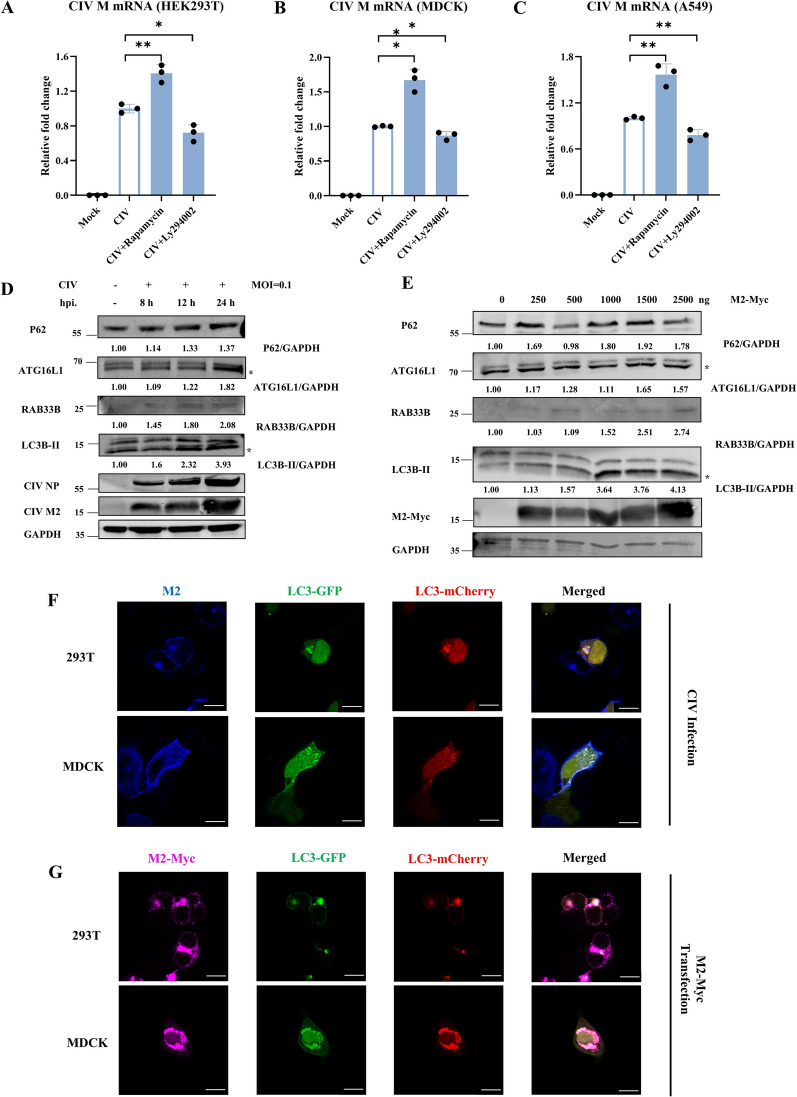
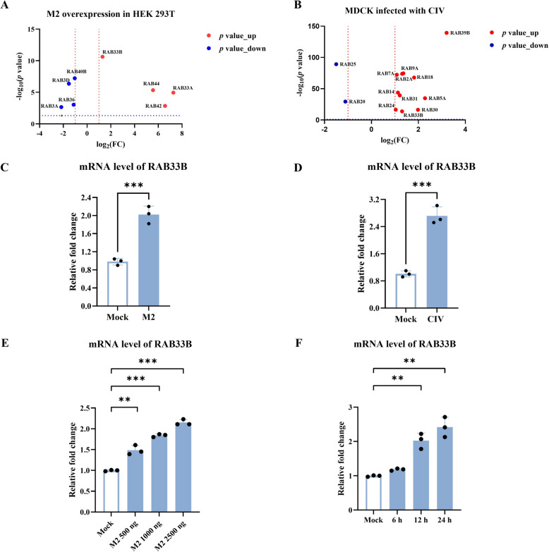
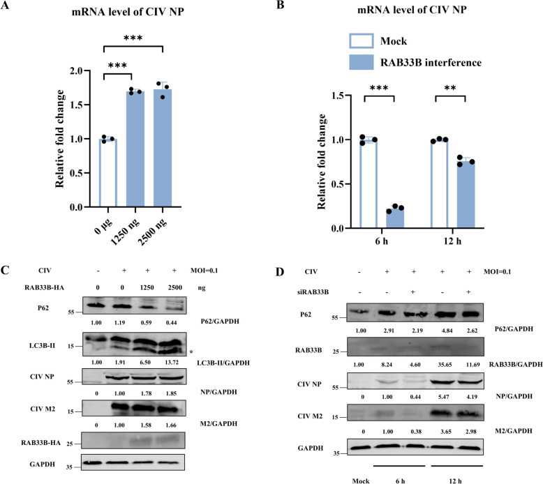
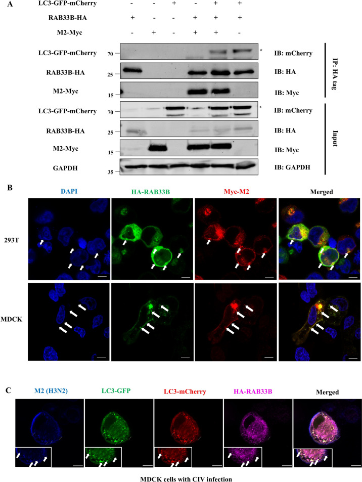
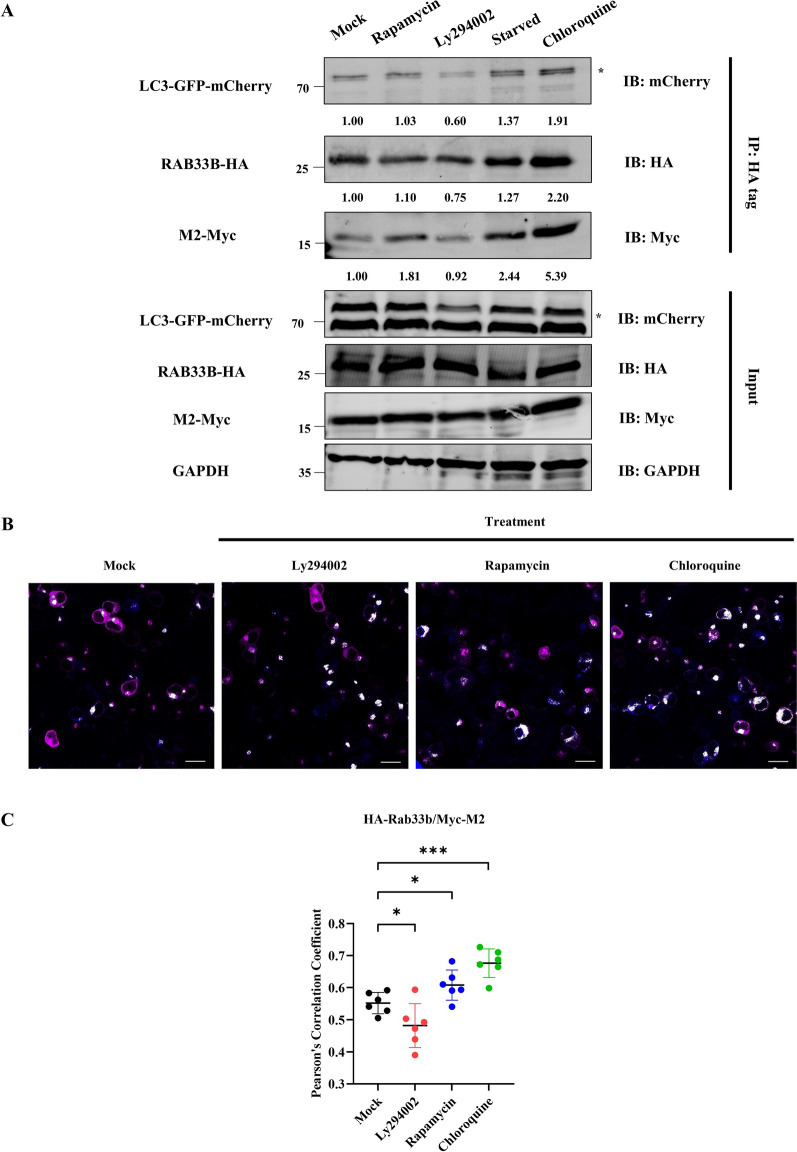
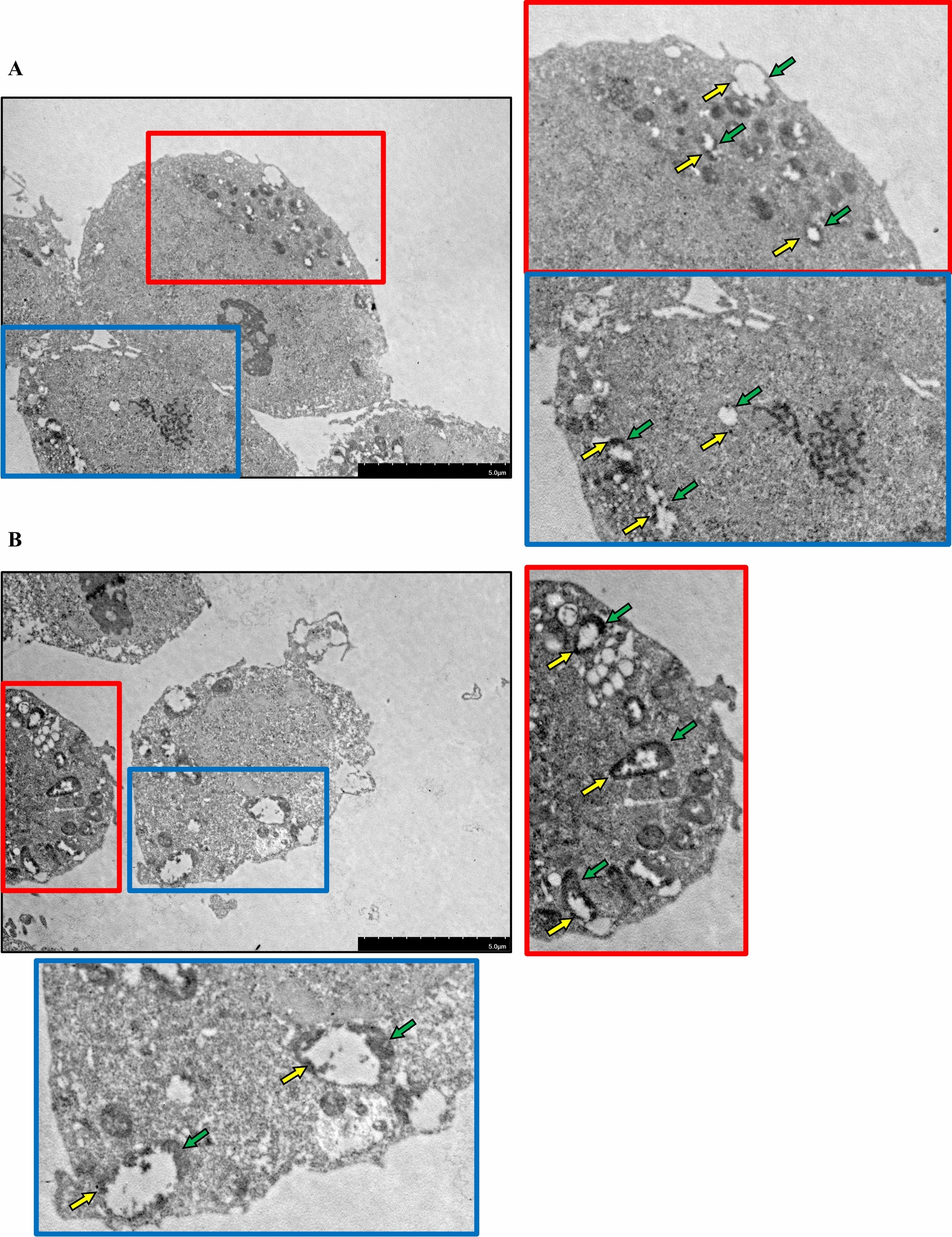
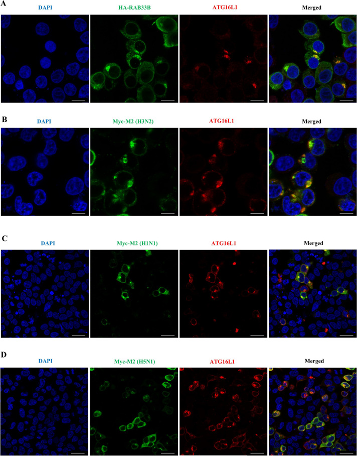
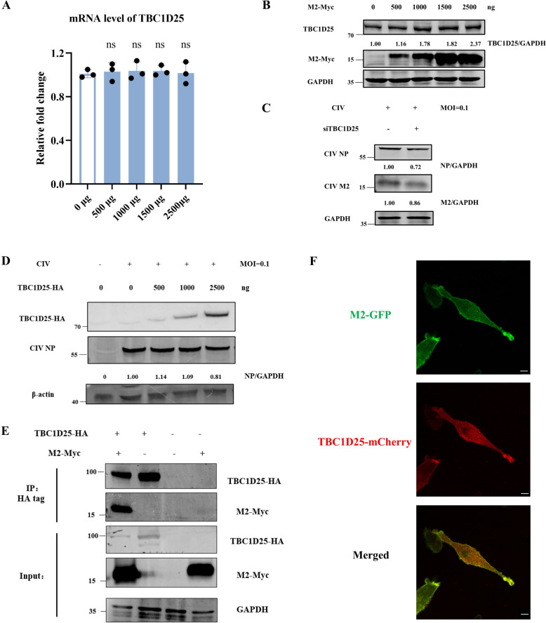
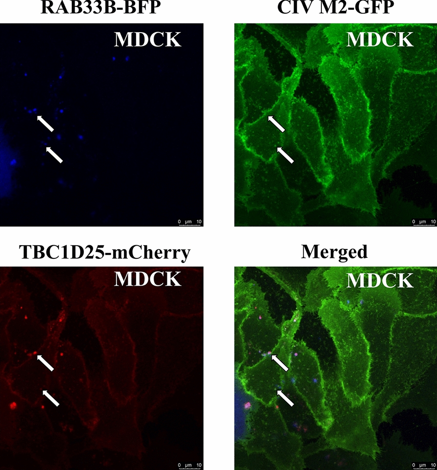
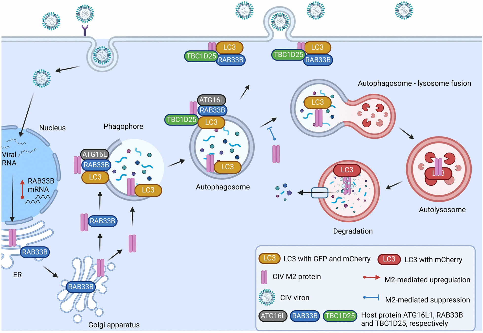
Influenza A virus (IAV) remains a major global health threat. Its M2 protein plays crucial roles in viral fusion, transportation, assembly, and release. Recent studies have shown that IAV impairs host autophagy flux to enhance viral replication. However, the precise mechanisms by which IAV M2 manipulates host cellular autophagy during virus replication remain unclear. In this study, we analysed cellular transcriptional responses of cells to IAV M2 overexpression and identified RAB GTPase protein RAB33B as a key factor. RAB33B was significantly up-regulated by IAV M2 and promoted IAV replication by enhancing autophagy. We further found that autophagy regulates the interaction of IAV M2, RAB33B, and LC3, facilitating M2 membrane trafficking through autophagic-like vesicles. In addition, ATG16L1 (an effector of RAB33B) and TBC1D25 (a GTPase-activating protein for RAB33B) contributed to IAV M2-induced autophagy, thereby affecting viral replication. Collectively, our findings reveal a novel mechanism in which RAB33B is essential for IAV M2 trafficking to the plasma membrane, facilitating viral replication through enhanced autophagy. These insights shed new light on the autophagy-based cellular transport mechanisms of IAV M2 and highlight potential antiviral targets.

Keywords: IAV; M2 protein; RAB33B; autophagy; host–pathogen interaction; membrane trafficking.


Conflict of interest statement

Declarations. Competing interests: The authors declare that they have no competing interests.

Figures

Figure 1
Figure 1
CIV infection induces autophagy. A CIV infection was regulated by the autophagy-modulation drug rapamycin and LY294002 in HEK293T cells, respectively. B CIV infection was regulated by autophagy-modulation drug rapamycin and LY294002 in MDCK cells, respectively. C CIV infection was regulated by autophagy-modulation drug rapamycin and LY294002 in A549 cells, respectively. D HEK293T cells were infected with CIV at MOI = 0.1 for 24 h, and cell lysates were analysed using western blotting. * represents the indicated protein. E HEK293T cells were transfected with different doses of CIV M2 plasmids. Cell lysates were analysed by western blotting. * represents the indicated protein. F HEK293T and MDCK cells were transfected with LC3-GFP-mCherry plasmid for 24 h and then infected with CIV at MOI = 0.1 for 24 h and analysed for the co-localisation of CIV M2 and LC3. Scale bar: 10 μm. G HEK293T and MDCK cells were co-transfected with CIV M2 and LC3-GFP-mCherry plasmids for 24 h, respectively, and analysed for the co-localisation of CIV M2 and LC3. Scale bar: 10 μm.
Figure 2
Figure 2
CIV M2 protein regulates the expression of RAB33B. A Volcano plot visualisation of DEGs of RAB genes in HEK293T cells transfected with the CIV M2 plasmid. B Volcano plot visualisation of DEGs of RAB genes in MDCK cells infected with CIV. C The relative mRNA expression levels of RAB33B were verified for A. D The relative mRNA expression levels of RAB33B were verified for B. E HEK293T cells were transfected with different doses of CIV M2 plasmids for 24 h, and the relative mRNA expression levels of RAB33B were tested. F HEK293T cells were infected with CIV M2 at MOI = 0.1 for 24 h, and the relative mRNA expression levels of RAB33B were detected.
Figure 3
Figure 3
RAB33B facilitates the replication of CIV. A HEK293T cells were transfected with different doses of RAB33B plasmid for 24 h and then infected with CIV at MOI = 0.1 for 24 h. B HEK293T cells were transfected with siRNA of RAB33B for 24 h and then infected with CIV at MOI = 0.1 for 6 h and 12 h, respectively. C HEK293T cells were transfected with different doses of RAB33B plasmid for 24 h and then infected with CIV at MOI = 0.1 for 24 h. Cell lysates were analysed by western blotting. * represents the indicated protein. D HEK293T cells were transfected with siRNA of RAB33B for 24 h and then infected with CIV at MOI = 0.1 for 6 h and 12 h, respectively. Cell lysates were analysed by western blotting.
Figure 4
Figure 4
RAB33B interacts with CIV M2 and LC3 on vesicles. A HEK293T cells were co-transfected with RAB33B-HA, M2-Myc, and LC3-GFP-mCherry plasmids for 24 h. Cell lysates were subjected to co-immunoprecipitation and western blotting. B HEK293T cells were co-transfected with RAB33B-HA and M2-Myc plasmids and analysed for the co-localisation of CIV M2 and LC3. Scale bar: 6 μm. C HEK293T cells were co-transfected with RAB33B-HA and LC3-GFP-mCherry plasmids for 24 h and infected with CIV at MOI = 0.1 for 24 h. Cells were analysed for the co-localisation of CIV M2, RAB33B and LC3. Scale bar: 6 μm. The white arrow indicates the co-localisation.
Figure 5
Figure 5
The interaction of CIV M2, RAB33B and LC3 is regulated by the autophagy process. A HEK293T cells were co-transfected with RAB33B-HA, M2-Myc, and LC3-GFP-mCherry plasmids and then treated with rapamycin, LY294002, starvation treatment and chloroquine, respectively. Cell lysates were subjected to co-immunoprecipitation and western blotting. B HEK293T cells were co-transfected with RAB33B-HA and M2-Myc plasmids and then treated with rapamycin, LY294002 and chloroquine, respectively. The co-localisation between CIV M2 and RAB33B was analysed by Leica LAS X software. Scale bar: 20 μm. The white dots indicated the co-localisation between CIV M2 and RAB33B. C The Pearson’s correlation coefficient was analysed from six different fields of vision for B, respectively.
Figure 6
Figure 6
Immunogold transmission electron microscopy observation for the CIV-infected HEK293T cells. A represents the mock group. B represents the cells transfected with RAB33B plasmid. Scale bar: 5 μm. The yellow arrow indicated the CIV M2, and the green arrow indicated the autophagic-like vesicles.
Figure 7
Figure 7
The redistribution of ATG16L1 induced by RAB33B and M2, respectively. A HEK293T cells were transfected with RAB33B-HA. Scale bar: 10 μm. B HEK293T cells were transfected with H3N2 CIV M2 plasmid. Scale bar: 10 μm. C HEK293T cells were transfected with H1N1 IAV M2 plasmid. Scale bar: 20 μm. D HEK293T cells were transfected with the H5N1 subtype of IAV M2 plasmid. Scale bar: 20 μm.
Figure 8
Figure 8
GTPase-activating protein TBC1D25 of RAB33B regulates the CIV replication. A HEK293T cells were transfected with CIV M2 plasmid for 24 h and were analysed by qPCR. B HEK293T cells were transfected with CIV M2 plasmid for 24 h. Cell lysates were analysed by western blotting. C HEK293T cells were transfected with siRNA of TBC1D25 for 24 h and then infected with CIV at MOI = 0.1 for 24 h. Cell lysates were analysed by western blotting. D HEK293T cells were transfected with different doses of TBC1D25 plasmid for 24 h and then infected with CIV at MOI = 0.1 for 24 h. Cell lysates were analysed by western blotting. E HEK293T cells were co-transfected with TBC1D25-HA and M2-Myc plasmids for 24 h. Cell lysates were subjected to co-immunoprecipitation and western blotting. F MDCK-TetOn-M2 cells were transfected with TBC1D25-mCherry and activated to express CIV M2-GFP by doxycycline for 48 h. Cells were analysed for the co-localisation of CIV M2 and TBC1D25. Scale bar: 10 μm.
Figure 9
Figure 9
Interaction among RAB33B, TBC1D25 and CIV M2. MDCK-TetOn-M2 cells were transfected with RAB33B-BFP and TBC1D25-mCherry and activated to express CIV M2-GFP by doxycycline for 48 h. Cells without fixation were directly observed by a confocal laser scanning microscope. The white arrow indicates the co-localisation. Scale bar: 10 μm.
Figure 10
Figure 10
Schematic diagram of proposed mechanisms underlying RAB33B-mediated Influenza A virus M2 trafficking.

Similar articles

References

    1. Joseph U, Su YCF, Vijaykrishna D, Smith GJD (2017) The ecology and adaptive evolution of influenza A interspecies transmission. Influenza Other Respi Viruses 11:74–84 - PMC - PubMed
    1. Wasik BR, Voorhees IEH, Parrish CR (2021) Canine and feline influenza. Cold Spring Harb Perspect Med 11:a038562 - PMC - PubMed
    1. Guo F, Roy A, Wang R, Yang J, Zhang Z, Luo W, Shen X, Chen R, Irwin DM, Shen Y (2021) Host adaptive evolution of avian-origin H3N2 canine influenza virus. Front Microbiol 12:655228 - PMC - PubMed
    1. Chen M, Lyu Y, Wu F, Zhang Y, Li H, Wang R, Liu Y, Yang X, Zhou L, Zhang M, Tong Q, Sun H, Pu J, Liu J, Sun Y (2023) Increased public health threat of avian-origin H3N2 influenza virus caused by its evolution in dogs. eLife 12:e83470 - PMC - PubMed
    1. Manzoor R, Igarashi M, Takada A (2017) Influenza A virus M2 protein: roles from ingress to egress. Int J Mol Sci 18:2649 - PMC - PubMed

LinkOut - more resources