. 2025 Jun 20;53(12):gkaf642.
doi: 10.1093/nar/gkaf642.

Mechanistic insights into direct DNA and RNA strand transfer and dynamic protein exchange of SSB and RPA

Affiliations

Mechanistic insights into direct DNA and RNA strand transfer and dynamic protein exchange of SSB and RPA

Tapas Paul et al. Nucleic Acids Res. .

Abstract

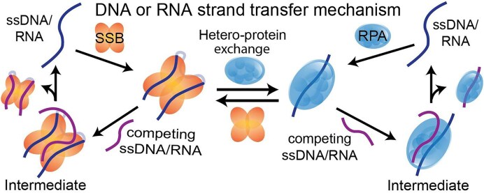
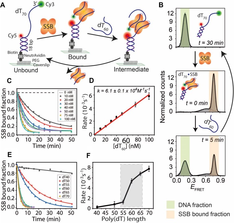
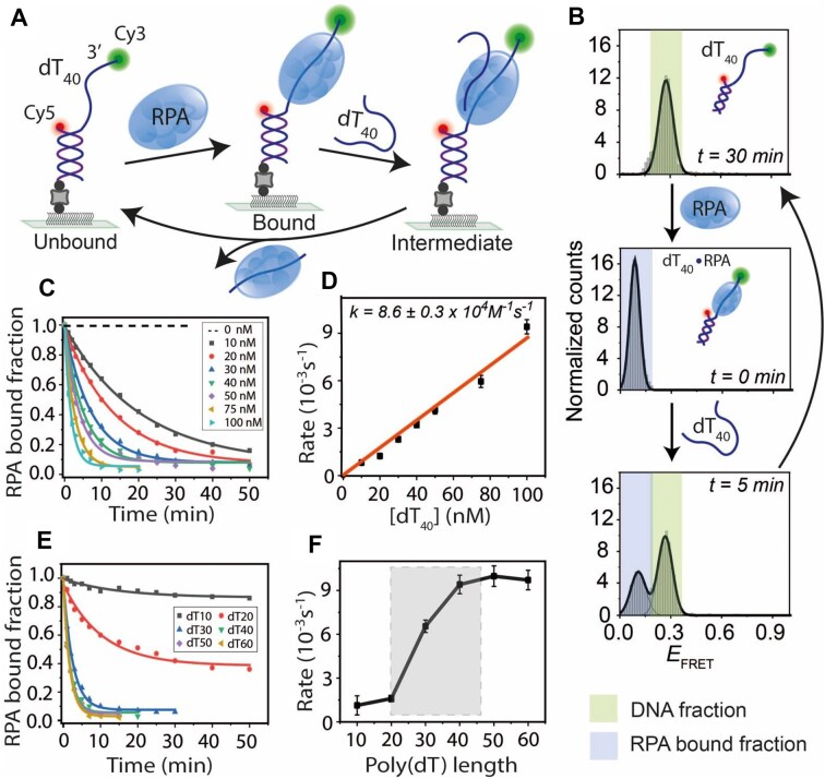
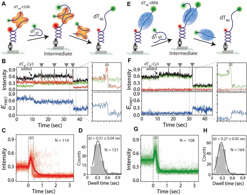
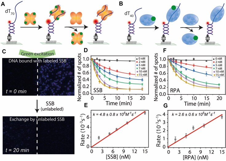
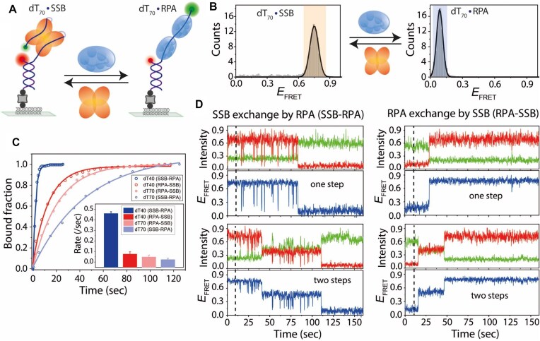
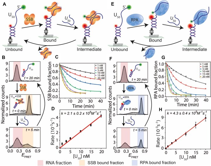
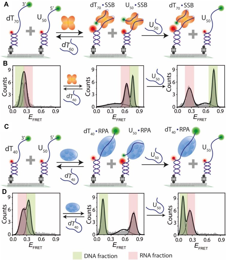
Single-stranded DNA-binding proteins (SSBs) are essential for genome stability, facilitating replication, repair, and recombination by binding single-stranded DNA (ssDNA), recruiting other proteins, and dynamically relocating in response to cellular demands. Using single-molecule fluorescence resonance energy transfer assays, we elucidated the mechanisms underlying direct strand transfer from one locale to another, protein exchange, and RNA interactions at high resolution. Both bacterial SSB and eukaryotic replication protein A (RPA) exhibited direct strand transfer to competing ssDNA, with rates strongly influenced by ssDNA length. Strand transfer proceeded through multiple failed attempts before a successful transfer, forming a ternary intermediate complex with transient interactions, supporting a direct transfer mechanism. Both proteins efficiently exchanged DNA-bound counterparts with freely diffusing molecules, while hetero-protein exchange revealed that SSB and RPA could replace each other on ssDNA, indicating that protein exchange does not require specific protein-protein interactions. Additionally, both proteins bound RNA and underwent strand transfer to competing RNA, with RPA demonstrating faster RNA transfer kinetics. Competitive binding assays confirmed a strong preference for DNA over RNA. These findings provide critical insights into the dynamic behavior of SSB and RPA in nucleic acid interactions, advancing our understanding of their essential roles in genome stability, regulating RNA metabolism, and orchestrating nucleic acid processes.


Conflict of interest statement

None declared.

Figures

Graphical Abstract
Graphical Abstract
Figure 1.
Figure 1.
SSB binding and length-dependent direct transfer kinetics on ssDNA. (A) Schematic of smFRET constructs showing a partial DNA duplex with a 70-nt poly(dT) overhang (dT70). Sequential addition of SSB binds to the ssDNA overhang, and competing free ssDNA (dT60) facilitates strand transfer to regenerate the tethered DNA. The binding and transfer reactions were conducted in a buffer containing 300 mM NaCl. (B) FRET histograms of dT70 before (top), after SSB binding (middle), and during SSB transfer (bottom) upon dT60 addition at the indicated time points. Time t = 30 min (top histogram) represents the complete strand transfer of bound SSB. (C) Single-exponential fitting of the SSB-bound fraction at varying concentrations of competing dT60. (D) Linear fit of SSB transfer rates at different dT60 concentrations. (E) Single-exponential fitting of SSB-bound fractions with competing ssDNA of different lengths (dT40 to dT70 at 100 nM). (F) SSB transfer rates plotted against competing ssDNA length, with a significant rate jump highlighted by the shaded box.
Figure 2.
Figure 2.
Length-dependent binding kinetics and direct transfer of budding yeast RPA on ssDNA. (A) Schematic of smFRET constructs with a partial duplex containing a 40-nt poly(dT) overhang (dT40). RPA binds to dT40 and competing free ssDNA (dT40) induces strand transfer to regenerate the tethered DNA. The binding and transfer reactions were conducted in a buffer containing 100 mM NaCl. (B) FRET histograms of dT40 before (top), after RPA binding (middle), and during RPA transfer (bottom) upon dT40 addition at indicated times. Time t = 30 min (top histogram) represents the complete strand transfer of bound RPA. (C) Single-exponential fitting of RPA-bound fractions at different dT40 concentrations. (D) Linear fit of RPA transfer rates at varying dT40 concentrations. (E) Single-exponential fitting of RPA-bound fractions with competing ssDNA of different lengths (dT10 to dT60 at 100 nM). (F) RPA transfer rates plotted against competing ssDNA length, with a significant rate jump highlighted by the shaded box.
Figure 3.
Figure 3.
Real-time direct transfer mechanism of SSB and RPA in between ssDNA. Schematics showing SSB (A) or RPA (E) transferring from tethered DNA to Cy3-labeled competing ssDNA. The transfer reactions were conducted in a buffer containing 100 mM NaCl. Real-time smFRET traces showing SSB (B) and RPA (F) transfer events. Arrows indicate Cy3–ssDNA flow; spikes represent transfer attempts (triangles), and boxed regions show successful transfers with intensity and FRET efficiency changes. The background intensity increases after addition of competing Cy3-labeled ssDNA. Traces represent total intensity, Cy5 intensity, Cy3 intensity, and FRET efficiency, respectively. Two arrows at the zoomed box represent the binding to successful transfer. (C, G) Combined time traces of successful transfers synchronized at the binding event, the moment of intensity increase. The bold line is the average of individuals. Gaussian fits of dwell time histograms for successful transfers of SSB (D) and RPA (H), with corresponding dwell times (Δt) indicated (calculated from the zoomed box, time difference between two arrows).
Figure 4.
Figure 4.
DNA-bound protein exchange dynamics of SSB and RPA. Schematic representation of SSB (A) and RPA (B) protein–protein exchange. Labeled proteins bound to DNA are replaced by unlabeled proteins. The binding and exchange reactions were conducted in a buffer containing 300 mM NaCl for SSB and 100 mM NaCl for RPA. (C) Representative field of view showing labeled SSB at t = 0 min (top) and after exchange by unlabeled protein at t = 20 min (bottom) under green laser excitation. Single-exponential fits of labeled protein disappearance at different unlabeled protein concentrations for SSB (D) and RPA (F). Linear fits of protein exchange rates at varying unlabeled protein concentrations for SSB (E) and RPA (G).
Figure 5.
Figure 5.
Hetero-protein exchange dynamics between SSB and RPA on ssDNA. (A) Schematic smFRET model of hetero-protein exchange between SSB and RPA. The binding and exchange reactions were conducted in a buffer containing 300 mM NaCl. (B) FRET histograms of dT70 showing SSB and RPA binding and their exchange. (C) Single-exponential fits of hetero-protein exchange (SSB to RPA and vice versa) on dT40 and dT70 at 100 nM protein concentrations. (D) Real-time smFRET traces showing SSB-to-RPA and RPA-to-SSB exchanges on dT70, with distinct FRET transition behaviors. Dashed lines indicate the addition of the competing protein.
Figure 6.
Figure 6.
RNA binding and strand transfer dynamics of SSB and RPA. (A, E) Schematics of smFRET constructs with a partial duplex containing a 50-nt poly-uracil overhang (U50). SSB (A) or RPA (E) binds U50, followed by transfer to competing RNA (U50). The binding and transfer reactions were conducted in a buffer containing 100 mM NaCl. FRET histograms of U50 before (top) and after (middle) SSB (B) or RPA (F) binding, and during SSB or RPA transfer (bottom) upon U50 addition at indicated times. Time t = 20 min (top histogram) represents the complete strand transfer of bound SSB or RPA. Single-exponential fits of protein-bound fractions during RNA-induced transfer at different U50 concentrations for SSB (C) and RPA (G). Linear fits of transfer rates at varying U50 concentrations for SSB (D) and RPA (H).
Figure 7.
Figure 7.
Binding competition between DNA and RNA for SSB and RPA. (A, C) Schematic of smFRET constructs with co-immobilized DNA (dT70/dT40) and RNA (U50). SSB (A) or RPA (C) binds to both DNA and RNA, with subsequent transfer to competing ssDNA (dT60 for SSB, dT40 for RPA) or U50. The binding and transfer reactions were conducted in a buffer containing 100 mM NaCl. FRET histograms showing DNA and RNA before and after SSB (B) or RPA (D) binding. Competing ssDNA facilitates complete transfer of protein from RNA and DNA, whereas competing RNA transfers only RNA-bound protein. Colors represent respective bound fractions.

Update of

Similar articles

References

    1. Dueva R, Iliakis G Replication protein A: a multifunctional protein with roles in DNA replication, repair and beyond. NAR Cancer. 2020; 2:zcaa022. 10.1093/narcan/zcaa022. - DOI - PMC - PubMed
    1. Broderick S, Rehmet K, Concannon C et al. Eukaryotic single-stranded DNA binding proteins: central factors in genome stability. Subcell Biochem. 2010; 50:143–63. 10.1007/978-90-481-3471-7_8. - DOI - PubMed
    1. Antony E, Lohman TM Dynamics of E. coli single stranded DNA binding (SSB) protein–DNA complexes. Semin Cell Dev Biol. 2019; 86:102–11. 10.1016/j.semcdb.2018.03.017. - DOI - PMC - PubMed
    1. Meyer RR, Laine PS The single-stranded DNA-binding protein of Escherichia coli. Microbiol Rev. 1990; 54:342–80. 10.1128/mr.54.4.342-380.1990. - DOI - PMC - PubMed
    1. Wang Y-R, Guo T-T, Zheng Y-T et al. Replication protein A plays multifaceted roles complementary to specialized helicases in processing G-quadruplex DNA. iScience. 2021; 24:102493. 10.1016/j.isci.2021.102493. - DOI - PMC - PubMed

MeSH terms

LinkOut - more resources