. 2025 Jun 20;53(12):gkaf584.
doi: 10.1093/nar/gkaf584.

A-to-I mRNA editing in bacteria can affect protein sequence, disulfide bond formation, and function

Affiliations

A-to-I mRNA editing in bacteria can affect protein sequence, disulfide bond formation, and function

Liron Didi et al. Nucleic Acids Res. .

Abstract

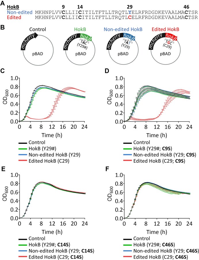
Adenosine-to-inosine (A-to-I) mRNA editing can alter genetic information at the RNA level and is known to affect protein sequence and function in eukaryotes. However, the ability of A-to-I mRNA editing to recode protein sequences in bacteria was never shown at the protein level. Furthermore, the effect of A-to-I mRNA editing itself-not by A-to-G DNA-mimicking mutations-on protein function was never demonstrated in bacteria. Here, we show at the RNA and protein levels that A-to-I mRNA editing directly recodes a tyrosine to a cysteine residue in the toxin HokB (part of the HokB/sokB toxin-antitoxin system in Escherichia coli). Consequently, the toxicity of edited HokB increases, inducing bacterial death or early entrance into the stationary phase, depending on its expression level. Furthermore, we demonstrate that in vivo disulfide bond formation underlies the effect of A-to-I mRNA editing on HokB function, suggesting that A-to-I mRNA editing constitutes a novel mechanism to regulate disulfide bond formation in bacteria. Finally, we observe that A-to-I mRNA editing of hokB is conserved in pathogenic bacteria, supporting functional importance with possible clinical relevance. Our work reveals that A-to-I mRNA editing can constitute a novel mechanism to regulate protein sequence, disulfide bonds, and function in bacteria.


Conflict of interest statement

None declared.

Figures

Graphical Abstract
Graphical Abstract
Figure 1.
Figure 1.
A-to-I mRNA editing can affect protein sequence and function in bacteria as evidenced by the case of HokB. (A) Adenosine is deaminated to inosine, which is similar to guanosine in its base-pair properties. (B) A-to-I mRNA editing by TadA in E. coli occurs in 80%–90% of endogenously expressed hokB transcripts at the logarithmic phase, and is assumed to recode a tyrosine to a cysteine codon at position 29 of HokB. (C) Growth analysis of E. coli (Top10-DH10B) co-expressing mCherry (control, black) or mCherry-HokB (green), with either GFP-TadA (left panel) or GFP (right panel). The mean and standard error of three biological replicates conducted on different days (N = 3), each with 21 technical replicates, are shown. The expression of mCherry and HokB was induced from the beginning of the experiment (time point “0”) with 0.2% arabinose from a pBAD vector. Expression of GFP-TadA or GFP was induced with 1 mM IPTG from a pME6032 vector. (D) A-to-I mRNA editing identification by next-generation sequencing (NGS; Illumina; amplicon-seq) of plasmid-borne hokB RNA (cDNA) co-overexpressed with GFP-TadA or with GFP alone. Minimum observed reads coverage per sample that passed our quality filters ≥17 515 391. Statistical analysis was conducted using Student’s t-test; P-value <.0001 (****). (E) A-to-I mRNA editing validation by Sanger sequencing of plasmid-borne DNA and RNA (cDNA) of hokB when co-overexpressed with GFP-TadA or with GFP. The sequence above the chromatograms represents the gene (DNA) sequence. A black arrow marks the double peak of A and G(I) that was observed only in the cDNA (complementary DNA) samples. Note that the G(I) peak is higher than the A peak when overexpressing GFP-TadA and vice versa when overexpressing only GFP. (F) MS/MS spectrum of non-edited (Y29; top) and edited (C29; bottom) HokB peptides found in strains co-overexpressing HokB with GFP (top) or GFP-TadA (bottom). Black arrows mark identified peptides and their mass in the MS/MS spectra that show a mass shift corresponding to tyrosine or cysteine at the edited site. The gray arrow marks an example of a peptide and its mass in the MS/MS spectra that does not include the edited site (same mass). All peptides were discovered with false discovery rate (FDR) ≤ 0.01. The peaks weight, font size, and axis were adjusted from the original figure for better visualization and comparison. A comprehensive mass distribution and the original MS/MS spectra and data can be found in Supplementary Tables S2–S4.
Figure 2.
Figure 2.
DNA-encoded cysteine residues are essential for the toxicity of edited HokB. (A) The protein sequence of non-edited and edited HokB according to their respective transcript. DNA-coded cysteines are shown in bold. (B) A description of the different plasmids containing different versions of HokB used in the growth assay is presented. (C) Growth analysis of E. coli (Top10-DH10B) WT strain expressing the HokB (Y29#, green), non-edited HokB (Y29, blue), and edited HokB (C29, red) fused to mCherry reporter protein (N-terminus) from the plasmid shown in panel (B). As a reference control, a plasmid harboring only mCherry was used (black). As previously reported [37], when highly expressed, edited HokB (C29) induces the highest level of toxicity. (D) Growth analysis as in panel (C), with all three versions of HokB having the C9S substitution. (E) Growth analysis as in panel (C), with all three versions of HokB having the C14S substitution. (F) Growth analysis as in panel (C), with all three versions of HokB having the C46S substitution. In all growth experiments, protein expression was induced from the beginning of the experiment (time point “0”) with 0.2% arabinose from a pBAD vector. The mean and standard error of three biological replicates conducted on different days (N = 3), each with 21 technical replicates, are shown.
Figure 3.
Figure 3.
In vivo disulfide bond formation is essential for the toxicity of the edited HokB. (A) Growth analysis of an E. coli ΔdsbA strain that expresses one of three versions of HokB, fused to mCherry from an inducible plasmid. As a reference control, we used a plasmid encoding only mCherry. (B) Growth analysis as in panel (A), but with overexpressing DsbA from a second plasmid (pME6032). (C) Growth analysis, as in panel (B), using an empty plasmid (pME6032 with no dsbA insert).
Figure 4.
Figure 4.
Western blot analysis supports that A-to-I mRNA editing mediates an intramolecular disulfide bond between C29 and C46 in HokB. (A) Western blot of membrane enriched protein fraction of E. coli (Top10-DH10B) WT strain expressing either mCherry only (control; black) or the HokB (Y29#, green), non-edited HokB (Y29, blue), and edited HokB (C29, red) fused to mCherry (N-terminus) from the plasmid shown in Fig. 2B. (B) Same as panel (A) but with the C9S substitution in the different expressed HokB versions. (C) Same as panel (A) but with the C14S substitution in the different expressed HokB versions. (D) Same as panel (A) but with the C46S substitution in the different expressed HokB versions.
Figure 5.
Figure 5.
High levels of edited HokB induce bacterial death. (A) Strains were allowed to grow without induction for 4 h. Then, arabinose was added to induce HokB protein expression (the black arrow marks the induction time). Presented are OD values over time. The expression of mCherry and HokB was induced with 0.2% arabinose from a pBAD vector. The mean and standard errors of three biological replicates conducted on different days (N = 3), each with 21 technical replicates, are shown. Black and white triangles correspond to sampling times for panel (B). (B) CFU (colony forming unit) counts before and after 1 h of induction (CFUs at 4 and 5 h). Only 0.3% of viable bacteria survived after 1 h of edited HokB induction (corresponding to the death of 99.7% of viable bacteria). The mean and standard error of four biological replicates conducted on different days (N = 4) are shown. Statistical analysis was conducted using Student’s paired t-test followed by Benjamini–Hochberg FDR correction: P-value ≤.05 (*).
Figure 6.
Figure 6.
Lower levels of edited HokB induce early entrance to the stationary phase. (A) Growth analysis of WT E. coli as described in Fig. 2C with 1:1000 lower arabinose concentration. The expression of mCherry and HokB was induced from the beginning of the experiment (time point “0”) with 0.0002% arabinose from a pBAD vector. Black and white triangles correspond to sampling times for panel (B). (B) CFU counts at 5 and 6 h of the beginning of growth. Notice that there are fewer CFUs when edited HokB is expressed, with similar numbers at 5 and 6 h after growth. The mean and standard error of four biological replicates conducted on different days (N = 4) are shown. Statistical analysis was conducted using Student’s paired t-test followed by Benjamini–Hochberg FDR correction: P-value ≤.05 (*).
Figure 7.
Figure 7.
A-to-I mRNA editing of hokB is conserved in pathogenic E. coli and Shigella strains. Sanger sequencing of the endogenous hokB gene and its mRNA from the same sample of non-pathogenic E. coli (used throughout this work), enterohemorrhagic E. coli, enteropathogenic E. coli, uropathogenic E. coli, and Shigella sonnei. A black arrow marks the double peak of A and G(I) observed only in the cDNA samples. Note that the G(I) peak (black) is higher than the A peak (green) in most samples. Sequences were aligned to the E. coli reference genome (NC_000913.3) and positions 1491982–1491990 are shown. See Supplementary Fig. S10 for exact genomic coordinates of the full-length hokB gene in each species.

References

    1. Picardi E, D’Erchia AM, Lo Giudice C et al. REDIportal: a comprehensive database of A-to-I RNA editing events in humans. Nucleic Acids Res. 2017; 45:D750–7. 10.1093/nar/gkw767. - DOI - PMC - PubMed
    1. Oakes E, Anderson A, Cohen-Gadol A et al. Adenosine deaminase that acts on RNA 3 (ADAR3) binding to glutamate receptor subunit B pre-mRNA inhibits RNA editing in glioblastoma. J Biol Chem. 2017; 292:4326–35. 10.1074/jbc.M117.779868. - DOI - PMC - PubMed
    1. Paz N, Levanon EY, Amariglio N et al. Altered adenosine-to-inosine RNA editing in human cancer. Genome Res. 2007; 17:1586–95. 10.1101/gr.6493107. - DOI - PMC - PubMed
    1. Eisenberg E, Levanon EY A-to-I RNA editing—immune protector and transcriptome diversifier. Nat Rev Genet. 2018; 19:473–90. 10.1038/s41576-018-0006-1. - DOI - PubMed
    1. Ramaswami G, Li JB RADAR: a rigorously annotated database of A-to-I RNA editing. Nucleic Acids Res. 2014; 42:D109–13. 10.1093/nar/gkt996. - DOI - PMC - PubMed

MeSH terms

LinkOut - more resources