. 2025 Jun 20;53(12):gkaf572.
doi: 10.1093/nar/gkaf572.

Differential oligomerization regulates PHF13 chromatin affinity and function

Affiliations

Differential oligomerization regulates PHF13 chromatin affinity and function

Francesca Rossi et al. Nucleic Acids Res. .

Abstract

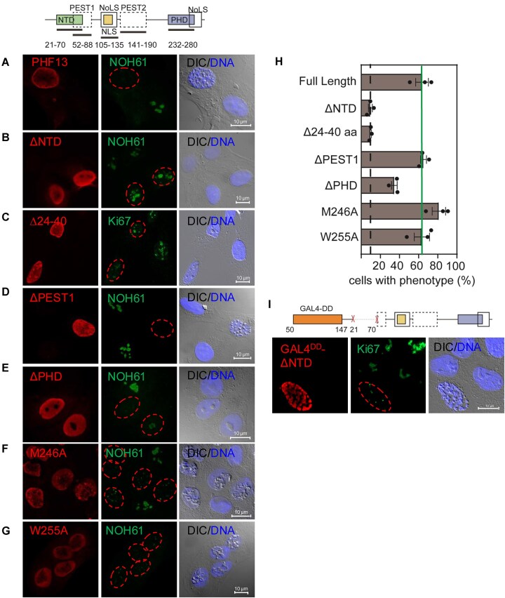
PHF13 is a H3K4me3 epigenetic reader that modulates key chromatin processes including transcription, DNA damage response, and chromatin architecture. PHF13 is found aberrantly regulated in different cancers and its misexpression alters the epigenetic landscape of key transcription factors that regulate epithelial-to-mesenchymal transition. In this study, we sought to understand how PHF13's chromatin affinity and diverse chromatin functions are intrinsically regulated. Our results show that PHF13 can oligomerize via conserved ordered regions in its N- and C- terminus increasing its chromatin valence and avidity, promoting polymer-polymer phase separation (PPPS) and chromatin inaccessibility. Impressively, a ∼3- to 5-fold overexpression of PHF13 was sufficient to globally compact chromatin visible by optical microscopy, dependent on its ordered dimerizing regions and oligomerization potential. Unexpectedly, we discovered that PHF13 can self-associate independent of its ordered domains via intrinsically disordered regions, which conversely reduced PHF13's chromatin affinity, formed liquid-liquid phase separated (LLPS) condensates, and differentially impacted gene expression. Our findings support that there is an intrinsic balance between PHF13's ordered and disordered regions and that PHF13 can phase transition between polymer-polymer and liquid-liquid phase separation states to impact chromatin structure and function.


Conflict of interest statement

D.H. is a founder and scientific advisor of Nuage Therapeutics.

Figures

Graphical Abstract
Graphical Abstract
Figure 1.
Figure 1.
PHF13 represses gene expression in an NTD-dependent manner. (A) Schematic overview of PHF13 domain structure. (B–  E) Luciferase assays: Luciferase reporter gene expression in H1299 cells from a GAL4-responsive promoter (B and C) after transfection with GAL4 alone (pM2) or GAL4 fusion proteins (PHF13, PHF13ΔNTD, PHF13ΔPHD, or E1B) or from p53 regulated cyclin G and Mdm2 promoters (D and E) that were co-transfected with p53 and the indicated pcDNA-4TO expression vectors for 24 h. E1B served as a positive control. Immunoblots of the expressed proteins are shown above each plot (D and E). All values were normalized to Renilla expression (B–E).
Figure 2.
Figure 2.
PHF13‘s NTD mediates PHF13 homo-dimerization. (A and B) In silico analysis: Merged PONDR and Tango plots of PHF13 measuring disorder and order, respectively (A). Red and black dotted lines represent the cut offs for calling disorder and aggregation potential, respectively (A). AlphaFold2_advanced analysis of PHF13 dimers (shown as a string model and contact map). The confidence of the called structure is colour coded (B). (C and D) Schematic of constructs used in IP:WB. Flag-IP:WB of cells co-expressing EGFP-PHF13 and Flag-PHF13 full-length or 1–150, 100–200, or 150–300 deletion proteins (C). Flag IP: WB from lysates co-expressing Flag-PHF13 with YFP, YFP-PHF13, YFP-ΔNTD, YFP-NTD, or YFP-Δ24–20 (D). (E) GST-pulldowns with purified recombinant GST, GST-PHF13, GST-PHF13-ΔNTD, and GST-PHF13-ΔPHD incubated with nuclease digested chromatin lysates and immunoblotted for GST and H3K4me3. (F) WB of fractionated lysates from cells expressing PHF13, PHF13ΔNTD, and PHF13-GAL4DDΔNTD and detected with PHF13 (rat mAb ID3), Tubulin, and H3 antibodies.
Figure 3.
Figure 3.
PHF13 oligomerizes via N- and C-terminal dimerization. (A) GFP-Trap:WB from cells co-expressing Flag-PHF13 with YFP, YFP-PHF13, or C-terminal YFP-PHF13 mutant proteins (Δ7;150–300, Δ7-ΔPHD, Δ7-M246A, and Δ-W255A). (B) FACS-FRET analysis of HEK 293 cells co-transfected with YFP and CFP fusion proteins. 10% FRET signal was arbitrarily defined as the minimum threshold and is denoted by a dotted line. (C) Graphical representation and immunodotblot of the size exclusion elution profile (Superose 6 10/300) from GST-cleaved recombinant PHF13 full-length (FL) and deletion proteins (ΔPHD, ΔNTD and Δ24–40). Graphs represent quantified signals obtained by dot blotting of individual fractions and quantification by Image quant.
Figure 4.
Figure 4.
PHF13 drives global chromatin condensation. (AH) IF: Phase contrast (A–C and F) and DIC imaging (D) of U2OS (A and C–H) or U2OS H2B-GFP (B) cells transfected with PHF13 (A–F), YFP-PHF13 (G), or CFP-PHF13 (H) for 24 h and paraformaldehyde fixed (A–E and G–H) or pre-extracted prior to fixation (F). PHF13 was detected with rabbit peptide Ab CR53 (A and F) or rat mAb 6F6 (B–E) and DNA was stained with Dapi (A, B, and F) or with DRAQ5 (D). Images were captured on a Zeiss Axiophot Phase Fluorescence microscope (A, B, andF), a Zeiss Z1 Observer (E, G, and H) or a Zeiss ConfoCor 2 LSM5 microscope (D). (I) Graphical depiction of the penetrance of the chromosome condensation phenotype for low (1- to 5-fold) and high (5- to 15-fold) PHF13-expressing cells in comparison to nontransfected endogenous PHF13-expressing cells. Dotted line represents background signal in nontransfected cells.
Figure 5.
Figure 5.
PHF13 oligomerization is required for chromatin compaction. (AG and I) IF staining of paraformaldehyde fixed U2OS cells, transfected with PHF13 (A), PHF13ΔNTD (B), PHF13Δ24-40 (C), PHF13ΔPEST1 (D), PHF13ΔPHD (E), PHF13-M246A (F), PHF13-W225A (G), and PHF13-GAL4DDΔNTD (I). PHF13 was stained with rat mAb 6F6 (A and E–G) or 1D3 (B–D and I) and DNA was stained with DRAQ5. Nucleoli were detected with NOH61 or Ki67 antibodies. Images were captured on a Zeiss Confocor 2 LSM5 microscope. (H) Graphical depiction of the penetrance of the chromosome condensation phenotype for PHF13 full-length, PHF13ΔNTD, PHF13Δ24-40, PHF13ΔPHD, PHF13-M246A, PHF13-W225A, and PHF13ΔPEST1 overexpressing mutants in U2OS cells. Black dotted line represents background signal in nontransfected cells.
Figure 6.
Figure 6.
Molecular dynamic simulations and model of PHF13 induced condensation. (A) PHF13 is modeled as 3 beads representing the NTD, NLS, and PHD, which mediates PHF13 self-interaction and the interactions with chromatin and with chromatin interaction partners. (B and C) IP:WB GFP-TRAP from U2OS cells expressing YFP-tagged proteins and probed for the co-precipitation of endogenous cohesin proteins (B) or endogenous PRC1 proteins (C). (D and E) Polymer folding dynamics from initially randomly open configurations is monitored by the gyration radius (D). When all PHF13 interactions are enabled and no indirect C-terminal dimerization with a chromatin partner is considered, the polymer stably folds into compacted structures (E) and the gyration radius sharply decreases (PHF13, red curve; D). Deletion of the PHD (ΔPHD) or the NTD domain (ΔNTD) prevent polymer compaction, which remains in an open conformation (D and E). (F and G) Polymer model end states of cohesin/PRC1-only (F) or PHF13 + cohesin/PRC1 (G). (H) Model: PHF13 is able to oligomerize via direct N-terminal dimers. This oligomerization creates a PHF13 polymer with alternating DNA binding (cation–π interactions) and H3K4me1/2/3 binding (PHD domain) regions allowing it to spread along chromatin fiber and facilitate linear compaction which may be coupled to cohesin or PRC1 (gray ovals) long distance looping, to promote global chromosome condensation.
Figure 7.
Figure 7.
PHF13 oligomerizes via its IDRs. (A) Overlap of PONDR and TANGO plots depicting deletion (X) of TANGO aggregation regions. (B and C) FlagIP:WB of Flag-PHF13 co-expressed with different YFP-PHF13 full-length and C-terminal (Δ7;150–300) mutant proteins. Flag-IPs were probed by immunoblot for their ability to co-IP the YFP-proteins, detected with GFP and Flag antibodies. (D) IF: U2OS cells expressing YFP-PHF13, YFP-PHF13Δ24-40, YFP-PHF13Δ24-40_Δ272-280 and imaged on a Zeiss Z1 Observer to evaluate protein localization. (E) WB of fractionated lysates (Cytoplasm-Cy, Nucleoplasm-Nu, and Chromatin-Chr) and quantification (% of PHF13/fraction) of cells expressing YFP-PHF13, YFP-PHF13Δ24-40_Δ232-238, and YFP-PHF13Δ24-40_Δ272-280 and detected with GFP, Vinculin, and SMC3 antibodies. (F) Luciferase reporter gene expression in H1299 cells from p53 regulated cyclin G and MDM2 co-transfected with p53 and the indicated pcDNA-4TO expression vectors for 24 h.
Figure 8.
Figure 8.
PHF13 phase separates in a PEST2 (IDR3) dependent manner. (A and B) In vitro phase condensate forming assays, demonstrate that recombinant GST-PHF13 can turn a PEG-salt solution turbid (A) and form condensates visible by light microscopy (B) in contrast to equimolar amounts of GST-only (A and B). (C and D) IF: imaging of endogenous PHF13 under wild-type (C and D) or treated conditions (0.5 μg/ml AMD- 4 h or 40 mM LiCl-6 h) (C) showing PHF13’s ability to form polymers or condensates, under different cellular conditions. Cells were imaged on a Leica Stellaris in confocal mode (D) or on a Zeiss Axiophot Phase Fluorescence microscope (C). PHF13 was stained with rat mAb 6F6 (D and F, lower panel) or rabbit polyclonal CR53 (C). (E) WB of fractionated lysates (Cytoplasm-Cy, Nucleoplasm-Nu, and Chromatin-Chr) and quantification (% PHF13/fraction) from cells expressing YFP-PHF13, YFP-PHF13ΔPEST1, and YFP-PHF13ΔPEST2 and detected with GFP, Vinculin, SMC3, and H3 antibodies. (F) Direct and indirect microscopy of YFP-PHF13ΔPEST1, YFP-PHF13ΔPEST2, and 4TO-PHF13 ΔPEST2. (G) Quantification of the penetrance of condensation of 4TO-PHF13, 4TO-PHF13ΔPEST1, 4TO-PHF13ΔPEST2, and PHF13Δ8 (ΔIDR5). Values are derived from three biological replicates.
Figure 9.
Figure 9.
PHF13 PPPS and LLPS states differentially impact chromatin accessibility and gene expression. (A and B) PCA of the ATAC-seq (A) and RNA-seq expression profiles (B) of U2OS cells expressing various PHF13 variants. (C) Heatmap and line plot representation of ATAC-Seq read densities of all samples centered at TSS within 3 kb window around. The signal represents the read counts normalized to the spike-in. (D) Bar plot showing the number of DEGs when tested against wild-type control sample (left) and tested against to EYFP control sample (right). (E) Heatmap and clustering analysis of RNA-Seq data. Cluster numbers are indicated on the left. (F) Gene ontology enrichment analysis heatmap for biological process of heatmap clusters for top 10 GOs for each cluster. (G) Genome tracks for ATAC-seq and RNA-seq data of two down-regulated genes in WT samples compared to EYFP control (left) and two up-regulated (right). The samples are color-coded as described above. (H). Comparison of z-scores across genes related to Pol-I/nucleoli and Pol II regulation in both ATAC-seq and RNA-seq.

Similar articles

References

    1. Chung HR, Xu C, Fuchs A et al. PHF13 is a molecular reader and transcriptional co-regulator of H3K4me2/3. eLife. 2016; 5:e10607. 10.7554/eLife.10607. - DOI - PMC - PubMed
    1. Hofmann S, Dehn S, Businger R et al. Dual role of the chromatin-binding factor PHF13 in the pre- and post-integration phases of HIV-1 replication. Open Biol. 2017; 7:2395–413. 10.1098/rsob.170115. - DOI - PMC - PubMed
    1. Kuderna AK, Reichel A, Tillmanns J et al. Discovery of a novel antiviral effect of the restriction factor SPOC1 against Human Cytomegalovirus. Viruses. 2024; 16:363. 10.3390/v16030363. - DOI - PMC - PubMed
    1. Schreiner S, Kinkley S, Burck C et al. SPOC1-mediated antiviral host cell response is antagonized early in human adenovirus type 5 infection. PLoS Pathog. 2013; 9:e1003775. 10.1371/journal.ppat.1003775. - DOI - PMC - PubMed
    1. Mund A, Schubert T, Staege H et al. SPOC1 modulates DNA repair by regulating key determinants of chromatin compaction and DNA damage response. Nucleic Acids Res. 2012; 40:11363–79. 10.1093/nar/gks868. - DOI - PMC - PubMed

LinkOut - more resources