

The Impact of VCAM-1 expression on left ventricular performance following acute coronary syndromes
- PMID: 40599142
- PMCID: PMC12207695
- DOI: 10.25122/jml-2025-0083
The Impact of VCAM-1 expression on left ventricular performance following acute coronary syndromes
Abstract
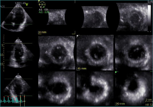
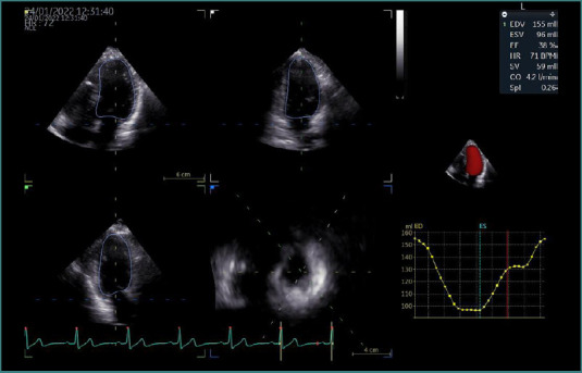
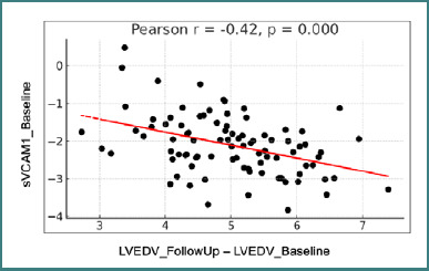
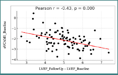
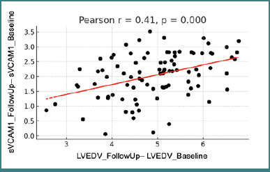
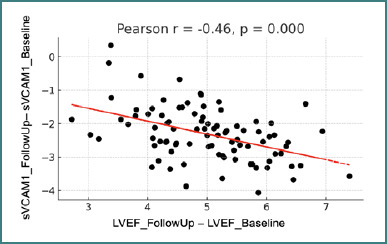
Inflammatory pathways, particularly those involving vascular cell adhesion molecule-1 (VCAM-1), play a central role in post-ischemic myocardial remodeling. However, its relationship with left ventricle (LV) performance or volumetric changes has not been systematically examined. This study aimed to investigate the association between circulating VCAM-1 levels, LV volumes, and LV ejection fraction (LVEF), quantified using three-dimensional echocardiography (3-DE), in patients with acute coronary syndrome (ACS). All patients underwent comprehensive clinical evaluation and 3-DE assessment within the first 24 hours of hospital admission. Concurrently, a full panel of locally available laboratory tests was performed, including serum sampling for VCAM-1 analysis. A follow-up evaluation, comprising repeated biological and echocardiographic measurements, was conducted two months after the index event. A total of 90 patients with ACS (mean age 54 ± 9 years; 75 males) were included in the analysis. Among these, 30 patients (33.3%) had a ≥10% increase in left ventricular end-diastolic volume (LVEDV) at follow-up, indicative of adverse left ventricular remodeling. Baseline VCAM-1 levels were significantly correlated with subsequent changes in LVEDV and LVEF from admission to follow-up (r = -0.42, P < 0.05, and r = -0.43, P < 0.05, respectively). Furthermore, the dynamic changes in VCAM-1 between assessments also showed significant correlations with changes in LVEDV and LVEF (r = 0.41, P < 0.05; r = -0.46, P < 0.05). This study suggests that VCAM-1, an inflammatory biomarker, may be a prognostic indicator of LV remodeling and dysfunction in patients with acute coronary syndromes. The findings support further exploration of VCAM-1 for risk stratification and therapy.
Keywords: 3-DE: 3-Dimensional Echocardiography; 3D echocardiography; ACS: Acute Coronary Syndrome; AMI: Acute Myocardial Infarction; CMR: Cardiac Magnetic Resonance; CV: Cardiovascular; EF: Ejection Fraction; LV: Left Ventricle; LVEDV: Left Ventricular End-Diastolic Volume; LVEF: Left Ventricular Ejection Fraction; PCI: Percutaneous Coronary Intervention; SPSS: Statistical Package for the Social Sciences; STEMI: ST-Elevation Myocardial Infarction; Svcam-1: Soluble(Serum) Vascular Cell Adhesion Molecule-1; VCAM-1; VCAM-1: Vascular Cell Adhesion Molecule-1; acute coronary syndrome; left ventricular remodeling.
© 2025 The Author(s).
Conflict of interest statement
The authors declare no conflict of interest.
Figures
References
-
- Bossowska A, Kiersnowska-Rogowska B, Bossowski A, Galar B, Sowiński P. Ocena stężenia wybranych cząsteczek adhezyjnych (sICAM-1, sVCAM-1, sE-selektyny) w stabilnej i niestabilnej chorobie niedokrwiennej serca oraz w zawale mięśnia sercowego. Przegl Lek. 2003;60(7):445–450. - PubMed
-
- Mashru MR, Shah VK, Soneji SL, Loya YS, Vasvani JB, Payannavar S, Walvalkar A, Mithbawkar SS, Mokal R, Kudalkar K, Abraham A, Thakur PK, Shalia KK. Soluble levels of cell adhesion molecules (CAMs) in coronary artery disease. Indian Heart J. 2010 Jan-Feb;62(1):57–63. - PubMed
MeSH terms
Substances
LinkOut - more resources
Full Text Sources
Miscellaneous