

Potential applications of focused ultrasound for spinal cord diseases: a narrative review of preclinical studies
- PMID: 40599219
- PMCID: PMC12212115
- DOI: 10.1016/j.bas.2025.104298
Potential applications of focused ultrasound for spinal cord diseases: a narrative review of preclinical studies
Abstract
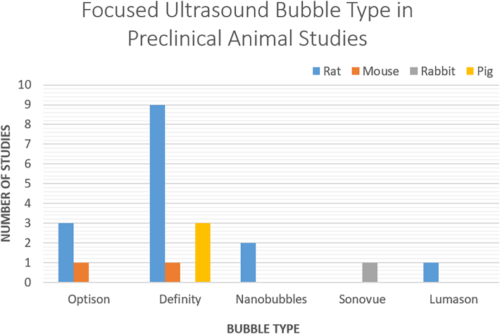
Focused ultrasound (FUS) technology provides unique advantages as a therapy targeting the central nervous system (CNS). We aimed to investigate and summarize the potential applications of FUS in the context of spinal cord diseases. Search strategies were created using a combination of keywords and standardized index terms. Searches were run in April 2025 in the Ovid Cochrane Central Register of Controlled Trials, EBSCO MegaFILE, Ovid Embase, Ovid Medline, Ovid PsycINFO, Scopus, and Web of Science Core Collection to retrieve all relevant studies from inception until 2024. A narrative and comprehensive summary of the current body of evidence was performed. Current preclinical studies indicate the potential use of spinal cord FUS in blood-spinal cord barrier (BSCB) disruption, neuromodulation, and inflammatory regulation following spinal cord injury. Targeted CNS drug delivery with BSCB disruption using FUS has proven promising in the context of neuro-oncology and neurotrauma. Additionally, FUS has been explored for neuromodulation in managing neuropathic pain and spasticity. FUS to the spinal cord may also provide anti-inflammatory effects and alter the local cellular response to injury. While therapeutic FUS ablation of brain structures has been extensively studied, research on similar applications within the spinal cord was less prevalent and faces multiple challenges. FUS is a highly promising technique with multiple advantages and potential applications in the treatment of spinal cord diseases. Current research efforts have shifted focus towards translational studies, while human trials are currently limited.
Keywords: Blood-brain barrier; Blood-spinal cord barrier; Focused ultrasound; High-intensity focused ultrasound; Low-intensity focused ultrasound.
© 2025 The Authors.
Conflict of interest statement
None of the authors declare any conflict of interest.
Figures


Similar articles
-
Technological aids for the rehabilitation of memory and executive functioning in children and adolescents with acquired brain injury.Cochrane Database Syst Rev. 2016 Jul 1;7(7):CD011020. doi: 10.1002/14651858.CD011020.pub2. Cochrane Database Syst Rev. 2016. PMID: 27364851 Free PMC article.
-
Low-intensity focused ultrasound of the spine in the treatment of chronic pain and movement disorder: a scoping review.Front Pain Res (Lausanne). 2025 Jun 17;6:1606672. doi: 10.3389/fpain.2025.1606672. eCollection 2025. Front Pain Res (Lausanne). 2025. PMID: 40599994 Free PMC article.
-
Therapeutics for treating mpox in humans.Cochrane Database Syst Rev. 2023 Mar 14;3(3):CD015769. doi: 10.1002/14651858.CD015769. Cochrane Database Syst Rev. 2023. PMID: 36916727 Free PMC article.
-
Steroids for acute spinal cord injury.Cochrane Database Syst Rev. 2012 Jan 18;1(1):CD001046. doi: 10.1002/14651858.CD001046.pub2. Cochrane Database Syst Rev. 2012. PMID: 22258943 Free PMC article.
-
Systemic pharmacological treatments for chronic plaque psoriasis: a network meta-analysis.Cochrane Database Syst Rev. 2021 Apr 19;4(4):CD011535. doi: 10.1002/14651858.CD011535.pub4. Cochrane Database Syst Rev. 2021. Update in: Cochrane Database Syst Rev. 2022 May 23;5:CD011535. doi: 10.1002/14651858.CD011535.pub5. PMID: 33871055 Free PMC article. Updated.
References
Publication types
LinkOut - more resources
Full Text Sources
Miscellaneous
