Review
. 2025 Jul 1;6(7):e70245.
doi: 10.1002/mco2.70245. eCollection 2025 Jul.

Ten-Eleven Translocation Family Proteins: Structure, Biological Functions, Diseases, and Targeted Therapy

Affiliations
Review

Ten-Eleven Translocation Family Proteins: Structure, Biological Functions, Diseases, and Targeted Therapy

Junzhi Liang et al. MedComm (2020). .

Abstract

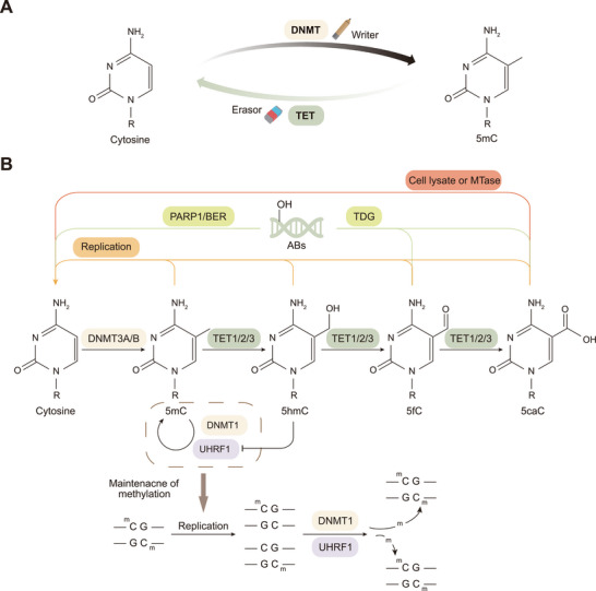
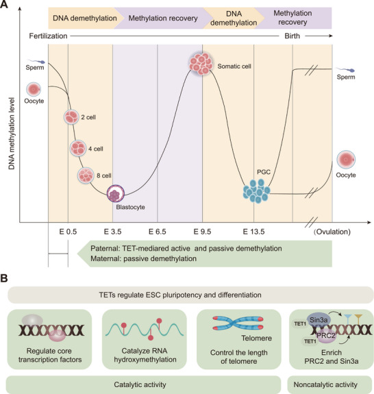
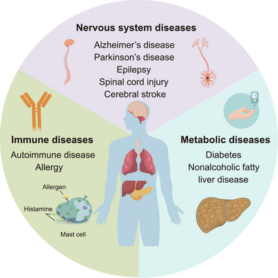
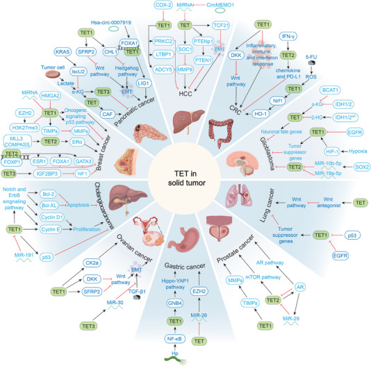
Ten-eleven translocation (TET) family proteins are Fe(II)- and α-ketoglutarate-dependent dioxygenases, comprising three family members: TET1, TET2, and TET3. These enzymes drive DNA demethylation by sequentially oxidizing 5-methylcytosine to 5-hydroxymethylcytosine, 5-formylcytosine, and 5-carboxylcytosine. Through these reactions, TET proteins remodel the epigenetic landscape and interact with transcription factors and RNA polymerase II to regulate gene expression, cell lineage specification, and embryonic development. Mutations and dysregulation of TETs have been associated with the pathogenesis of various diseases, including the nervous system, immune system, and metabolic diseases, as well as cancers. Therapeutic modulation of TETs may be an effective strategy for the treatment of these diseases. Here, we provide a comprehensive overview of the mechanisms by which TET proteins mediate DNA demethylation and detail their biological functions. Additionally, we highlight recent advances in understanding the molecular mechanisms linking TET dysregulation to disease pathogenesis and explore their potential as therapeutic targets. This review supplements the current understanding of the critical role of epigenetic regulation in disease pathogenesis and further facilitates the rational design of targeted therapeutic agents for diseases associated with mutations and dysregulation of TETs.

Keywords: Biological functions; Demethylation; Diseases; Ten‐eleven translocation; Therapeutic strategies.


Conflict of interest statement

The authors declare no competing interests.

Figures

FIGURE 1
FIGURE 1
Mechanisms of DNA methylation and TET‐dependent demethylation. (A) DNMT acts as a DNA methylation writer to add methyl groups to cytosine, and TET proteins act as methylation erasers involved in the precise regulation of the DNA methylation landscape. (B) Cytosine on DNA is converted to 5mC under the catalytic action of DNMT3A/B/C (DNMT3L is involved in regulation). DNMT1 and its accessory factor UHRF1 maintain the methylation of double strands in DNA replication. The demethylation process consists mainly of passive and active forms. Passive demethylation refers to the failure of maintenance of methylation by DNA replication, which eventually manifests itself as 5mC dilution, whereas active demethylation is an ongoing process of research. We summarize the current understanding of the mechanism of three DNA demethylation pathways mediated by TET proteins, including: (1) TDG converts these 5fC and 5caC bases to transient abasic sites and DNA single strand breaks, followed by DNA damage repair by PARP1 in concert with BER proteins; (2) TET‐catalyzed generation of 5hmC inhibits UHRF1 and DNMT1, leading to a failure in maintenance of methylation. This results in DNA breaks, followed by repair of DNA damage by PARP1 in concert with BER proteins; (3) cell lysate and MTases are involved in the direct decarboxylation of 5caC. Abbreviations: DNMT, DNA methyltransferase; TET, ten‐eleven translocation; 5mC, 5‐methylcytosine; MTases, S‐adenosylmethionine‐dependent DNA methyltransferases; PARP1, poly(ADP‐ribose) polymerase 1; BER, base excision repair; ABs, abasic sites; TDG, thymine DNA glycosylase; DNMT3A/B/C, DNA methyltransferase 3A/B/C; DNMT3L, DNA methyltransferase 3‐like protein; 5hmC, 5‐hydroxymethylcytosine; 5fC, 5‐formylcytosine; 5caC, 5‐carboxylcytosine; UHRF1, ubiquitin‐like containing PHD and RING finger domains 1.
FIGURE 2
FIGURE 2
Biological functions of TET. (A) TET proteins are involved in two DNA methylation reprogramming processes. In the first reprogramming, which occurs during the early stages of fertilization, the paternal and maternal genomes undergo independent demethylation processes. The paternal genome undergoes active and then passive demethylation, whereas the maternal genome is passively demethylated in a DNA replication‐dependent manner until the level of DNA methylation is minimized at the blastocyst stage. The second reprogramming occurs during the establishment of PGCs. Methylation gradually recovers after the blastocyst stage. The restored somatic cells are demethylated at the E9.5–E13.5 stage, mediated by TET1 and TET2, and reach the lowest level of DNA methylation around day E13.5. PGCs differentiate to form sperm or eggs in different sexes and undergo different methylation regulation during differentiation. (B) TET proteins are able to regulate ESC pluripotency and differentiation through both catalytic and noncatalytic functions. TET proteins rely on their catalytic activity to regulate ESC pluripotency and differentiation by regulating core transcription factors, catalyzing hydroxylation of RNA, and controlling chromosome length. Meanwhile, TET also enriches PRC2 and Sin3a to participate in the regulation of ESCs through noncatalytic functions. Abbreviations: E, embryonic day; ESC, embryonic stem cell; PGC, primordial germ cell; PRC2, polycomb repressive complex; TET, ten‐eleven translocation.
FIGURE 3
FIGURE 3
Dysregulation of TET proteins are widely involved in the development of noncancer diseases, including nervous system, immune, and metabolic diseases. Although TET predominantly acts as a cancer suppressor, it can also act as an oncogenic factor. Abbreviation: TET, ten‐eleven translocation.
FIGURE 4
FIGURE 4
The role of TET proteins in cancer is heterogeneous. Dysregulation of TET involved in the development of a variety of cancers, including breast, pancreatic, lung, prostate, gastric, and ovarian cancers as well as cholangiocarcinoma, HCC, CRC, and glioblastomas. Abbreviations: TET, ten‐eleven translocation; HCC, hepatocellular carcinoma; CRC, colorectal cancer; MMP, matrix metalloproteinase; TIMPs, tissue inhibitors of metalloproteinases; ERα, estrogen receptor alpha; HMGA2, high mobility group AT‐hook 2; SFRP2, secreted frizzled‐related protein 2; EMT, epithelial–mesenchymal transition; α‐KG, α‐ketoglutarate; CAF, cancer‐associated fibroblast; DKK, Dikkopf; ROS, reactive oxygen species; 5‐FU, 5‐fluorouracil; 2‐HG, 2‐hydroxyglutarate; IDH, isocitrate dehydrogenase; HIF‐1, hypoxia‐inducible factor‐1; BCAT1, branched‐chain amino acid transaminase 1; AR, androgen receptor; Hp, helicobacter pylori; GNB4, guanine nucleotide‐binding protein subunit beta‐4; SFRP2, secreted Fzd receptor protein 2; CK2α, casein kinase II subunit alpha.
FIGURE 5
FIGURE 5
TET2 and its concomitant mutations cause HSPCs to enter the clonal hematopoiesis of indeterminate potential (CHIP, a precancerous lesion) state. TET2 has different mutation rates in CHIP and different hematologic cancers. However, mutations in TET2 alone are not sufficient to cause cancer but are driven by co‐mutations in TET2 and multiple genes, leading to the development of hematologic cancers, which include myeloid neoplasms, MPN, MDS, AML, MDS/MPN, B cell lymphomas, and T cell lymphomas. The high‐frequency and important concomitant mutations in these cancers are further discussed. Abbreviations: HSPC, hematopoietic stem and progenitor cell; TET, ten‐eleven translocation; CHIP, clonal hematopoiesis of indeterminate potential; MPN, myeloproliferative neoplasm; MDS, myelodysplastic syndrome; AML, acute myeloid leukemia.
FIGURE 6
FIGURE 6
Development timeline of TET protein agonists and inhibitors. Since 2009, when TET proteins were first found to catalyze 5mC demethylation, there has been a growing recognition of the critical role of TET in physiology and pathology (especially cancer). Since then, agonists and inhibitors targeting TET have received extensive attention in clinical and basic research. Among them, vitamin C has been widely studied for its potential therapeutic role in hematologic tumors due to its safety and accessibility. In recent years, the development of agonists and inhibitors targeting TET has notably accelerated. Abbreviations: TET, ten‐eleven translocation; 5hmC, 5‐hydroxymethylcytosine; DMOG, dimethyloxallyl glycine; AA6, (S)‐2‐[(2,6‐dichlorobenzoyl) amino] succinic acid; TETi76, 2‐methelene and 4‐hydroxy; AML, acute myeloid leukemia; T‐ALL, T‐cell acute lymphoblastic leukemia.

Similar articles

References

    1. Waddington C. H., “The epigenotype. 1942,” International Journal of Epidemiology 41 (2012): 10–13. - PubMed
    1. Wu Y.‐L., Lin Z.‐J., Li C.‐C., et al., “Epigenetic regulation in metabolic diseases: mechanisms and advances in clinical study,” Signal Transduction and Targeted Therapy 8 (2023): 98. - PMC - PubMed
    1. Ito S., D'alessio A. C., Taranova O. V., Hong K., Sowers L. C., and Zhang Y., “Role of Tet Proteins in 5mC to 5hmC Conversion, ES‐cell Self‐renewal and Inner Cell Mass Specification,” Nature 466 (2010): 1129–1133. - PMC - PubMed
    1. Parry A., Rulands S., and Reik W., “Active Turnover of DNA Methylation During Cell Fate Decisions,” Nature Reviews Genetics 22 (2021): 59–66. - PubMed
    1. Fu T., Liu L., Yang Q.‐L., et al., “Thymine DNA Glycosylase Recognizes the Geometry Alteration of minor Grooves Induced by 5‐formylcytosine and 5‐carboxylcytosine,” Chemical Science 10 (2019): 7407–7417. - PMC - PubMed

LinkOut - more resources