

A Combined Transtemporal and High-parietal Approach for Large Intraventricular Trigone Meningioma: A Case Series and Review of the Literature
- PMID: 40599307
- PMCID: PMC12208790
- DOI: 10.2176/jns-nmc.2025-0031
A Combined Transtemporal and High-parietal Approach for Large Intraventricular Trigone Meningioma: A Case Series and Review of the Literature
Abstract
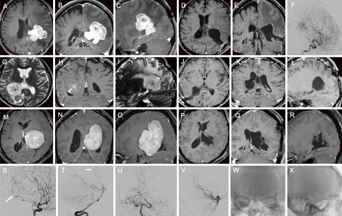
The trigone of the lateral ventricle is deep and surrounded by eloquent gyri and subcortical fibers. Resection of intraventricular trigone tumors has therefore been challenging, and the optimal surgical approach to the trigone of the lateral ventricle remains controversial. Three patients with large intraventricular trigone meningioma (≥4 cm in diameter) underwent surgical excision using a combined transtemporal and high-parietal approach at Osaka City University Hospital between July 2016 and January 2021. Clinical and imaging studies, as well as surgical complications, were retrospectively reviewed based on medical records from our institution. We also reviewed 153 patients with intraventricular trigone meningioma from 11 reports in the literature and assessed pre- and postoperative symptoms. Gross total resection of the tumor was achieved in all cases. None of the patients showed deterioration of neurological symptoms at 3 months after tumor resection, although one patient experienced transient language dysfunction several weeks after surgery. No cases showed recurrence or required additional therapy. According to our literature review, postoperative visual field defects are more likely to persist than postoperative language dysfunction at 3 months postoperatively. In conclusion, combining the transtemporal and high-parietal approaches appears to be useful for treating large intraventricular trigone meningioma. Postoperative language dysfunction after resection of intraventricular trigone meningioma may tend to resolve more rapidly than postoperative visual field defect.
Keywords: high-parietal approach; meningioma; transtemporal approach; trigone.
© 2025 The Japan Neurosurgical Society.
Conflict of interest statement
All authors have no conflicts of interest.
Figures
Similar articles
-
Systemic pharmacological treatments for chronic plaque psoriasis: a network meta-analysis.Cochrane Database Syst Rev. 2021 Apr 19;4(4):CD011535. doi: 10.1002/14651858.CD011535.pub4. Cochrane Database Syst Rev. 2021. Update in: Cochrane Database Syst Rev. 2022 May 23;5:CD011535. doi: 10.1002/14651858.CD011535.pub5. PMID: 33871055 Free PMC article. Updated.
-
Systemic pharmacological treatments for chronic plaque psoriasis: a network meta-analysis.Cochrane Database Syst Rev. 2017 Dec 22;12(12):CD011535. doi: 10.1002/14651858.CD011535.pub2. Cochrane Database Syst Rev. 2017. Update in: Cochrane Database Syst Rev. 2020 Jan 9;1:CD011535. doi: 10.1002/14651858.CD011535.pub3. PMID: 29271481 Free PMC article. Updated.
-
Does Augmenting Irradiated Autografts With Free Vascularized Fibula Graft in Patients With Bone Loss From a Malignant Tumor Achieve Union, Function, and Complication Rate Comparably to Patients Without Bone Loss and Augmentation When Reconstructing Intercalary Resections in the Lower Extremity?Clin Orthop Relat Res. 2025 Jun 26. doi: 10.1097/CORR.0000000000003599. Online ahead of print. Clin Orthop Relat Res. 2025. PMID: 40569278
-
What Are the Recurrence Rates, Complications, and Functional Outcomes After Multiportal Arthroscopic Synovectomy for Patients With Knee Diffuse-type Tenosynovial Giant-cell Tumors?Clin Orthop Relat Res. 2024 Jul 1;482(7):1218-1229. doi: 10.1097/CORR.0000000000002934. Epub 2023 Dec 28. Clin Orthop Relat Res. 2024. PMID: 38153106 Free PMC article.
-
Drugs for preventing postoperative nausea and vomiting in adults after general anaesthesia: a network meta-analysis.Cochrane Database Syst Rev. 2020 Oct 19;10(10):CD012859. doi: 10.1002/14651858.CD012859.pub2. Cochrane Database Syst Rev. 2020. PMID: 33075160 Free PMC article.
References
-
- Staneczek W, Jänisch W. Epidemiologic data on meningiomas in East Germany 1961-1986: incidence, localization, age and sex distribution. Clin Neuropathol. 1992;11(3):135-41. - PubMed