. 2025 Jun 18:52:511-528.
doi: 10.1016/j.bioactmat.2025.06.017. eCollection 2025 Oct.

Biosynthesis of a dual growth factors (GFs) functionalized silk sericin hydrogel to promote chronic wound healing in diabetic mice

Affiliations

Biosynthesis of a dual growth factors (GFs) functionalized silk sericin hydrogel to promote chronic wound healing in diabetic mice

Hanxin Deng et al. Bioact Mater. .

Abstract

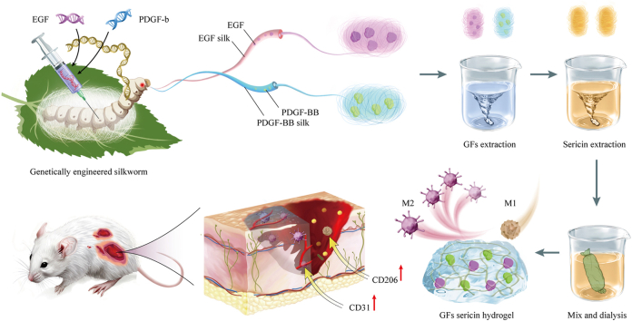
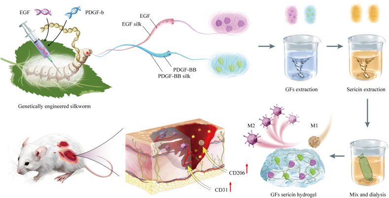
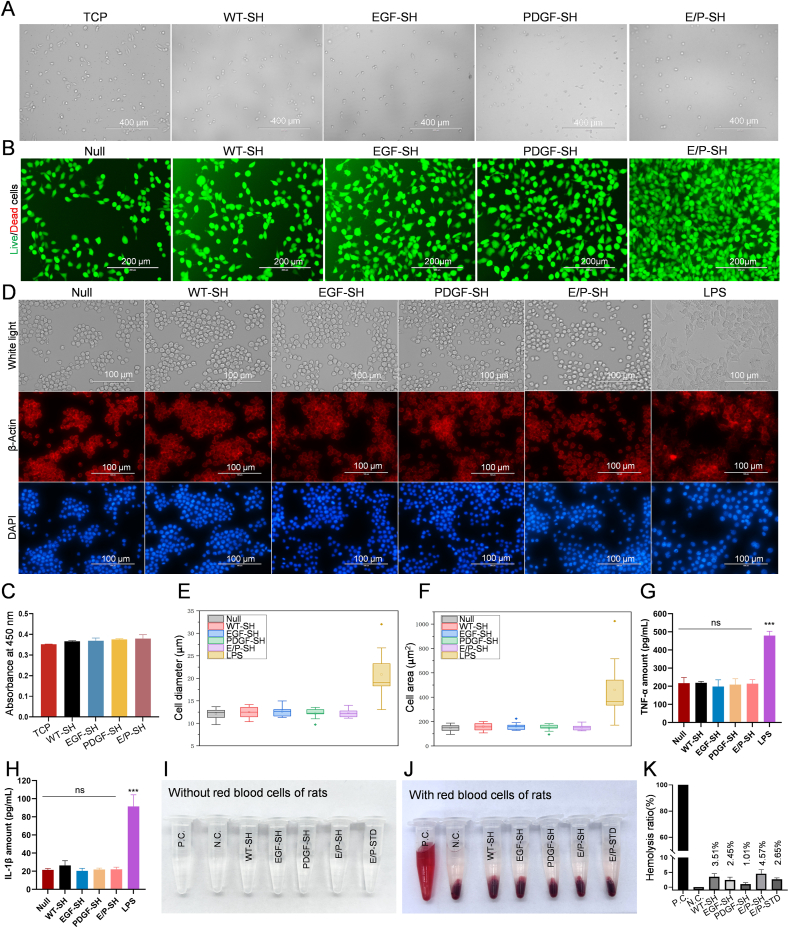
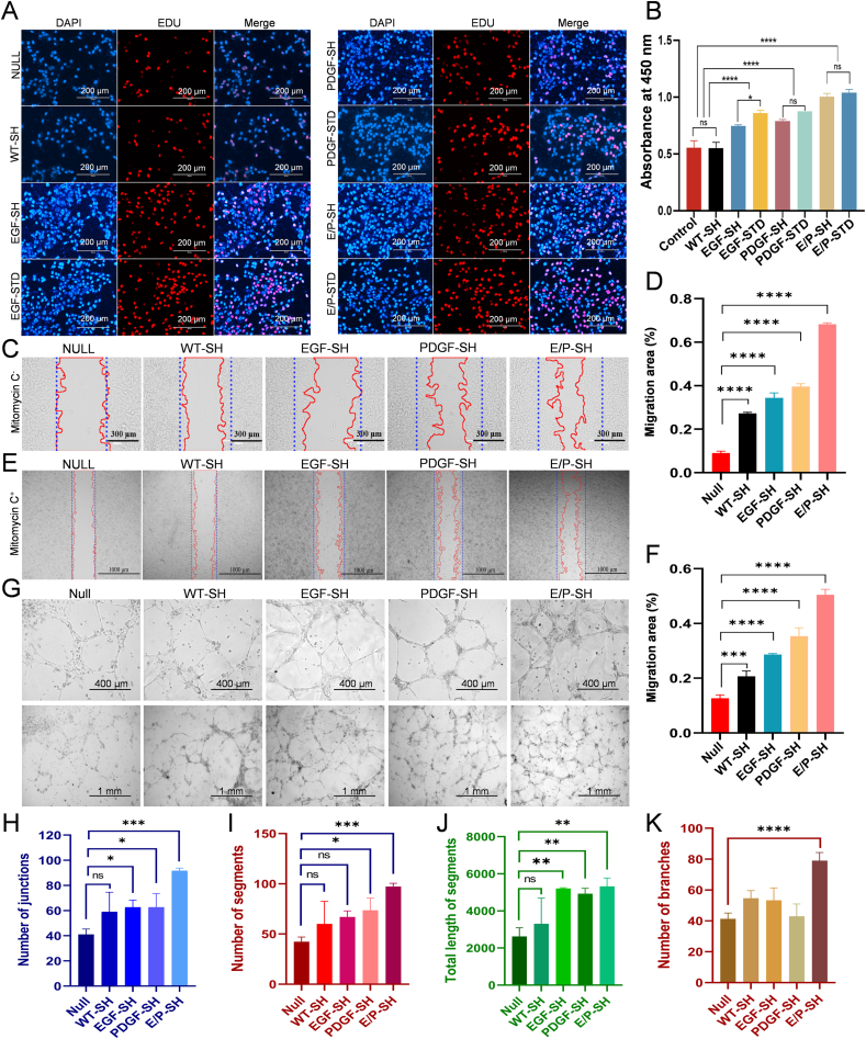
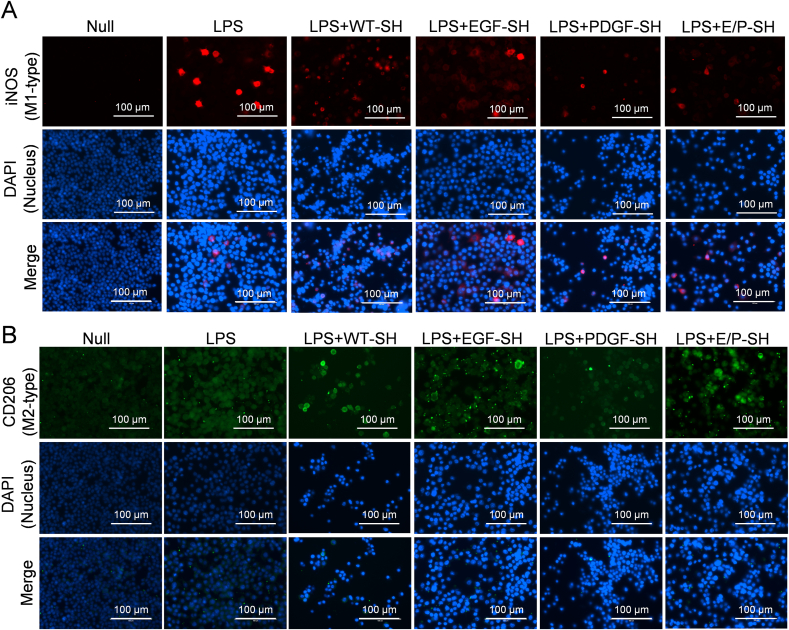
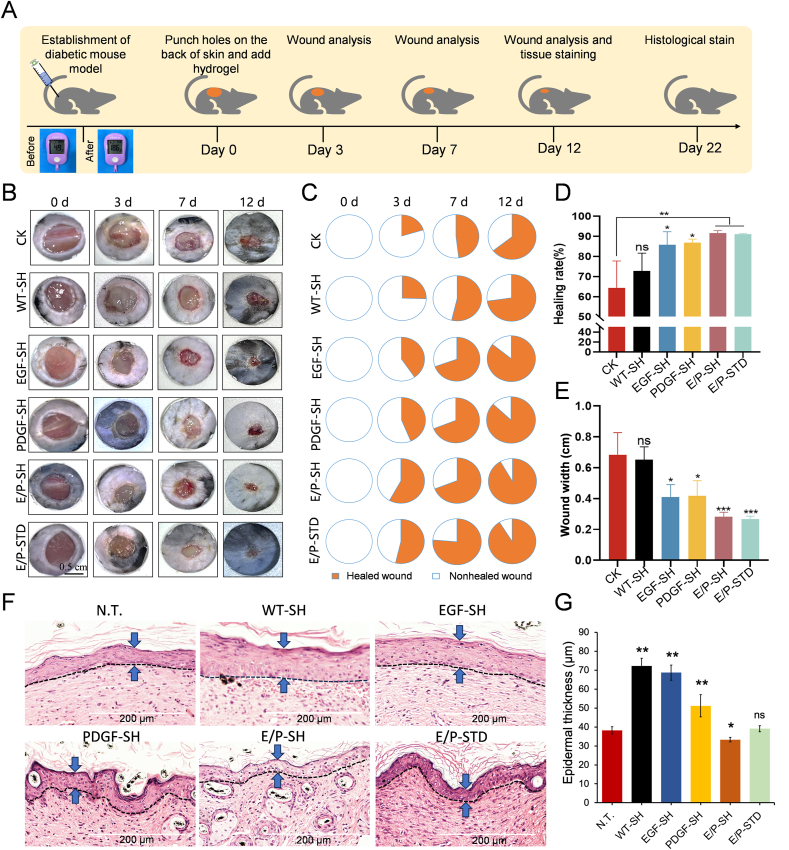
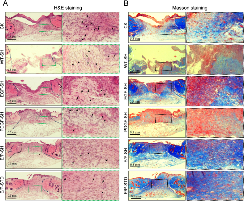
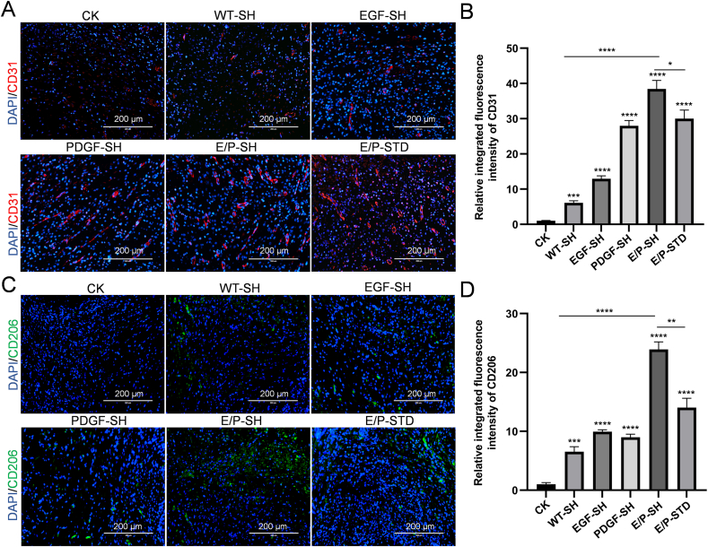
Chronic non-healing wounds, such as diabetic foot, pressure sores and bedsores have seriously affected the life quality of patients worldwide. GFs provide a potential solution to promote chronic wound healing by promoting cell proliferation and differentiation. However, limited resource, high cost, and instability in vivo greatly hindered their clinical applications. In present study, two silk gland bioreactor silkworm stains were generated to successfully synthesize functional silk fibers incorporating high expressions of EGF and PDGF-BB. The two GFs functionalized silk raw materials were used to fabricate a dual GFs sericin hydrogel (E/P-SH) delivering system with tunable material performances for better cell adhesion and biocompatibility, sustainable release of the dual GFs to synergistically promote cell proliferation and migration, which realized the significant healing of chronic full-thickness skin wound in diabetic mice within 12 days with more organized collagen arrangement and better epithelialization degree by reducing inflammatory response and promoting vascularization. These findings demonstrated that the biosynthesized dual GFs-SH delivering system provides an opportunity to broaden the wide utility of GFs in clinical treatment of diabetic wound healing.

Keywords: Biosynthesis; Cell proliferation; Chronic diabetic wound healing; Growth factors; Sericin hydrogels.


Conflict of interest statement

The authors declare no conflict of interest.

Figures

Image 1
Graphical abstract
Scheme 1
Scheme 1
The present study illustrates the genetical engineering of transgenic silkworms for synthesizing the EGF and PDGF-BB silk fibers, which were subsequently used as raw materials to fabricate a sericin hydrogel based dual GFs delivering system (E/P-SH). The developed E/P-SH was successfully used to treat the chronic non-healing wounds in diabetic mice within 12 days with significant therapeutic effects by reducing inflammatory response and promoting vascularization.
Fig. 1
Fig. 1
Design and fabrication of sericin hydrogels with tunable material performances to meet the demand of diabetic non-healing wound. (A) Digital photographs of the silk sericin hydrogels fabricated from different extraction bath ratios, right inset represents the good mechanical properties of the silk sericin hydrogel (5 % extraction ratio) which maintained good structural integrity of material. Scale bar is 1 cm. (B) SEM images of freeze-dried sericin hydrogel materials fabricated using different extraction ratios. Scale bar is 200 μm. (C), (D) and (E) represents the porosity, pore size, and swelling behavior of the lyophilized sericin hydrogels fabricated using different extraction ratios, respectively. (F) Optical microscope images of the cell adhesion on the surfaces of different fabricated sericin hydrogel for 4 h. Scale bar is 200 μm. (G) CCK-8 assay of the cell adhesion on the different hydrogel surfaces after 4 h. Analysis of viscoelasticity (H) and amplitude scanning (I) of silk sericin hydrogels with different extraction ratios. Data are presented as mean ± standard deviation (n = 3).
Fig. 2
Fig. 2
Generation of transgenic silkworm strains for cost effective synthesis of the EGF and the PDGF-BB silk fibers. (A) Schematic of transgenic vector for construction of transgenic EGF silkworm. 3 × P3DsRed represents the genetic marker to screen transgenic silkworms. hr3CQ represents an enhancer from BmNPV to improve the transcriptional activity of the promoter. Ser1P represents the basal promoter region of the Sericin-1 (Ser1) gene. Ser1PA each represents the transcriptional terminal signal isolated from the sericin-1 gene. ITR represents the sequences of the piggyBac left and right arms mediating the integration of transgene into the silkworm genome. (B) Images of the G1 eggs of positive transgenic PDGF-BB silkworm individuals exposed under white light, red fluorescence and EGFP fluorescence, respectively. Scale bar is 200 μm. (C) Schematic of transgenic vector for construction of transgenic PDGF-BB silkworm. (D) Images of the G1 moth of positive transgenic PDGF-BB silkworm individuals exposed under white light, red fluorescence and EGFP fluorescence, respectively. Scale bar is 5 mm. (E) SDS-Page and western blotting of the EGF expression in the silk fibers of the transgenic silkworm. Black triangle indicates the EGF recombinant protein. (F) Quantitative analysis of the EGF recombinant proteins in the silk fibers through gray-scale comparison of western blotting with commercial EGF standard. Black triangle indicates the EGF recombinant protein. Hollow triangle indicates the commercial EGF protein. (G) SDS-Page and western blotting of the PDGF-BB expression in the silk fibers of the transgenic silkworm. Black triangle indicates the PDGF-BB recombinant protein. (H) Quantitative analysis of the PDGF-BB proteins in the silk fibers through gray-scale comparison of western blotting with commercial PDGF-BB standard. Black triangle indicates the PDGF-BB recombinant protein. (I) SEM images of the raw silk fibers from the WT, transgenic EGF and transgenic PDGF-BB silkworms under different magnifications. Scale bars are 200 μm for the above images and 50 μm for the below images, respectively. (J) Calculation and comparison of the diameters of the different raw silk fibers. Significance differences are indicated as ns, ∗P < 0.05, ∗∗P < 0.01, ∗∗∗P < 0.001, and ∗∗∗∗P < 0.0001, respectively.
Fig. 3
Fig. 3
Physicochemical characterization of functional GFs sericin hydrogels. (A) Preparation process (∼3–4 days) of GFs sericin hydrogel with bioactivity, dose and combination adjustability. (B) Digital photos of the prepared GFs sericin hydrogel material. Scale bar: 1 cm. (C) SEM analysis of the four freeze-dried sericin hydrogel materials. Scale bar: 200 μm. (D) In vitro degradation curves of the four sericin hydrogel materials within 28 days. (E) Release behavior of the EGF from the EGF-SH and the E/P-SH within 28 days. (F) Release behavior of PDGF-BB from the PDGF-SH and the E/P-SH within 28 days. (G) FTIR absorbance curves of the four fabricated sericin hydrogels scanned from 1100 cm−1 to 2000 cm−1. (H–K) each represent the FTIR absorption spectrum of the amide I peak for the WT-SH (H), EGF-SH (I), PDGF-SH (J), and E/P-SH (K), respectively. Wavelength numbers of 1615–1630 cm−1 and 1695-1705 cm−1 as β-sheet structure; 1631-1655 cm−1 as random-coil structure; 1650-1660 cm−1 as α-helix bands; and 1660-1695 cm−1 as β-turn. (L) Chart summarizing the contents of secondary structure of the four types of sericin hydrogels in amide I bands by FTIR.
Fig. 4
Fig. 4
Biocompatibility and inflammation analysis of the GFS sericin hydrogels. (A) White light images of HaCaT cells seeding on the surface of different sericin hydrogels after 4 h to reflect the adhesion assay of the GFs sericin hydrogel. Scale bar is 400 μm. (B) Live/Dead cell staining assay to analyze the in vitro toxicity of the four fabricated sericin hydrogels. Live cells are stained by green fluorescence; the dead cells are stained by red fluorescence. Scale bars are 200 μm. (C) CCK-8 assay of the cell adhesion on the four different hydrogel surfaces after 4 h. (D) The morphology of Raw264.7 cells after coculturing with different hydrogels and LPS for 24 h. Cytoskeleton and nucleus were stained with red fluorescence and blue fluorescence, respectively. Scale bars are 100 μm. (E) Diameters of Raw264.7 cells post co-cultivation with the different sericin hydrogels and LPS for 24 h. (F) Cell areas of the Raw264.7 cells post co-cultivation with the different sericin hydrogels and LPS for 24 h. ELISA analysis of the proinflammatory factors, TNF-α (G) and IL-1β (H) released from the RAW264.7 cells post treatment by different samples for 1 day. (I) Digital photo of the in vitro hemolysis assay without the blood cells. (J) Digital photo of the in vitro hemolysis assay with the blood cells. (K) Hemolysis rates of different fabricated sericin hydrogels. P.C. represents the positive control. Data are presented as mean ± standard deviation (n = 6) on a scale of 100 μm.
Fig. 5
Fig. 5
Effects of the GFs sericin hydrogel on promoting cell proliferation and migration. (A) Cell proliferation was detected by EdU incorporation after 24 h co-culture of different sericin hydrogels with HaCaT cells. The nucleus and proliferating cells are stained with blue and red fluorescence, respectively. Scale bar is 200 μm. (B) CCK-8 assay of the cell growth conditions in the presence of different sericin hydrogels. (C) Cell scratch tests for evaluating the cell migration promoted by different sericin hydrogels in absence of mitomycin C. Blue dotted line indicates the initial wound edge, red line indicates the migrated cell area. The scale bars are 300 μm. (D) Quantitative and statistics analysis of migrated cell areas after treatments of different sericin hydrogels in absence of mitomycin C. (E) Cell scratch tests for evaluating the cell migration promoted by different sericin hydrogels in presence of mitomycin C. Blue dotted line indicates the initial wound edge, red line indicates the migrated cell area. The scale bars are 1000 μm. (F) Quantitative and statistics analysis of migrated cell areas after treatments of different sericin hydrogels in presence of mitomycin C. (G) In vitro vascularization by co-culturing HUVECs with different sericin hydrogels on Matrigel at different magnifications. The scale bars are 400 μm and 1 mm, respectively. (H–K) Quantitative and statistics analysis of total tube length, number of reticular pores, number of junctions, and number of segments in HUVECs compared to the control group. Data are presented as mean ± standard deviation (n = 3). Significance differences are indicated as ns, ∗p < 0.05, ∗∗p < 0.01, ∗∗∗p < 0.001, and ∗∗∗∗p < 0.0001.
Fig. 6
Fig. 6
In vitro anti-inflammatory activity of the GFs-loaded sericin hydrogels. Immunofluorescence of the iNOS (A) and the CD206 (B) in the LPS-induced Raw 264.7 macrophages treated with different WT-SH and GFs-sericin hydrogels for 24 h. Scale bars are 200 μm.
Fig. 7
Fig. 7
The GFs sericin hydrogels promoted the healing of full-thickness wound in diabetic mice. (A) Schedule to generate a diabetic mouse model for the wound healing analysis of the GFs sericin hydrogels. (B–C) Representative pictures of mouse wounds in different treatment groups on days 0, 3, 7, and 12. Scale bar is 0.5 cm. Statistics of wound healing areas (D) and wound width (E) in different treatment groups on the 12th days. (F) H&E staining of completely healed tissue of diabetic mouse treated by different hydrogels on the 22nd day. Epidermal thickness is indicated by the blue arrows. Scale bar: 200 μm. (G) Statistical analysis of the epidermal thickness after complete healing. Data are presented as mean ± standard deviation (n = 3), scale bar is 200 μm.
Fig. 8
Fig. 8
Histological and immunofluorescence analysis of wound healing on the 12th day. (A) H&E staining of the healing tissues of diabetic mouse treated by different hydrogels on the 12th day. The black dashed lines indicated the new epidermal tissue. Black arrows indicated the segmented neutrophil. Scale bar: 0.5 cm. (B) Masson staining of the healing tissues of diabetic mouse treated by different hydrogels on the 12th day. The white dashed lines indicated the newly formed epidermal tissues. Scale bar: 0.5 cm.
Fig. 9
Fig. 9
Immunofluorescence analysis of the diabetic mice wound tissue healed on the 12th day. (A) Representative immunofluorescent images of CD31 staining in the wound tissue of mice treated by different samples. Blue: DAPI; Red: CD31. Scale bar: 200 μm. (B) Quantitative analysis of the CD31 expression according to the positive-stained areas in (A). (C) Representative immunofluorescent images of CD201 staining in the wound tissues of mice treated by different samples. Blue: DAPI; Green: CD206. Scale bar: 200 μm. (D) Quantitative analysis of the CD206 expression according to the positive-stained areas in (C). Significance differences are indicated as ns, ∗P < 0.05, ∗∗P < 0.01, ∗∗∗P < 0.001, and ∗∗∗∗P < 0.0001, respectively.

Similar articles

References

    1. Sun H., Saeedi P., Karuranga S., Pinkepank M., Ogurtsova K., Duncan B.B., Stein C., Basit A., Chan J.C.N., Mbanya J.C., Pavkov M.E., Ramachandaran A., Wild S.H., James S., Herman W.H., Zhang P., Bommer C., Kuo S., Boyko E.J., Magliano D.J. IDF Diabetes Atlas: global, regional and country-level diabetes prevalence estimates for 2021 and projections for 2045. Diabetes Res. Clin. Pract. 2022;183 - PMC - PubMed
    1. Heald A.H., Stedman M., Davies M., Livingston M., Alshames R., Lunt M., Rayman G., Gadsby R. Estimating life years lost to diabetes: outcomes from analysis of National Diabetes Audit and Office of National Statistics data. Cardiovasc Endocrinol Metab. 2020;9(4):183–185. - PMC - PubMed
    1. Zhang X., Wu Y., Gong H., Xiong Y., Chen Y., Li L., Zhi B., Lv S., Peng T., Zhang H. A multifunctional herb-derived glycopeptide hydrogel for chronic wound healing. Small. 2024 - PubMed
    1. Xiong Y., Feng Q., Lu L., Qiu X., Knoedler S., Panayi A.C., Jiang D., Rinkevich Y., Lin Z., Mi B., Liu G., Zhao Y. Metal-organic frameworks and their composites for chronic wound healing: from bench to bedside. Adv. Mater. 2024;36(2) - PubMed
    1. Burgess J.L., Wyant W.A., Abdo Abujamra B., Kirsner R.S., Jozic I. Diabetic wound-healing science. Medicina (Kaunas) 2021;57(10) - PMC - PubMed

LinkOut - more resources