

Intramural Hematoma of the Duodenum in a Four-Year-Old Child Following a Blunt Bicycle Handlebar Injury: A Case Report
- PMID: 40599522
- PMCID: PMC12208807
- DOI: 10.7759/cureus.85113
Intramural Hematoma of the Duodenum in a Four-Year-Old Child Following a Blunt Bicycle Handlebar Injury: A Case Report
Abstract
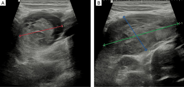
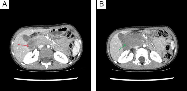
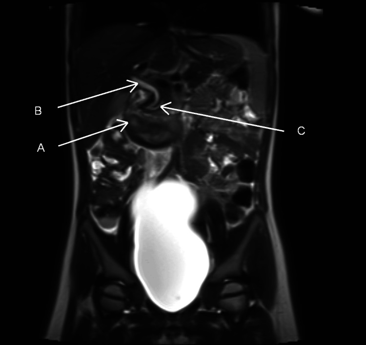
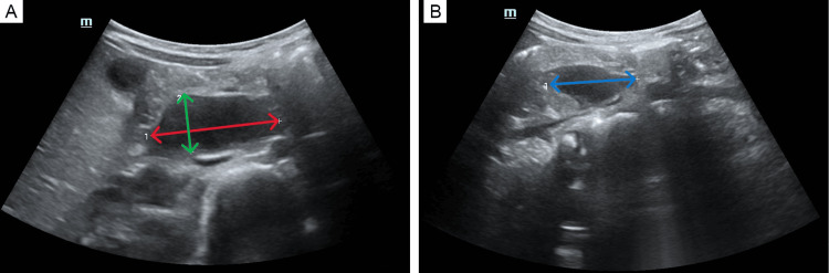
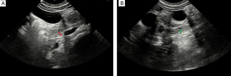
Duodenal hematomas are an uncommon occurrence in the pediatric population, most frequently resulting from blunt abdominal trauma. The preferred treatment modality is a non-operative management, which typically includes bowel rest, nasogastric tube, parenteral nutrition, serial laboratory evaluations, and follow-up imaging. Surgical intervention is rare and generally reserved for cases with complications or failure of conservative therapy. Clinical presentation often includes abdominal pain, nausea, and vomiting. Due to the retroperitoneal location of the duodenum, physical examination findings are typically non-specific. Furthermore, imaging studies may yield false-negative results, contributing to delayed diagnosis and increased risk of complications. In this case report, we present a four-year-old patient diagnosed with a large intramural duodenal hematoma and successfully managed with a conservative, non-operative approach.
Keywords: blunt injury; hematoma of duodenum; non-operative; pediatric; trauma.
Copyright © 2025, Gryszkiewicz et al.
Conflict of interest statement
Human subjects: Consent for treatment and open access publication was obtained or waived by all participants in this study. Conflicts of interest: In compliance with the ICMJE uniform disclosure form, all authors declare the following: Payment/services info: All authors have declared that no financial support was received from any organization for the submitted work. Financial relationships: All authors have declared that they have no financial relationships at present or within the previous three years with any organizations that might have an interest in the submitted work. Other relationships: All authors have declared that there are no other relationships or activities that could appear to have influenced the submitted work.
Figures
References
-
- A 12-year retrospective analysis of non-operative management for paediatric duodenal hematomas caused by trauma at a single center. Shen Q, Li Y, Wang D, Wang L, Li S, Chen L, Liu T. ANZ J Surg. 2024;94:1990–1994. - PubMed
-
- Management of duodenal injuries in children. Clendenon JN, Meyers RL, Nance ML, Scaife ER. J Pediatr Surg. 2004;39:964–968. - PubMed
-
- Management of traumatic duodenal hematomas in children. Peterson ML, Abbas PI, Fallon SC, Naik-Mathuria BJ, Rodriguez JR. J Surg Res. 2015;199:126–129. - PubMed
-
- Systematic review and meta-analysis of emergency ultrasonography for blunt abdominal trauma. Stengel D, Bauwens K, Sehouli J, Porzsolt F, Rademacher G, Mutze S, Ekkernkamp A. Br J Surg. 2001;88:901–912. - PubMed
Publication types
LinkOut - more resources
Full Text Sources