. 2025 Apr-Jun;10(2):87-98.
doi: 10.1097/CP9.0000000000000122. Epub 2025 Jun 24.

Rats subject to extracorporeal membrane oxygenation have improved cardiac function following anticoagulation and reversal with factor IXa aptamer-antidote oligonucleotide pair

Affiliations

Rats subject to extracorporeal membrane oxygenation have improved cardiac function following anticoagulation and reversal with factor IXa aptamer-antidote oligonucleotide pair

Shahid M Nimjee et al. Cardiol Plus. 2025 Apr-Jun.

Abstract

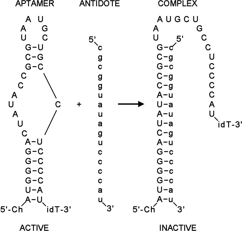
Background and purpose: Unfractionated heparin (UFH) is the most commonly utilized rapid-onset anticoagulant, valued for its potency and reversibility with protamine. However, UFH and protamine are associated with significant side effects, including increased morbidity and mortality, and concerns about sustainability due to the environmental impact of large-scale pig farming for heparin production. This study evaluates an alternative anticoagulant strategy using a factor IXa (FIXa) aptamer paired with a matched oligonucleotide antidote, comparing its efficacy and safety to heparin-protamine in a rat extracorporeal membrane oxygenation (ECMO) model.

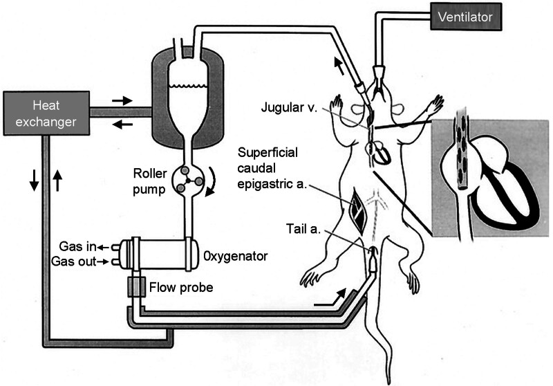
Methods: Twenty-four Sprague-Dawley rats were randomized into two groups: one receiving heparin (600 IU/kg) and protamine (1 mg/100 IU heparin), and the other receiving a cholesterol-modified FIXa aptamer (10 mg/kg) and its antidote (50 mg/kg). Coagulation parameters, platelet counts, inflammatory markers, cardiac function, and histopathology were assessed during and after 60 minutes of ECMO.

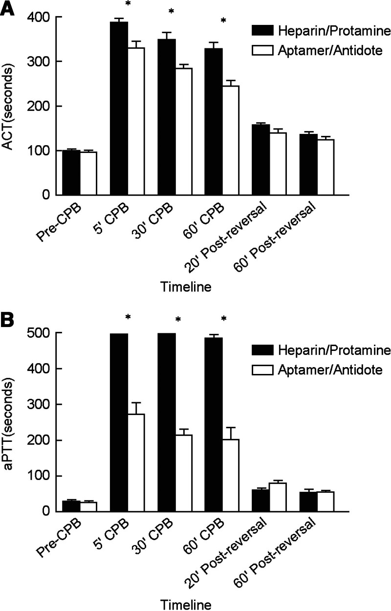
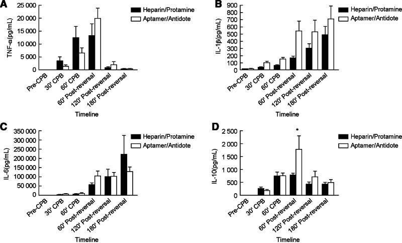
Results: The FIXa aptamer effectively maintained circuit patency without clot formation, comparable to heparin. The antidote rapidly reversed the aptamer's anticoagulant activity, similar to protamine's reversal of heparin. Notably, the aptamer-antidote group demonstrated superior outcomes, including improved mean arterial pressure (58 ± 6 mmHg vs. 54 ± 3 mmHg at 30 minutes; 59 ± 8 mmHg vs. 51 ± 5 mmHg at 3 hours post-ECMO) and cardiac function (shortening fraction: 60 ± 16% vs. 42 ± 8%; P = 0.01). Additionally, the aptamer group exhibited better platelet preservation (platelet count decrease: -288,000 ± 121,000/μL vs. -404,000 ± 89,000/μL; P = 0.03). Inflammatory profiles were similar between groups, except for a transient increase in interleukins 10 (IL-10) in the aptamer group. Histopathological analysis revealed no significant differences in myocardial lesions.

Conclusions: The antidote-controlled anti-FIXa aptamer represents an alternative anticoagulant strategy that may prove useful for managing patients with a history of heparin-induced thrombocytopenia (HIT) and myocardial dysfunction associated with protamine administration.

Keywords: Anticoagulant; Aptamer; Cardiopulmonary bypass; Extracorporeal membrane oxygenation; Heparin.


Conflict of interest statement

Duke University has submitted patent applications on anticoagulant aptamers and Drs. Shahid M. Nimjee, George A. Pitoc, and Bruce A. Sullenger are inventors on such applications.

Figures

Figure 1.
Figure 1.
A schematic representation of the rat extracorporeal membrane oxygenation circuit. Adapted from Cao et al.[24]
Figure 2.
Figure 2.
Depiction of factor (F)IXa aptamer sequence and secondary structure and antidote oligonucleotide sequence. The factor IX aptamer (left) is shown in capitals. The pyrimidines in the aptamer are modified with 2’F-nucleotides while the purines are composed of 2’OH-nucleotides. The 5’-end of the aptamer contains a cholesterol, and the 3’-end is blocked by an idT. The antidote oligonucleotide (middle) is shown in lower case and is comprised of 2’OMe nucleotides. It binds to the active aptamer via standard base-pairs to form an inactive complex (right). idT: inverted deoxy-thymidine.
Figure 3.
Figure 3.
Heparin- and FIXa aptamer-treated animals showed increases in clotting parameters and equivalent sustained return to baseline after antidote administration. A, ACT (n = 10). Factor IXa aptamer administration (10 mg/kg, bolus injection) prolongs clotting time measured by ACT. Antidote oligonucleotide (50 mg/kg, bolus injection) efficiently reversed anticoagulation after 60 min of ECMO/CPB. Heparin administration (600 IU/kg, bolus injection) prolongs clotting time measured by ACT. Protamine administration (1 mg/100 IU heparin, bolus injection) efficiently reversed anticoagulation after 60 min of ECMO/CPB; B, aPTT (n = 10). Factor IXa aptamer administration (10 mg/kg, bolus injection) prolongs clotting time measured by aPTT. Antidote oligonucleotide (50 mg/kg, bolus injection) efficiently reversed anticoagulation after 60 min of ECMO/CPB. Heparin administration (600 IU/kg, bolus injection) prolongs clotting time measured by aPTT. Protamine administration (1 mg/100 IU heparin, bolus injection) efficiently reversed anticoagulation after 60 min of ECMO/CPB. Solid black bars represent heparin/protamine group, open boxes represent aptamer/antidote group. Error bars represent SD. In animals treated with heparin, the aPTT values during ECMO/CPB exceeded the upper limit of the assay and were noted as 500 s. As no absolute maximum value could be determined at the 5- and 30-min times, no error bars are presented for the heparin samples at these time points. *P < 0.05 compared to heparin group at indicated time points. ACT: activated clotting time; aPTT: activated partial thromboplastin time; CPB: cardiopulmonary bypass; ECMO: extracorporeal membrane oxygenation; FXIa: factor IXa; SD: standard deviation.
Figure 4.
Figure 4.
Heparin- and FIXa aptamer-treated animals showed similar cytokine responses with respect to TNF-α, IL-1β, and IL-6, and a transient, differential response of IL-10 during the course of the experiment that returned to equivalent levels at the end of the experiment. A, TNF-α (n = 10). Levels in both heparin- and FIXa aptamer-treated groups returned to near baseline 180 min after reversal of ECMO/CPB (P = 0.97); B, IL-1β (n = 10). Levels in both heparin- and FIXa aptamer-treated groups increased equivalently throughout experiment (P = 0.34); C, IL-6 (n = 10) reflected similar trend as IL-1β, where both heparin- and FIXa aptamer-treated groups increased equivalently throughout experiment (P = 0.47); D, For IL-10 (n = 10), a statistically significant difference between the heparin- and aptamer-treated groups (P < 0.05) largely due to a high IL-10 level in the aptamer-treated group 60 min post-reversal ECMO/CPB. The level of IL-10 at the end of the experiment, however, was similar in both groups (P = 0.68).Solid black bars represent heparin/protamine group, open boxes represent aptamer/antidote group. Error bars represent SD. *P < 0.05. CPB: cardiopulmonary bypass; ECMO: extracorporeal membrane oxygenation; IL-1β: interleukin 1-beta; IL-6: interleukin 6; IL-10: interleukin 10; SD: standard deviation; TNF-α: tumor necrosis factor alpha.
Figure 5.
Figure 5.
Echocardiography analysis. Three hours after the cessation of ECMO/CPB, an estimate of the SF was obtained in all rats using ultrasonography. A transthoracic cross-sectional view of the left ventricle was obtained at papillary level. The EDD and ESD were measured and the SF calculated (EDD − ESD/EDD × 100) for both groups of rats. Rat heart rates remained > 200 bpm during the analyses. The SF was 42% ± 8% and 60% ±16% in heparin group and FIXa aptamer group, respectively (P = 0.01). bpm: beats per minute; ECMO/CPB: extracorporeal membrane oxygenation/cardiopulmonary bypass; EDD: end-diastolic diameter; ESD: end-systolic diameter; FXIa: factor IXa; SF: shortening fraction.

Similar articles

References

    1. Hirsh J, Warkentin TE, Shaughnessy SG, et al. Heparin and low-molecular-weight heparin mechanisms of action, pharmacokinetics, dosing, monitoring, efficacy, and safety. Chest 2001;119(1 Suppl):64S–94S. doi:10.1378/chest.119.1_suppl.64s. - PubMed
    1. Zang LX, Zhu HM, Wang K, et al. Not just anticoagulation-new and old applications of heparin. Molecules 2022;27(20):6968. doi:10.3390/molecules27206968. - PMC - PubMed
    1. Izaguirre G, Swanson R, Raja SM, et al. Mechanism by which exosites promote the inhibition of blood coagulation proteases by heparin-activated antithrombin. J Biol Chem 2007;282(46):33609–33622. doi:10.1074/jbc.M702462200. - PubMed
    1. Chabata CV, Frederiksen JW, Olson LB, et al. Combining heparin and a FX/Xa aptamer to reduce thrombin generation in cardiopulmonary bypass and COVID-19. Nucleic Acid Ther 2022;32(3):139–150. doi:10.1089/nat.2021.0077. - PMC - PubMed
    1. Weitz JI, Hudoba M, Massel D, et al. Clot-bound thrombin is protected from inhibition by heparin-antithrombin III but is susceptible to inactivation by antithrombin III-independent inhibitors. J Clin Invest 1990;86(2):385–391. doi:10.1172/JCI114723. - PMC - PubMed

LinkOut - more resources