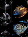
Aortic dilation might be a risk factor for early cardiac perforation after interventional closure of patent foramen ovale: a case report
- PMID: 40599604
- PMCID: PMC12209831
- DOI: 10.1093/ehjcr/ytaf189
Aortic dilation might be a risk factor for early cardiac perforation after interventional closure of patent foramen ovale: a case report
Abstract
Background: Patent foramen ovale (PFO) is present in ∼20% of adults and is associated with cryptogenic stroke, recurrent transient neurological deficit, decompression illness, and migraines. In patients with high probability of a PFO-related ischaemic event, the preferred management strategy is the endovascular closure of the PFO, whenever feasible. Cardiac erosion or perforation is an unusual event after the interventional closure of a PFO, but its occurrence portends a life-threatening potential. Therefore, clinicians should be aware of this serious complication when faced with acute cardiac tamponade in a patient with a history of PFO closure.
Case summary: We report the case of a 57-year-old man, with a recent PFO-related ischaemic stroke, who presented for elective endovascular closure of the PFO. Patent foramen ovale and surrounding structures seemed amenable for closure, and recommended a 25-mm Amplatzer device. Isolated dilation of the aortic bulb was noted by transoesophageal echocardiography (TEE), but with normal valve morphology and no aortic regurgitation. The procedure was performed without complications, and 24 h echo follow-up showed no impingement of the device on the surrounding structures. However, 36 h after the procedure, the patient developed sudden chest pain and cardiac tamponade. Emergency cardiac surgery with intra-operative TEE guidance revealed right atrial and aortic bulb perforation caused by the larger right disc of the occluder. The device was removed, and the defects were sutured. The patient was discharged within 14 days after the event, with no further complications.
Discussion: Perforation of the aortic root is a very rare complication after interventional PFO closure. This complication usually occurs at a long distance after the procedure, and is associated with oversized devices, deficient or absent aortic rim, or misalignment of the defect with the aorta. Our patient presented none of the above, but a moderate dilation of the aortic bulb, which might have triggered the rapid erosion and perforation.
Keywords: Amplatzer device complication; Aortic dilation; Aortic perforation; Case report; PFO interventional closure.
© The Author(s) 2025. Published by Oxford University Press on behalf of the European Society of Cardiology.
Conflict of interest statement
Conflict of interest. None declared.
Figures


References
-
- Verma SK, Tobis JM. Explantation of patent foramen ovale closure devices: a multicenter survey. JACC Cardiovasc Interv 2011;4:579–585. - PubMed
-
- Hakman EN, Cowling KM. Paradoxical Embolism. Treasure Island, FL: StatPearls Publishing; 2022. - PubMed
-
- Saremi F, Emmanuel N, Wu PF, Ihde L, Shavelle D, Go JL, et al. Paradoxical embolism: role of imaging in diagnosis and treatment planning. Radiographics 2014;34:1571–1592. Erratum in: Radiographics. 2014 Nov-Dec;34(7):10A. Wu, Philip F [corrected to Wu, Phil F]. - PubMed
-
- Pristipino C, Sievert H, D'Ascenzo F, Mas JL, Meier B, Scacciatella P, et al. European position paper on the management of patients with patent foramen ovale. General approach and left circulation thromboembolism. Eur Heart J 2019;40:3182–3195. Erratum in: Eur Heart J. 2021 May 7;42(18):1807. - PubMed
Publication types
LinkOut - more resources
Full Text Sources
