. 2025 Jun 27:18:8527-8539.
doi: 10.2147/JIR.S506056. eCollection 2025.

IFNα/JAK/STAT1 Axis-Induced FBXO4 Modulates Muscle Cell Differentiation via β-Catenin Degradation in Dermatomyositis

Affiliations

IFNα/JAK/STAT1 Axis-Induced FBXO4 Modulates Muscle Cell Differentiation via β-Catenin Degradation in Dermatomyositis

Liguo Yin et al. J Inflamm Res. .

Abstract

Purpose: Dermatomyositis (DM) is an inflammatory myopathy characterized by chronic muscle inflammation and damage. Although the pathogenesis of DM has been widely reported to be related to chronic inflammation, the role of ubiquitin E3 ligases in DM remains unclear. In the current study, we aimed to investigate the biological roles of ubiquitin E3 ligase in DM.

Methods: Deseq2 was used to screen the differential express genes in DM public datasets. Quantitative real time PCR and Western blot were used to examine the mRNA and protein levels. Co-immunoprecipitation assays were used to investigate the protein interactions between proteins. Dual-luciferase reporter assays were applied to investigate the regulation between transcription factors and targets.

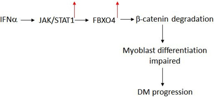
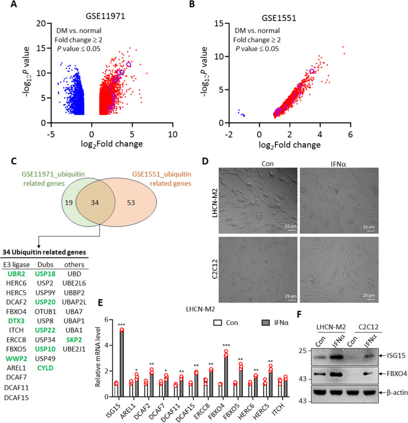
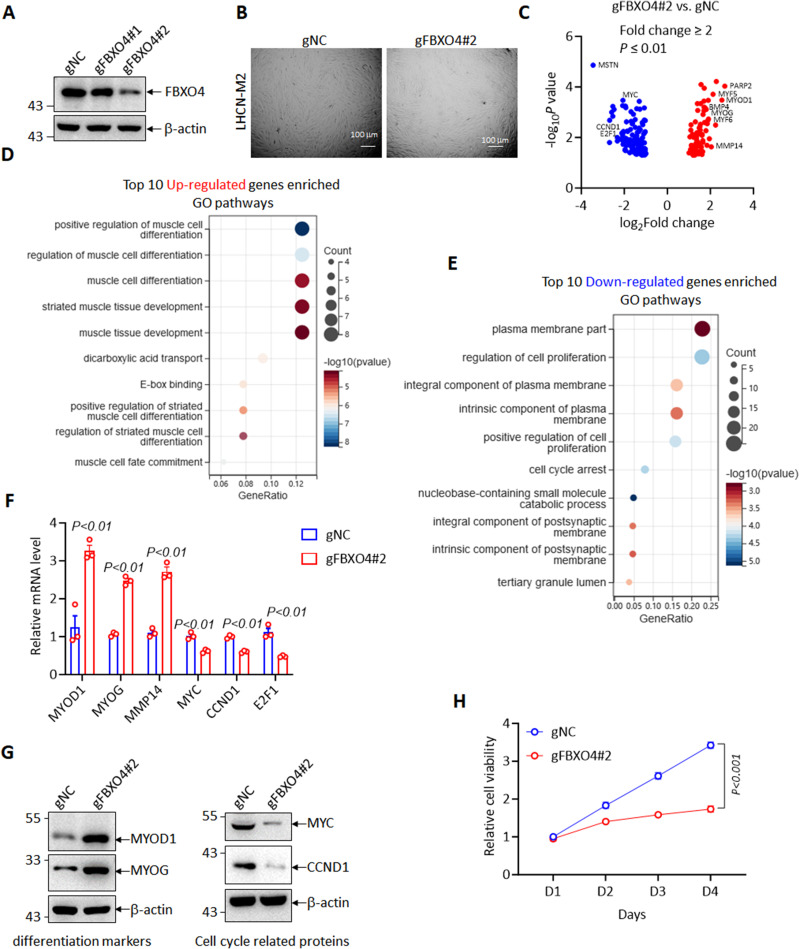
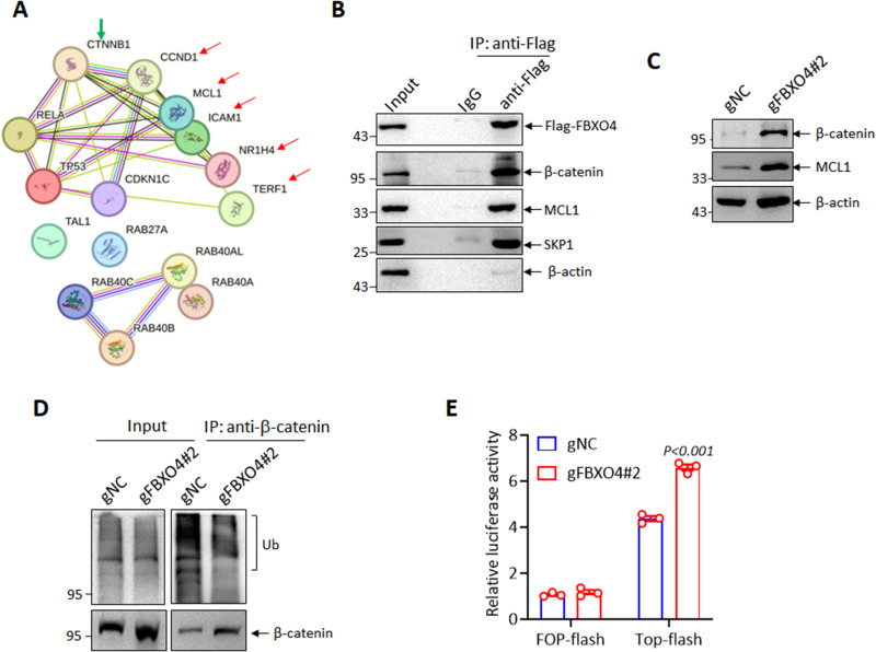
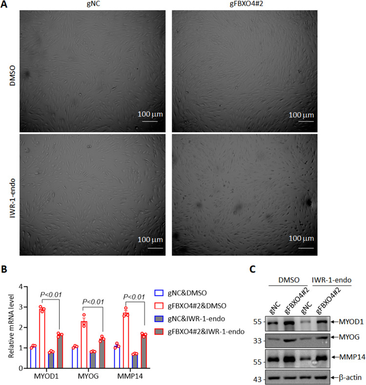
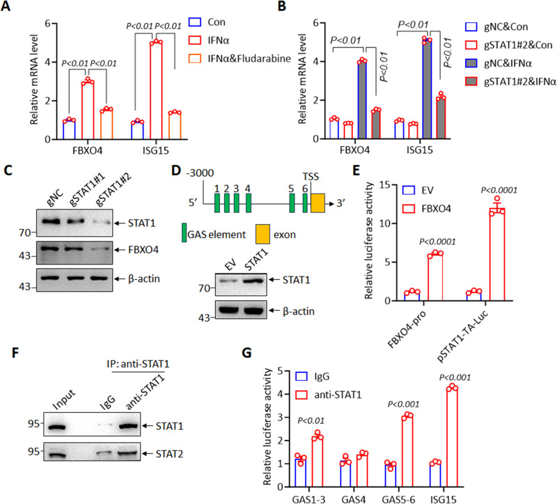
Results: In the current study, we screened public DM-related datasets and focused on ubiquitin-proteasome-related enzymes. Ultimately, we identified the ubiquitin E3 ligase FBXO4. FBXO4 was significantly upregulated in DM muscle tissues compared to normal controls. In human muscle cells (LHCN-M2), FBXO4 knockout led to significant upregulation of genes related to muscle cell differentiation and significant downregulation of genes enriched in cell cycle pathways, as revealed by RNA-seq. These results suggest that FBXO4 knockout promotes muscle cell differentiation. Mechanistic studies showed that FBXO4 ubiquitinates and degrades β-catenin, thereby inhibiting the Wnt/β-catenin signaling pathway and suppressing muscle cell differentiation. On the other hand, FBXO4 may promote muscle cell apoptosis in DM by degrading MCL1. Additionally, we found that FBXO4 is regulated by the IFNα/JAK/STAT1 signaling pathway in DM and identified FBXO4 as a direct target of STAT1.

Conclusion: In conclusion, our findings suggest that IFNα/JAK/STAT1 signaling pathway elevates the expression of FBXO4 in DM and then it contributes to muscle atrophy by inhibiting differentiation and promoting apoptosis. Targeting FBXO4 may offer a novel therapeutic approach for DM.

Keywords: FBXO4; IFNα/JAK/STAT1 axis; Wnt/β-catenin signaling; dermatomyositis; muscle cell differentiation.


Conflict of interest statement

The authors declare that they have no conflicts of interest in this work.

Figures

None
Graphical abstract
Figure 1
Figure 1
Screening of Ubiquitin E3 Ligases in Dermatomyositis Muscle Tissues. (A and B) The volcano plot illustrates the differential gene expression analysis comparing dermatomyositis (DM) muscle tissues to normal muscle tissues using the GSE11971 (A) and GSE1551 (B) datasets. Genes with a fold change > 2 and P value < 0.01 were considered significantly differentially expressed. Red and blue plots indicate significant up- and down-regulated genes respectively. Purple plots indicate ubiquitin related genes. (C) Venn diagram showing the overlap of ubiquitination-related proteins significantly upregulated in both datasets, with 34 common proteins identified. Green fonts indicate previously reported ubiquitin-related genes. (D) Cell morphology of indicated cells treated with 1000 U/mL IFNα for 24 hours. (E) qRT-PCR analysis of selected E3 ligases and ISG15 in LHCN-M2 cells treated with 1000 U/mL IFNα. ISG15 served as a positive control, confirming the effectiveness of IFNα treatment. * Indicates P < 0.05, ** indicates P <0.01, *** indicates P <0.001. (F) Western blot analysis showing the protein levels of FBXO4 in LHCN-M2 and C2C12 cells following 1000 U/mL IFNα treatment.
Figure 2
Figure 2
FBXO4 Knockout Promotes Myocyte Differentiation. (A) Confirmation of FBXO4 knockout in LHCN-M2 cells using two gRNA constructs, with gFBXO4#2 showing higher efficiency. (B) Morphological changes observed in FBXO4 knockout LHCN-M2 cells compared to control, with cells becoming larger and more elongated, indicative of differentiation. (C) The volcano plot illustrates the differential gene expression analysis comparing gFBXO4#2 LHCN-M2 cells to control. Genes with a fold change > 2 and P value < 0.01 were considered significantly differentially expressed. Red and blue plots indicate significant up- and down-regulated genes respectively. (D) Gene ontology enrichment analysis of upregulated genes, highlighting pathways related to muscle cell differentiation. (E) Gene ontology enrichment analysis of downregulated genes, showing enrichment in cell proliferation and cell cycle pathways. (F and G) qRT-PCR (F) and Western blot (G)analysis confirming increased mRNA levels of differentiation markers (MYOD1, MYOG, MMP14) and decreased levels of cell proliferation markers (MYC, CCND1, E2F1). (H) CCK8 assay demonstrating impaired cell proliferation following FBXO4 knockout in LHCN-M2 cells.
Figure 3
Figure 3
FBXO4 Regulates β-Catenin Ubiquitination and Degradation. (A) The string analysis of the UbiBrowser 2.0 tool predicted potential substrates of FBXO4, with arrows highlighting those that have been previously reported as FBXO4 substrates. (B) Immunoprecipitation (IP) assays in LHCN-M2 cells expressing Flag-FBXO4, confirming the interaction between FBXO4 and β-catenin. MCL1 and SKP1 were used as positive controls. (C) Western blot analysis showing that FBXO4 knockout increases β-catenin protein levels in LHCN-M2 cells. (D) Ubiquitination assay showing reduced levels of ubiquitinated β-catenin in FBXO4 knockout cells compared to controls. (E) Luciferase reporter assays demonstrating that FBXO4 depletion enhances Top-flash luciferase activity, indicating activation of Wnt/β-catenin signaling.
Figure 4
Figure 4
Wnt/β-Catenin Pathway Mediates FBXO4’s Effect on Muscle Cell Differentiation. (A) Morphological analysis of FBXO4 knockout and control LHCN-M2 cells treated with Wnt signaling inhibitor IWR-1-endo or DMSO, showing that IWR-1-endo reduces cell size in FBXO4 knockout cells. (B and C) qRT-PCR (B) and Western blot (C) analysis of differentiation markers MYOD and MYOG in FBXO4 knockout and control cells treated with IWR-1-endo or DMSO, demonstrating rescue of differentiation.
Figure 5
Figure 5
FBXO4 Modulates Apoptosis in Muscle Cells. Apoptosis analysis in FBXO4 knockout and control LHCN-M2 cells under 1000 U/mL IFNα treatment, showing that FBXO4 depletion inhibits cell apoptosis.
Figure 6
Figure 6
FBXO4 Is Regulated by IFNα/STAT1 Pathway in Dermatomyositis. (A) Western blot analysis showing that Fludarabine, a STAT1 signaling inhibitor, reverses the increase in FBXO4 levels induced by IFNα treatment. (B) qRT-PCR (B) and Western blot (C) analysis of FBXO4 mRNA and protein levels in STAT1 knockout LHCN-M2 cells under IFNα treatment. (D) Promoter analysis of FBXO4 revealing six STAT1 binding sites (GAS sites, top panel); immunoblotting analysis of STAT1 in HEK-293T overexpression of STAT1 and control. (E) Dual-luciferase assay results demonstrate that STAT1 enhances FBXO4 promoter activity under 1000 U/mL IFNα treatment. (F and G) Immunoblotting analysis assessed the immunoprecipitation efficiency of the STAT1 antibody, with STAT2 serving as a positive control (F). qRT-PCR demonstrated STAT1 binding to the FBXO4 promoter in HEK-293T cells following IFNα treatment, using ISG15 as a positive control.

Similar articles

References

    1. Suárez-Calvet X, Gallardo E, Pinal-Fernandez I, et al. RIG-I expression in perifascicular myofibers is a reliable biomarker of dermatomyositis. Arthritis Res Ther. 2017;19(1):174. doi: 10.1186/s13075-017-1383-0 - DOI - PMC - PubMed
    1. DeWane ME, Waldman R, Lu J. Dermatomyositis: clinical features and pathogenesis. J Am Acad Dermatol. 2020;82(2):267–281. doi: 10.1016/j.jaad.2019.06.1309 - DOI - PubMed
    1. Waldman R, DeWane ME, Lu J. Dermatomyositis: diagnosis and treatment. J Am Acad Dermatol. 2020;82(2):283–296. doi: 10.1016/j.jaad.2019.05.105 - DOI - PubMed
    1. Lundberg IE, Fujimoto M, Vencovsky J, et al. Idiopathic inflammatory myopathies. Nat Rev Dis Primers. 2021;7(1):86. doi: 10.1038/s41572-021-00321-x - DOI - PubMed
    1. Kim H. Updates on interferon in juvenile dermatomyositis: pathogenesis and therapy. Curr Opin Rheumatol. 2021;33(5):371–377. doi: 10.1097/bor.0000000000000816 - DOI - PMC - PubMed

LinkOut - more resources