Review
. 2025 Jun 24:2025:7990876.
doi: 10.1155/av/7990876. eCollection 2025.

The Possible Mechanistic Basis of Individual Susceptibility to Spike Protein Injury

Affiliations
Review

The Possible Mechanistic Basis of Individual Susceptibility to Spike Protein Injury

Matthew Halma et al. Adv Virol. .

Abstract

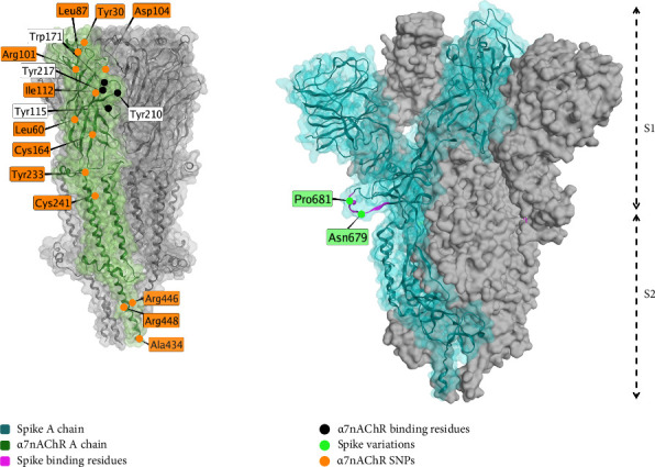
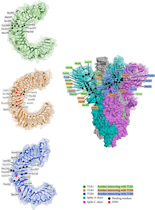
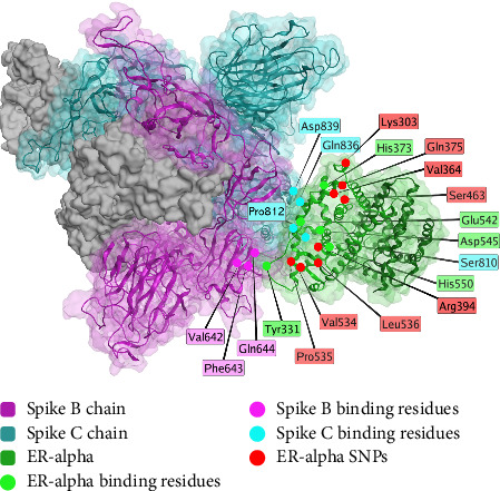
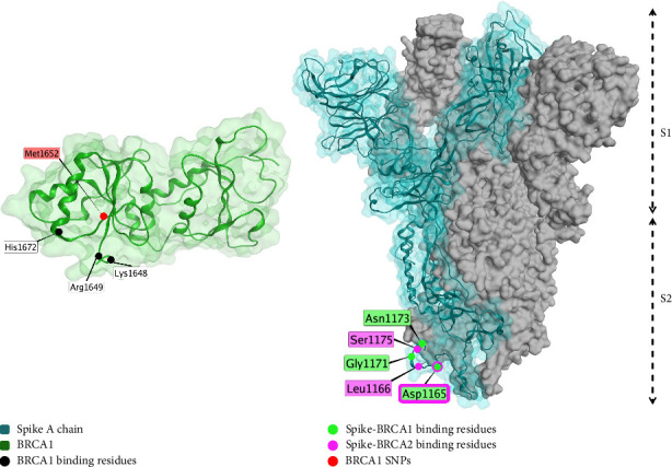
Injury from spike protein, whether induced by COVID-19 infection or vaccination, constitutes a significant health concern for numerous individuals. Considerable heterogeneity exists in individual responses to both COVID-19 infection and vaccination, despite the latter being principally more controlled and consistent than the wide variety of infection circumstances. This review explores the possible mechanisms by which the spike protein contributes to cellular and systemic damage, highlighting the importance of understanding these processes for developing effective diagnostics and treatments.


Conflict of interest statement

The authors declare no conflicts of interest.

Figures

Figure 1
Figure 1
A model for long-term spike protein persistence in vaccine-injured individuals. This figure is an original figure created by the authors. A version of this figure appears in a preprint authored by the first author [56].
Figure 2
Figure 2
Pathophysiology related to spike protein. (a) Pathophysiological interactions of the spike protein. Slices show potential mechanisms (inside) and subsequent pathophysiology (outside). This figure has been reproduced from [95] under the permission of CC Attribution 4.0 international license (CC BY 4.0 DEED, https://creativecommons.org/licenses/by/4.0/). (b) Pathways and associated pathology. This figure panel is an original production by the authors.
Figure 3
Figure 3
The interaction interface between the spike protein receptor binding domain (RBD, blue) and the angiotensin-converting enzyme 2 (ACE-2) receptor (green) (PDB ID: 6LZG, X-ray diffraction, resolution 2.5 Å) [168]. Interacting residues are shown as black dots, and variable residues with SNPs are shown in violet for variation in the spike protein and orange for variation on the ACE2 receptor. This figure is an original figure created by the authors.
Figure 4
Figure 4
The interaction interface between the spike protein receptor binding domain (RBD, blue) and the CD-147 receptor (violet). The structural model represented here has been reproduced from the in silico model by Helal et al. [172], with permission from the authors. Variable residues on spike RBD are shown in orange, whereas variable residues on CD-147 are shown in green. Residues observed to interact are highlighted in black. This figure is an original figure created by the authors.
Figure 5
Figure 5
The interaction between spike (S) protein (blue, PDB ID: 7OAN [203], electron microscopy, resolution 3.0 Å) and functional α7 nicotinic acetylcholine receptor (α7nAChR, green, PDB ID: 7EKI [204], electron microscopy, resolution 3.18 Å). Interacting residues on α7nAChR are shown in black, where variable residues are shown in orange. Variable residues of the spike protein are shown in green. This figure is an original figure created by the authors.
Figure 6
Figure 6
Interaction of spike protein with human toll-like receptors (TLRs) and the spike (S) protein (PDB ID: 7OAN [203], electron microscopy, resolution 3.0 Å). TLR1 (PDB ID: 6NIH [240], X-ray diffraction, resolution 2.3 Å) is shaded in green, TLR4 (PDB ID: 3FXI [241], X-ray diffraction, resolution 3.1 Å) is shaded in brown, and TLR6 (PDB ID: 3A79 [242], X-ray diffraction, resolution 2.9 Å) is shaded in blue. Binding residues are shown in black, and variable residues shown in red. Spike protein residues interacting with TLR1 are shaded in green, residues interacting with TLR4 are shaded in orange, and residues interacting with TLR6 are shaded in blue. This figure is an original figure created by the authors.
Figure 7
Figure 7
Interaction between spike protein (purple shading for Chain B and cyan shading for Chain C) and the estrogen receptor alpha (ERα) protein (green). Binding residues are shown in green for residues of ERα, and violet and cyan for residues of spike protein chains B and C, respectively. SNPs of ERα are highlighted in red. The structural model represented here has been reproduced from the model by Solis et al. [119], with permission from the authors. This figure is an original figure created by the authors.
Figure 8
Figure 8
Interaction between spike protein (blue and gray, PDB ID: 7OAN [203], electron microscopy, resolution 3.0 Å) and BRCA1 (green, PDB ID: 3PXB [319], X-ray diffraction, resolution 2.5 Å). Binding residues of BRCA1 (interacting with spike protein) are highlighted in black. SNPs of BRCA1 are shown in red. BRCA1- and BRCA2-interacting residues on spike protein are highlighted green or violet, respectively. Asp1165 interacts with both BRCA1 and BRCA2. This figure is an original figure created by the authors.

Similar articles

References

    1. Kim K. Q., Burgute B. D., Tzeng S.-C., et al. N1-Methylpseudouridine Found Within COVID-19 mRNA Vaccines Produces Faithful Protein Products. Cell Reports . 2022;40(9):p. 111300. doi: 10.1016/j.celrep.2022.111300. - DOI - PMC - PubMed
    1. Halma M. T. J., Rose J., Lawrie T. The Novelty of mRNA Viral Vaccines and Potential Harms: A Scoping Review. D-J Series . 2023;6(2):220–235. doi: 10.3390/j6020017. - DOI
    1. Theoharides T. C., Conti P. Be Aware of SARS-CoV-2 Spike Protein: There is More Than Meets the Eye. Journal of Biological Regulators & Homeostatic Agents . 2021;35(3):833–838. doi: 10.23812/THEO_EDIT_3_21. - DOI - PubMed
    1. Cox F., Khalib K., Conlon N. PEG That Reaction: A Case Series of Allergy to Polyethylene Glycol. The Journal of Clinical Pharmacology . 2021;61(6):832–835. doi: 10.1002/jcph.1824. - DOI - PMC - PubMed
    1. Uddin M. N., Roni M. A. Challenges of Storage and Stability of mRNA-Based COVID-19 Vaccines. Vaccines . 2021;9:p. 1033. doi: 10.3390/vaccines9091033. - DOI - PMC - PubMed

LinkOut - more resources