. 2025 Jun 6:15:1536365.
doi: 10.3389/fonc.2025.1536365. eCollection 2025.

Optimizing breast cancer ultrasound diagnosis: a comparative study of AI model performance and image resolution

Affiliations

Optimizing breast cancer ultrasound diagnosis: a comparative study of AI model performance and image resolution

Yunqing Yin et al. Front Oncol. .

Abstract

Objectives: To determine the optimal combination of artificial intelligence (AI) models and ultrasound (US) image resolutions for breast cancer diagnosis and evaluate whether this combination surpasses the diagnostic accuracy of senior radiologists.

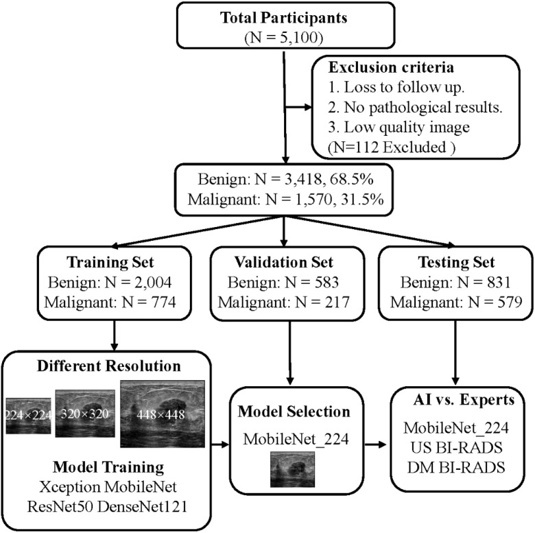
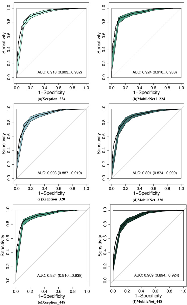
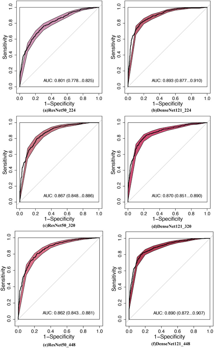
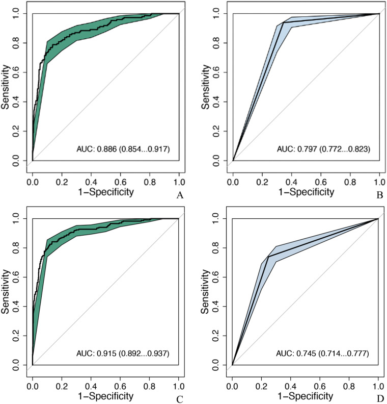
Materials and methods: We systematically compared lightweight (MobileNet, Xception) and dense neural networks (ResNet50, DenseNet121) using three image resolutions (224 × 224, 320 × 320, 448 × 448 pixels). A retrospective cohort of 4,998 patients was divided into training/validation (8:2 ratio, n = 3,578) and independent testing sets (n = 1,410). Diagnostic performance was assessed via AUC, sensitivity, specificity, and analysis speed, with direct comparisons against senior radiologists.

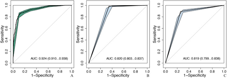
Results: MobileNet with 224 × 224 input achieved the highest AUC (0.924, 95% CI: 0.910-0.938) and accuracy (87.3%) outperforming senior US (AUC: 0.820, accuracy: 79.1%) and mammography doctors (AUC: 0.819, accuracy: 83.6%) (p < 0.05). After excluding BI-RADS 4c and 5 nodules, the diagnostic efficacy of MobileNet_224 is better than that of senior doctors (p < 0.05), can reduce 60.1% false positives of US, and 46.6% of mammography. MobileNet_224 and MobileNet_320 had the fastest analysis speed.

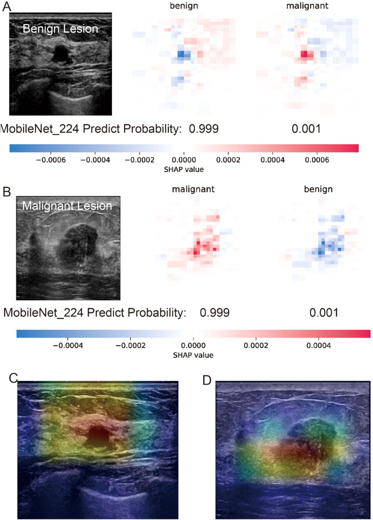
Conclusion: MobileNet_224 represents a novel, efficient AI framework for breast cancer diagnosis demonstrating superior accuracy and speed compared to both complex AI models and experienced clinicians. This work highlights the critical role of optimizing model architecture and resolution to enhance diagnostic workflows and reduce unnecessary biopsies.

Keywords: artificial intelligence; breast cancer; diagnosis; mammography; ultrasound.


Conflict of interest statement

The authors declare that the research was conducted in the absence of any commercial or financial relationships that could be construed as a potential conflict of interest.

Figures

Figure 1
Figure 1
Flow chart and results of this study. The optimal model: MobileNet_224, senior ultrasound doctors, senior mammography doctors. MobileNet_224, MobileNet with 224 × 224-pixel image input; US_BI-RADS, senior ultrasound doctors’ diagnostic results; DM_BI-RADS, senior mammography doctors’ diagnostic results.
Figure 2
Figure 2
Comparison of diagnostic efficacy between LW-CNNs in the testing set. AUC, area under the curve; 95% CI: 95% confidence interval. (a) Xception_224: Xception with 224 × 224-pixel image input, (b) MobileNet_224, (c) Xception_320, (d) MobileNet_320, (e) Xception_448, (f) MobileNet_448.
Figure 3
Figure 3
Comparison of diagnostic efficacy between DNNs in the testing set. AUC, area under the curve; 95% CI, 95% confidence interval. (a) ResNet50_224: ResNet50 with 224 × 224-pixel image input, (b) DenseNet121_224, (c) ResNet50_320, (d) DenseNet121_320, (e) ResNet50_448, (f) DenseNet121_448.
Figure 4
Figure 4
Interpretability analysis of MobileNet_224 predictions for benign and malignant breast lesions. (A) Benign lesion: prediction probability (0.999 for benign, 0.001 for malignant) with SHAP values highlighting key image regions contributing to the benign classification. (B) Malignant lesion: prediction probability (0.999 for malignant, 0.001 for benign) with SHAP values emphasizing tumor margin irregularity and microcalcifications. (C, D) Grad-CAM heatmaps for the benign (C) and malignant (D) lesions illustrating the model’s focus on clinically relevant anatomical features (e.g., smooth margins in benign vs. spiculated regions in malignant).
Figure 5
Figure 5
Comparison of diagnostic efficacy between the optimal model and senior doctors in the testing set. MobileNet_224, MobileNet with 224 × 224-pixel image input; AUC, area under the curve; 95% CI, 95% confidence interval. (A) The optimal model: MobileNet_224, (B) senior ultrasound doctors, (C) senior mammography doctors.
Figure 6
Figure 6
Comparison of diagnostic efficacy between the optimal model and senior doctors after excluding BIRADS 4c and 5 nodules. MobileNet_224, MobileNet with 224 × 224-pixel image input; AUC, area under the curve; 95% CI, 95% confidence interval. (A) MobileNet_224, (B) senior ultrasound doctors, (C) MobileNet_224, (D) senior mammography doctors.

Similar articles

References

    1. Chen W, Zheng R, Zhang S, Zeng H, Xia C, Zuo T, et al. Cancer incidence and mortality in China, 2013. Cancer Lett. (2017) 401:63–71. doi: 10.1016/j.canlet.2017.04.024 - DOI - PubMed
    1. Miller KD, Nogueira L, Devasia T, Mariotto AB, Yabroff KR, Jemal A, et al. Cancer treatment and survivorship statistics, 2022. CA: A Cancer J For Clinicians. (2022) 72:409–36. doi: 10.3322/caac.21731 - DOI - PubMed
    1. Makama M, Drukker CA, Rutgers EJT, Slaets L, Cardoso F, Rookus MA, et al. An association study of established breast cancer reproductive and lifestyle risk factors with tumour subtype defined by the prognostic 70-gene expression signature (MammaPrint®). Eur J Cancer (Oxford England: 1990). (2017) 75:5–13. doi: 10.1016/j.ejca.2016.12.024 - DOI - PubMed
    1. Jin Z-Q, Lin M-Y, Hao W-Q, Jiang H-T, Zhang L, Hu W-H, et al. Diagnostic evaluation of ductal carcinoma in situ of the breast: ultrasonographic, mammographic and histopathologic correlations. Ultrasound In Med Biol. (2015) 41:47–55. doi: 10.1016/j.ultrasmedbio.2014.09.023 - DOI - PubMed
    1. Su X, Lin Q, Cui C, Xu W, Wei Z, Fei J, et al. Non-calcified ductal carcinoma in situ of the breast: comparison of diagnostic accuracy of digital breast tomosynthesis, digital mammography, and ultrasonography. Breast Cancer (Tokyo Japan). (2017) 24:562–70. doi: 10.1007/s12282-016-0739-7 - DOI - PubMed

LinkOut - more resources