Review
. 2024 Dec 23;5(3):179-202.
doi: 10.1515/mr-2024-0064. eCollection 2025 Jun.

Immune cells in Alzheimer's disease: insights into pathogenesis and potential therapeutic targets

Affiliations
Review

Immune cells in Alzheimer's disease: insights into pathogenesis and potential therapeutic targets

Xiaofeng Fu et al. Med Rev (2021). .

Abstract

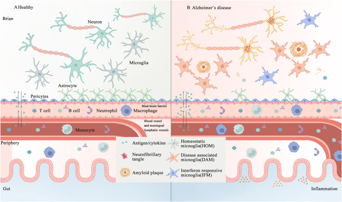
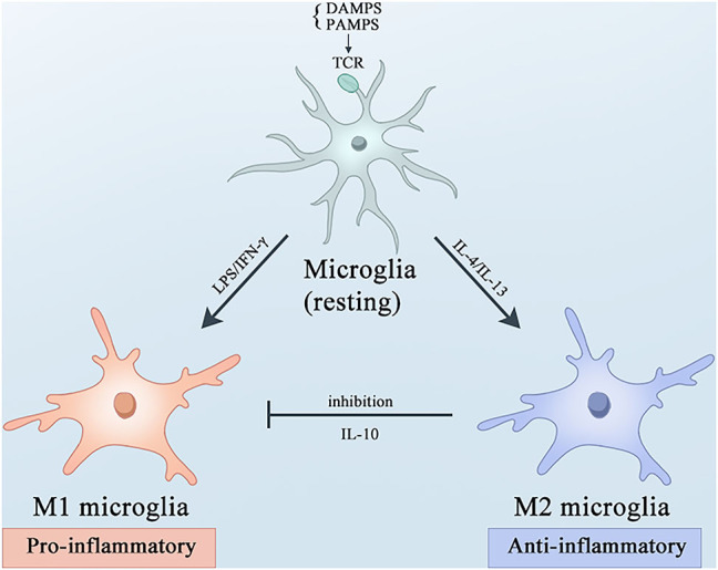
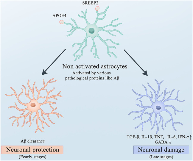
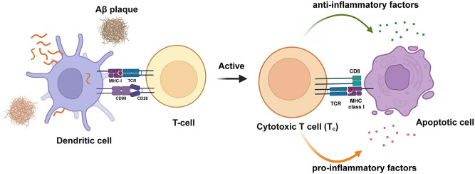
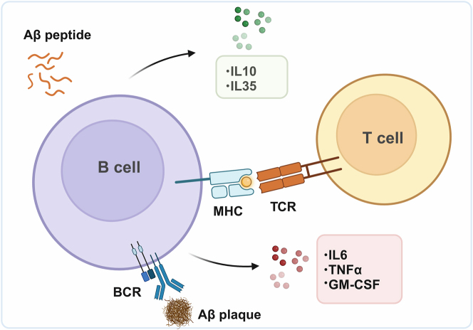
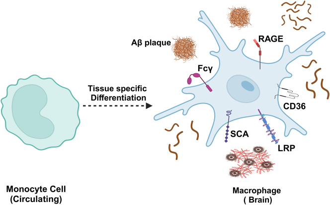
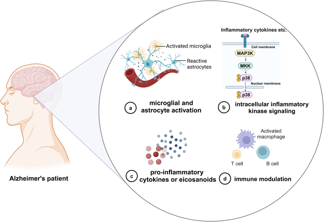
Alzheimer's disease (AD) is a chronic neurodegenerative disorder for which there are currently no effective treatment options. Increasing evidence suggests that AD is a systemic disease closely associated with the immune system, not merely a central nervous system (CNS) disorder. Immune cells play crucial roles in the onset and progression of AD. Microglia and astrocytes are the primary inflammatory cells in the brain that can sensitively detect changes in the internal environment and transform into different phenotypes to exert differing effects at various stages of AD. Peripheral immune cells, such as T cells, B cells, monocytes/macrophages, and neutrophils can also be recruited to the CNS to mediate the inflammatory response in AD. As such, investigating the role of immune cells in AD is particularly important for elucidating its specific pathogenesis. This review primarily discusses the roles of central innate immune cells, peripheral immune cells, and the interactions between central and peripheral immune cells in the development of neuroinflammation in AD. Furthermore, we listed clinical trials targeting AD-associated neuroinflammation, which may represent a promising direction for developing effective treatments for AD in the future.

Keywords: Alzheimer’s disease; immune cell; inflammation; therapeutics.


Conflict of interest statement

Conflict of interest: Authors state no conflict of interest.

Figures

Figure 1:
Figure 1:
The interplay between central and peripheral immune cells in the healthy and AD brain. A. In healthy individuals, microglial cells, astrocytes, and an intact BBB and meningeal lymphatic system collectively maintain the functionality of the brain. B. In AD, the aggregation of Aβ and formation of neurofibrillary tangles composed of hyperphosphorylated tau activate immune cells, thus disrupting the integrity of the BBB, and trigger inflammation both within and outside the brain, including in the gut. AD, Alzheimer’s disease; Aβ, amyloid β-protein; BBB, blood-brain barrier.
Figure 2:
Figure 2:
Microglial activation and polarization under basal states and amidst neuroinflammatory processes. Microglial cells undergo polarization to the M1 or M2 phenotype, accompanied by distinct immunoregulatory roles. Under resting conditions, microglia are activated by PAMPs or DAMPs via TLRs. Upon stimulation with LPS and IFN-γ, microglia polarize towards the M1 phenotype, leading to the secretion of pro-inflammatory cytokines and mediators, and NO. Conversely, IL-4 and IL-1β drive the alternative activation of microglia into the M2 phenotype, which dampens M1-associated functions by promoting the release of the anti-inflammatory cytokine IL-10. AD, Alzheimer’s disease; PAMPs, pathogen-associated molecular patterns; DAMPs, damage-associated molecular patterns; TLRs, Toll-like receptors; LPS, lipopolysaccharide; IFN-γ, interferon-gamma; NO, nitric oxide; IL-4, interleukin-4; IL-1β, interleukin-1β.
Figure 3:
Figure 3:
TREM2 activation and downstream signaling TREM2 binds to ligands such as Aβ, ApoE, HDL, and LDL, leading to its activation. Following activation, TREM2 interacts with the ITAMs on TYROBP, resulting in the phosphorylation of TYROBP and the recruitment of SYK. The activation of the PI3K - AKT pathway mediated by TYROBP/SYK recruits other signaling adapters, including PLCγ and MAPKs inducing an release of Ca2+. TREM2, triggering receptors expressed on myeloid cells 2; Aβ, amyloid β-protein; ApoE, apolipoprotein E; HDL, high-density lipoprotein; LDL, low-density lipoprotein; ITAMs, immunoreceptor tyrosine-based activation motifs; TYROBP, TYRO protein tyrosine kinase-binding protein; SYK, spleen tyrosine kinase; PI3K, phosphatidylinositol 3-kinase; PLCγ, phospholipase Cγ; MAPKs, mitogen-activated protein kinases. Created with BioRender.com.
Figure 4:
Figure 4:
The roles of astrocytes in different stages of AD. Microglia express APOE4 and SREBP2 on their surfaces. Upon activation by various pathological proteins, microglia can clear Aβ and exert neuroprotective effects in the early stages of AD. However, in the late stages of AD, they secrete large amounts of inflammatory factors such as TGFβ, IL-1β, TNF, IL-6, and IFNγ accompanied by disturbances in GABA metabolism, which exacerbate neuronal damage. AD, Alzheimer’s disease; APOE4, Apolipoprotein E4; SREBP2, Sterol Regulatory Element-Binding Protein 2; Aβ, amyloid β-protein; TGFβ, Transforming Growth Factor beta; IL-1β, Interleukin-1 beta; TNF, Tumor Necrosis Factor; IL-6, Interleukin-6; IFNγ, Interferon gamma.
Figure 5:
Figure 5:
Activation of T-cells in AD. Dendritic cells present antigens to T-cells, activating T-cells and releasing pro-inflammatory and anti-inflammatory cytokines.Aβ, amyloid β-protein. Created with BioRender.com.
Figure 6:
Figure 6:
Crosstalk between T-cells and microglia in AD. Aβ-specific CD4+ Th1 cells induce the expression of major histocompatibility MHCII on microglia, potentially mediated by the IFN-γ cytokine signaling pathway. Aβ, amyloid β-protein; Th1, type 1 T helper; MHCII, major histocompatibility complex class II; IFN-γ, interferon-gamma. Created with BioRender.com.
Figure 7:
Figure 7:
Functions of B-cells in AD. Mature B-cells can act as specialized antigen-presenting cells, presenting antigens to T-cells. On the other hand, B-cells release cytokines such as IL-6, TNF-α, and GM-CSF to promote inflammatory responses, while also secreting IL-10 and IL-35 to exert anti-inflammatory effects. Aβ, amyloid β-protein; IL, interleukin; TNF-α, tumor necrosis factor-alpha; GM-CSF, granulocyte-macrophage colony-stimulating factor. Created with BioRender.com.
Figure 8:
Figure 8:
Monocytes in the brain. Blood-derived monocytes entering the brain parenchyma express various receptors to clear Aβ, including Fcγ, SCA, CD36, RAGE, and LRP receptors.Aβ, amyloid β-protein; SCA, scavenger receptor A; CD36, cluster of differentiation 36; RAGE, receptor for advanced glycation end-products; LRP, low-density lipoprotein receptor-associated protein. Created with BioRender.com.
Figure 9:
Figure 9:
The potential role of peripheral immune cells in AD. In AD, the BBB is compromised, allowing peripheral immune cells located at the meningeal borders to be stimulated by antigens such as Aβ. T-cells release cell-derived cytokines such as IL-12, IL-4, TGF-β, and IL-6 to regulate the development of Th1, Th2, Th17, and Treg cells, which subsequently secrete anti or pro-inflammatory factors to modulate neurons. Antigens can also directly stimulate B-cells, which on the one hand produce antibodies to eliminate Aβ and on the other hand secrete inflammatory factors to regulate the immune response. Blood-derived monocytes can cross the BBB and differentiate into bone marrow-derived macrophages with microglia-like properties, which are more effective at clearing Aβ than resident microglia. Neutrophils migrate to the brain parenchyma and secrete inflammatory factors. AD, Alzheimer’s disease; Aβ, amyloid β-protein; BBB, blood-brain barrier; IL-12, Interleukin-12; IL-4, Interleukin-4; TGF-β, transforming growth factor beta; IL-6, Interleukin-6; Th1, T-helper 1 cells; Th2, T-helper 2 cells; Th17, T-helper 17 cells; Treg, regulatory T-cell. BBB, blood-brain barrier.
Figure 10:
Figure 10:
Clinical trials targeting neuroinflammation in AD. Clinical trials targeting neuroinflammation in AD can be divided into (a) microglial and astrocyte activation; (b) intracellular inflammatory kinase signaling; (c) pro-inflammatory cytokines or eicosanoids; and (d) immune modulation. AD, Alzheimer’s disease. Created with BioRender.com.

Similar articles

References

    1. Tanzi RE, Bertram L. Twenty years of the Alzheimer’s disease amyloid hypothesis: a genetic perspective. Cell. 2005;120:545–55. doi: 10.1016/j.cell.2005.02.008. - DOI - PubMed
    1. Krstic D, Knuesel I. Deciphering the mechanism underlying late-onset Alzheimer disease. Nat Rev Neurol. 2013;9:25–34. doi: 10.1038/nrneurol.2012.236. - DOI - PubMed
    1. Huimin C, Xiaofeng F, Shuiyue Q, Ziye R, Changbiao C, Longfei J. Amyloid-β-targeted therapies for Alzheimer’s disease: currently and in the future. Ageing Neurodegener Dis. 2023;3:13. doi: 10.20517/and.2023.16. - DOI
    1. Cribbs DH, Berchtold NC, Perreau V, Coleman PD, Rogers J, Tenner AJ, et al. Extensive innate immune gene activation accompanies brain aging, increasing vulnerability to cognitive decline and neurodegeneration: a microarray study. J Neuroinflammation. 2012;9:179. doi: 10.1186/1742-2094-9-179. - DOI - PMC - PubMed
    1. Guerreiro R, Wojtas A, Bras J, Carrasquillo M, Rogaeva E, Majounie E, et al. TREM2 variants in Alzheimer’s disease. N Engl J Med. 2013;368:117–27. doi: 10.1056/nejmoa1211851. - DOI - PMC - PubMed

LinkOut - more resources