. 2025 Jun 30:20:26331055251352807.
doi: 10.1177/26331055251352807. eCollection 2025.

Widespread neuronal activity related to bimanual coordination in non-human primates: evidence from Fos-like activation during bimanual versus unimanual motor task

Affiliations

Widespread neuronal activity related to bimanual coordination in non-human primates: evidence from Fos-like activation during bimanual versus unimanual motor task

Yu Liu et al. Neurosci Insights. .

Abstract

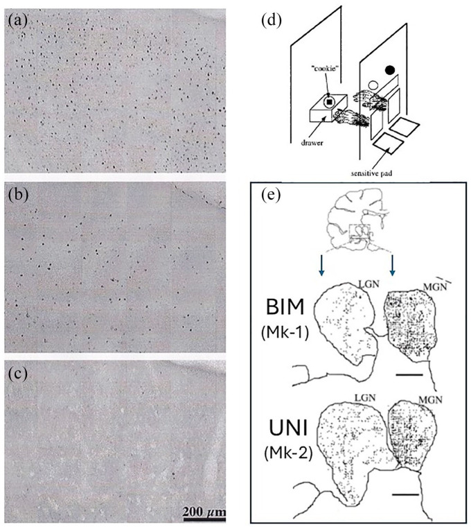
Electrophysiological data support the notion that spatial and temporal coordination between the forelimbs in primates takes place in a wide network of cortical and subcortical brain structures. However, single neuron electrophysiology is biased towards large, long distance projecting neurons. The aim of the present study was to assess whether the same neural network is involved when small and medium size neurons are considered. To address this issue, neuronal activity with cellular resolution was investigated and quantified using the c-fos mapping technique, targeting small and medium size diameter neurons, in adult non-human primates. Two male macaque monkeysi were trained to perform a reach and grasp drawer task, executed either bimanually (BIM) or unimanually (UNI). Extensive single unit electrophysiological recordings were conducted in these two monkeys over a two-year period, preceding a final terminal c-fos session during which one monkey (Mk-1) performed exclusively the BIM task, while the second monkey (Mk-2) performed the UNI task only (250 trials each). One additional monkey (control Mk-3) did not perform any task. Fos-like immunoreactivity (FLI) was significantly higher in both Mk-1 and Mk-2 in motor brain areas than in the control monkey, demonstrating that motor activity triggered c-fos. Although the overall muscle activity was roughly comparable in both tasks, Mk-1 (BIM) exhibited a clearly stronger FLI than Mk-2 all along the rostrocaudal axis of the primary, supplementary and cingulate motor cortices, as well as the striatum. In contrast, Mk-1 and Mk-2 displayed a comparable FLI in non-motor regions, such as the visual and auditory thalamus. The present study, a very rare c-fos mapping investigation conducted in macaques performing a complex behavioral task, suggests that small and medium size (local) neurons may also contribute to the specific neural activity responsible for precise interlimb coordination, within a network associating motor cortical areas and the basal ganglia.

Keywords: basal ganglia; bimanual coordination; c-fos; functional neuronal mapping; hand control; macaque monkeys; motor cortical areas; striatum.


Conflict of interest statement

The author(s) declared no potential conflicts of interest with respect to the research, authorship, and/or publication of this article.

Figures

Figure 1.
Figure 1.
Typical appearance of FLI on photomicrographs of histological sections in the cingulate motor cortex (CMA), in the form of dark dots representing Fos positive cells in Mk1 (a; bimanual task), in Mk-2 (b; unimanual task) and in Mk-3 (c; control - no task). The precise location of the cortical zone depicted in each panel is indicated by the squares pointed by the arrows in Figure 2a, in the right CMA of the 3 monkeys. In (d), the reach and grasp drawer task is represented schematically for a bimanual trial, with a vertical board facing the monkey, comprising two windows each with a sliding door, above two resting pads detecting the presence or absence of the hand on it. The monkey self-initiates a trial by placing each hand on the corresponding resting pad (left and right). Then, two LEDs higher up on the vertical board are used to indicate the monkey whether the coming trial is bimanual (2 LEDs turned on; both windows open), or unimanual left (left LED only turned on; left window open only), or unimanual right (right LED only turned on; right window open only), instructed in a pseudo-random order, as this was the case for the electrophysiological investigations conducted on these 2 monkeys., In contrast, in the present c-fos terminal session, Mk-1 performed only the bimanual task (250 trials), while Mk-2 performed the unimanual left task only (250 trials). A transparent rectangle window provides vision for the monkey to see a drawer placed on a second vertical board in the back. The drawer includes a hole in which a solid reward (“cookie”) was placed automatically by the system before each trial. In the bimanual task (Mk-1), after simultaneous opening of the two windows, the monkey reached the knob of the drawer, pulled it and maintained it open with the same left hand as the drawer was spring loaded; the right hand was used to grasp the reward inside the drawer and transport it back to the mouth. In the left unimanual task (Mk-2), after pulling with the left hand, the drawer was automatically fixed by the system in the open position (neutralizing the effect of the spring load) so that the same left hand was used to grasp the reward, while the right hand had to stay still at all time during the trial in contact with the right resting pad. See Kermadi et al. - for more details on the task, set-up, monkey training, etc. In panel E, reconstruction of individual frontal histological sections at the level of the thalamus (top drawing) in the zone comprising the lateral geniculate nucleus (LGN) and the medial geniculate nucleus (MGN) in Mk-1 and Mk-2. In LGN and MGN, positive Fos cells are indicated by dots. FLI was denser in MGN than in LGN. More importantly, the density of FLI in LGN and MGN was comparable in the 2 monkeys, irrespective of the task executed by Mk-1 (bimanual task) or Mk-2 (unimanual task). Scale bars = 1 mm.
Figure 2.
Figure 2.
Reconstruction of individual frontal histological sections taken at the level of the cingulate motor cortex (CMA, a) and striatum (b), showing the location and density of Fos positive neurons (black dots). Mk-1 (bimanual task) sections are on top of each panel, whereas Mk-2 (unimanual task) and Mk-3 (no task) are in the middle and in the bottom, respectively. Squares and rectangles represent specific zones in which quantification was conducted on consecutive sections along the whole rostro-caudal axis in order to produce the quantitative data shown in Figure 3. Cd = caudate nucleus; Put = putamen. Note the strongly different FLI density across monkeys, with a substantial decrease of FLI from Mk-1 (top) to Mk-2 (middle) and from Mk-2 (middle) to Mk-3 (bottom). The arrows in panel A point to the 3 zones illustrated in the form of photomicrographs in Figure 1 (a-c).
Figure 3.
Figure 3.
Quantitative data in the supplementary motor area (SMA), primary motor cortex (M1) and cingulate motor area (CMA), on the left hemisphere (left column) and on the right hemisphere (right column). The 2 solid lines curves represent the raw Fos data. The curves in the plot gives the density of FLI (ordinate) as a function of the rostro-caudal axis position of the corresponding individual sections (abscissa), going from rostral (left) to caudal (right). In M1 (hand area), the distance between 2 sections subjected to quantification was 0.7 mm, whereas it was 1.75 mm in SMA and CMA. In each plot, the top curve (close symbols in blue) is for Mk-1 (bimanual task) whereas the bottom curve (open symbols in brown) is for Mk-2 (unimanual task). The # symbols point to zones of the left SMA and CMA in Mk-2 in which a neuroanatomical tracer (BDA) was injected, partly and possibly obscuring the FLI. The vertical dashed lines in the top two plots represent the approximate limit between pre-SMA and SMA-proper, as defined in Liu et al. The stars on the right of each plot indicates the significance level of the differences between the 2 curves (Mk-1 vs Mk-2): * is for p < .05 and ** is for p < .01. The third curve in the bottom (brown crosses with dashed lines) represents the normalized data for Mk-2, after normalization using the LGN as a reference for comparison between Mk-1 and Mk-2 (see methods and results).
Figure 4.
Figure 4.
Quantitative data in the caudate nucleus (Cd) and putamen (Put), on the left hemisphere (left column) and on the right hemisphere (right column). The 2 solid lines curves represent the raw Fos data. The curves in the plot gives the density of FLI (ordinate) as a function of the rostro-caudal axis position of the corresponding individual sections (abscissa), going from rostral (left) to caudal (right). The distance between 2 sections subjected to quantification was 1.75 mm in Cd and Put. In each plot, the top curve (close symbols in blue) is for Mk-1 (bimanual task) whereas the bottom curve (open symbols in brown) is for Mk-2 (unimanual task). The stars on the right of each plot indicates the significance level of the differences between the 2 curves (Mk-1 vs Mk-2): * is for p < .05. The third curve in the bottom (brown crosses with dashed lines) represents the normalized data for Mk-2, after normalization using the LGN as a reference for comparison between Mk-1 and Mk-2 (see methods and results).

Similar articles

References

    1. Luppino G, Matelli M, Camarda RM, Gallese V, Rizzolatti G. Multiple representations of body movements in mesial area 6 and the adjacent cingulate cortex: an intracortical microstimulation study in the macaque monkey. J Comp Neurol. 1991;311:463-482. - PubMed
    1. Luppino G, Rizzolatti G. The Organization of the frontal motor cortex. News Physiol Sci Int J Physiol Prod Jointly Int Union Physiol Sci Am Physiol Soc. 2000;15:219–224. - PubMed
    1. Dum R, Strick P. Motor areas in the frontal lobe of the primate. Physiol Behav. 2002;77:677–682. - PubMed
    1. Lemon RN. Descending pathways in motor control. Ann Rev Neurosci. 2008;31:195–218. - PubMed
    1. Bufacchi RJ, Battaglia-Mayer A, Iannetti GD, Caminiti R. Cortico-spinal modularity in the parieto-frontal system: a new perspective on action control. Prog Neurobiol. 2023;231,102537:1-39. - PubMed

LinkOut - more resources