. 2025 Jul 1;18(7):dmm052148.
doi: 10.1242/dmm.052148. Epub 2025 Jul 16.

Lymphatic dysfunction correlates with inflammation in a mouse model of amyotrophic lateral sclerosis

Affiliations

Lymphatic dysfunction correlates with inflammation in a mouse model of amyotrophic lateral sclerosis

Akshaya Narayanan et al. Dis Model Mech. .

Abstract

Amyotrophic lateral sclerosis (ALS) is a rapidly progressive, ultimately fatal neurodegenerative disease, without effective modifying treatments. It affects both lower and upper motor neurons, causing skeletal muscle denervation and paralysis. Regardless of the mechanisms that initiate and drive ALS, chronic neuroinflammation and systemic immune system activation play key roles in disease progression. The lymphatic system is a network of vessels and organs essential for immune surveillance, tissue fluid balance and lipid absorption, critical for the resolution and progression of inflammation in the periphery. Its recent rediscovery in the central nervous system raises the possibility of it playing similar roles in neurological and neurodegenerative diseases featuring prominent neuroinflammation, such as ALS. We hypothesized that the structure and function of lymphatics are compromised in the most widely used murine model of ALS, the SOD1-G93A mouse. We found that these mice exhibit lymph transport dysfunction, diminished intrinsic lymphatic vessel tonic and phasic contractions, and an association between inflammation and lymphatic marker upregulation, despite absence of major structural changes in lymphatic network coverage in key affected tissues in the disease, skeletal muscle and spinal cord.

Keywords: Amyotrophic lateral sclerosis; Lymphangiogenesis; Lymphatics; Neuroinflammation; Systemic inflammation.


Conflict of interest statement

Competing interests The authors declare no competing or financial interests.

Figures

Fig. 1.
Fig. 1.
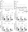
Lymph transport is delayed in the lower limbs of presymptomatic and postsymptomatic SOD1-G93A ALS mice. (A) Cartoon to the left shows the site of injection of FITC-Dextran in the shaved right leg paw and the area in the right hindlimb monitored under a fluorescent scope after that (boxed). The right panel shows time-lapse sequential images taken every 5 min after FITC-Dextran injection, showing the peripheral lymphatics (arrows) in the right hindlimb (outlined within dashed lines) of postnatal day (p)50 and p120 non-transgene-carrier (NC) control and amyotrophic lateral sclerosis (ALS) SOD1-G93A mice. (B) Average time taken to attain maximum fluorescence intensity, which was longer on average for the ALS mice, was measured and plotted for control and ALS groups. (C) Average fluorescence intensity in a standardized area was quantified in ImageJ at each time point up to 25 min for control and ALS groups. n=6/genotype/disease stage, all males. NC animals of the same age were used as controls for p50 and p120 ALS mice, respectively. *P<0.05. Values are presented as mean±s.e.m. Means were compared using Mann–Whitney test. Scale bars: 410 µm.
Fig. 2.
Fig. 2.
Lymphatic vessel contractile activity is impaired in postsymptomatic SOD1-G93A ALS mice. (A) Representative diameter traces over 30 s period in NC control and ALS lymphatic vessels at a transmural pressure of 1 cm. (B) Phasic contractile frequency. (C-E) Fractional pump flow (C), amplitude (D) and lymphatic tonic index (E) were calculated as described in the Materials and Methods. For NC, N=9 vessels from four animals; for ALS, N=14 vessels from four animals. *P<0.05, **P<0.01, ***P<0.001. Values are presented as mean±s.e.m. Means were compared using Mann–Whitney test.
Fig. 3.
Fig. 3.
Lymphatic gene marker mRNA induction is linked to inflammation in skeletal muscle of presymptomatic SOD1-G93A mice. (A-F) Quantitative real-time PCR of mRNA for lymphatic and inflammation gene markers for hindlimb skeletal muscles [extensor digitorum longus (EDL), gastrocnemius (Gastro), tibialis anterior (TA)] (A-C) and lumbar spinal cord (D-F) from p50 NC and ALS SOD1-G93A mice. n=6 males/genotype. Values are mean±s.e.m. *P<0.05, **P<0.01. Unpaired two-tailed t-test was used to compare means for all markers except for Ccl1 (lumbar SPC); Vegfr3, Cxcl1, Clcx10 (TA); Il10, Tnfa, Ifng, Ccl12, Cxcl1, Cxcl10, Cx3cl1 (Gastro); Pdpn, Ccl2, Ccl12 (EDL), for which means were compared using Mann–Whitney test. Ccl1 mRNA was undetectable in the EDL samples.
Fig. 4.
Fig. 4.
Lymphatic gene marker mRNA induction is linked to inflammation in skeletal muscle and spinal cord of postsymptomatic SOD1-G93A mice. (A-F) Quantitative real-time PCR of mRNA for lymphatic and inflammation gene markers for hindlimb skeletal muscles (EDL, Gastro, TA) (A-C) and lumbar spinal cord (D-F) from p120 NC and ALS SOD1-G93A mice. n=6 males/genotype. Values are mean±s.e.m. *P<0.05, **P<0.01. Unpaired two-tailed t-test was used to compare means for all markers except for Il6, Nos2, Tgfb, Csf1 (lumbar SPC); Ccl2, Cxcl1 (TA); Prox1, Il1b, Ifng, Cxcl1 (Gastro); Ccl2, Cxcl1, Cx3cl1 (EDL), for which means were compared using Mann–Whitney test.
Fig. 5.
Fig. 5.
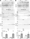
Western blots for p120 TA and EDL probed for Lyve1 and Pdpn. (A) p120 TA. (B) p120 EDL. Top panels show total loaded protein, 30 μg/lane, as stained with Revert 700 total protein stain (LI-COR). Arrows in middle panels point to expected bands for each protein, as predicted by their molecular mass in kDa. Other bands in Lyve1 blots are non-specific. Histograms in bottom panels show quantification of signal normalized to total protein/lane and expressed relative to control. The data points in the histograms represent individual muscles, each from different mice (n=6/genotype, males). Data presented as mean±s.e.m. *P<0.05, **P<0.01. Means for TA data were compared with Mann–Whitney test; means for EDL data were compared with unpaired two-tailed t-test.
Fig. 6.
Fig. 6.
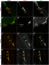
Detection of lymphatic vessels in skeletal muscle with antibodies against Lyve1 and Pdpn. (A) Maximal-intensity projection (MIP) of a representative cross-section of a p120 control NC TA double-labeled for Lyve1 and Pdpn, imaged in a confocal microscope and pseudocolored red (Lyve1) and green (Pdpn). Yellow arrowheads point to regions of staining overlap; green and red arrowheads point to Pdpn and Lyve1 staining, respectively, where there is no staining overlap. (A′) Grayscale Lyve1 staining. (A″) Grayscale Pdpn staining. (B) MIP of a representative cross-section of a p120 ALS TA double-labeled for Lyve1 and Pdpn, imaged and pseudocolored as in A, with two areas of partial colocalization of Lyve1 and Pdpn staining. (B′) Grayscale Lyve1 staining. (B″) Grayscale Pdpn staining. Owing to green autofluorescence of muscle fibers, here it can be better appreciated that the Lyve1/Pdpn staining is detected outside muscle fibers. (C) MIP of a representative longitudinal section of a p120 NC TA that was double-labeled for Lyve1 and Pdpn, imaged and pseudocolored as in A. Vessel-like structures, running parallel to the muscle fibers can be seen co-labeling for Lyve1 and Pdpn (yellow arrowheads) or singly labeled for each marker. Red arrowheads, Lyve1; green arrowheads, Pdpn. (C′) Grayscale Lyve1 staining. (C″) Grayscale Pdpn staining. (D) MIP of a representative longitudinal section of an p120 ALS TA that was double-labeled for Lyve1 and Pdpn, imaged and pseudocolored as in A. (D′) Grayscale Lyve1 staining. (D″) Grayscale Pdpn staining. Scale bars: 5 μm (A-B″); 50 μm (C-D″).
Fig. 7.
Fig. 7.
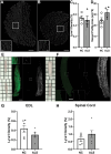
Lymphatic capillary coverage in lumbar spinal cord and EDL muscle appears largely unaltered in SOD1-G93A mice. (A) Representative cross-section of p120 NC EDL muscle stained for Lyve1. Scale bar: 200 μm. Inset: higher-magnification view. Scale bar: 50 μm. (B) Representative cross-section of p120 ALS EDL muscle stained for Lyve1. Scale bar: 200 μm. Inset: higher-magnification view. Scale bar: 50 μm. (C) Lyve1 density (Lyve1 staining area/area of cross-section×100%) (n=4 males/genotype; P=0.6875). (D) Vessel profile area (μm2). (E) Muscle tissue clearing before (top left) and after (bottom left) iDISCO protocol. Representative lightsheet slice of p126 NC EDL tissue labeled for Lyve1 (left, raw Lyve1 staining; middle, thresholded Lyve1 signal; right, MIP of thresholded Lyve1 image). Scale bar: 1 cm. (F) Representative lightsheet slice of p126 NC lumbar spinal cord tissue labeled for Lyve1 (left, raw Lyve1 staining; middle, thresholded Lyve1 signal; right, MIP of thresholded Lyve1 image). Scale bar: 1 cm. (G) Lyve1 density in thresholded lightsheet images in EDL tissue (n=6 males/genotype; P=0.1597). (H) Lyve1 density in thresholded lightsheet images in spinal cord tissue (n=6 males/genotype; P=0.2784). All means in C, D, G and H compared by unpaired two-tailed t-test.

References

    1. Abouelkheir, G. R., Upchurch, B. D. and Rutkowski, J. M. (2017). Lymphangiogenesis: fuel, smoke, or extinguisher of inflammation's fire? Exp. Biol. Med. 242, 884-895. 10.1177/1535370217697385 - DOI - PMC - PubMed
    1. Alitalo, K. (2011). The lymphatic vasculature in disease. Nat. Med. 17, 1371-1380. 10.1038/nm.2545 - DOI - PubMed
    1. Antila, S., Karaman, S., Nurmi, H., Airavaara, M., Voutilainen, M. H., Mathivet, T., Chilov, D., Li, Z., Koppinen, T., Park, J. H.et al. (2017). Development and plasticity of meningeal lymphatic vessels. J. Exp. Med. 214, 3645-3667. 10.1084/jem.20170391 - DOI - PMC - PubMed
    1. Antila, S., Chilov, D., Nurmi, H., Li, Z., Näsi, A., Gotkiewicz, M., Sitnikova, V., Jäntti, H., Acosta, N., Koivisto, H.et al. (2024). Sustained meningeal lymphatic vessel atrophy or expansion does not alter Alzheimer's disease-related amyloid pathology. Nat. Cardiovasc. Res. 3, 474-491. 10.1038/s44161-024-00445-9 - DOI - PMC - PubMed
    1. Appel, S. H., Beers, D. R. and Zhao, W. (2021). Amyotrophic lateral sclerosis is a systemic disease: peripheral contributions to inflammation-mediated neurodegeneration. Curr. Opin. Neurol. 34, 765-772. 10.1097/WCO.0000000000000983 - DOI - PubMed

MeSH terms

Substances