. 2025 Jul 4;11(27):eado1223.
doi: 10.1126/sciadv.ado1223. Epub 2025 Jul 2.

Nanosilicates promote angiogenesis through activation of ROS-mediated WNT/β-catenin pathway

Affiliations

Nanosilicates promote angiogenesis through activation of ROS-mediated WNT/β-catenin pathway

Giriraj Lokhande et al. Sci Adv. .

Abstract

Scaffold vascularization plays a pivotal role in the wound healing process, facilitating the recruitment of endogenous progenitor cells and delivering crucial signaling molecules that promote cellular invasion, angiogenesis, and neotissue formation. In this study, we introduce nanosilicates as proangiogenic biomaterials that can prime endogenous cells to expedite angiogenesis and graft vascularization in vivo. Characterized by their mineral-based two-dimensional structure and extensive surface area, nanosilicates are efficiently internalized by endothelial cells, leading to augmented cellular migration and the formation of tubular structures. Through whole transcriptome sequencing, we elucidated that nanosilicates activate the canonical Wnt pathway through hypoxia-induced ROS production. In vivo investigations further corroborate the proangiogenic efficacy of nanosilicate-loaded biomaterials. This study posits nanosilicates as a potential proangiogenic adjunct in the design of biomaterials for in situ tissue regeneration.


Figures

Fig. 1.
Fig. 1.. Characterization, cellular uptake, and proangiogenic effects of nanosilicates.
(A) Schematic representation of nanosilicates and their particle size distribution using dynamic light scattering. Dh, hydrodynamic diameter; PDI, polydispersity index. (B) Cytotoxicity of nanosilicates when exposed to HUVECs using MTT assay. IC50, half-maximal inhibitory concentration. (C) Alamar Blue cell proliferation assay of HUVECs treated with nanosilicates (10 and 50 μg/ml) for 3 days. (D) Cell cycle analysis of HUVECs when exposed to various nanosilicate concentrations for 72 hours with 10 μg/ml showing highest shift to the G0 cycle. DAPI, 4′,6-diamidino-2-phenylindole. (E) Increased ATP production in HUVECs treated with nanosilicates (10 μg/ml) for 24 hours. Nanosilicates showed least membrane depolarization in HUVECs, almost equivalent to untreated HUVECs, when treated with 10 μg/ml using JC-1 dye. (F) Confocal microscopy showing cellular uptake of rhodamine-tagged nanosilicates by HUVECs after 24 hours of treatment. (G) Transmission electron microscopy showing internalization of nanosilicates by HUVECs within vesicular structures after 24 hours of treatment. (H) Nanosilicate uptake analysis was conducted using endocytosis inhibitors which showed that nanosilicates were uptaken by clathrin-mediated endocytosis. a.u., arbitrary units. (I) CAM assay conducted on 3-day-old chicken embryo showing an increased number of vascular branches post–24-hour nanosilicate treatment. Statistical significance is denoted as follows: *P < 0.05, **P < 0.01, ***P < 0.001, and ****P < 0.0001.
Fig. 2.
Fig. 2.. Nanosilicates modulate gene expression and pathways linked to angiogenesis in endothelial cells.
(A) PCA of HUVEC treated with nanosilicates (HUVEC_nSi) based on mRNA expression obtained from RNA-seq (n = 3, technical replicates). Cells without nanomaterials are used as control (HUVEC). The PCA was done on the mRNA expression [log2 fragments per kilobase per million (FPKM)] of 50% of the most variable expressed genes across all samples (n = 5693). (B) MA (Bland-Altman) plot highlighting differentially regulated genes [false discovery rate (FDR)–adjusted P < 0.05] of the treatment groups against the untreated HUVEC and extent of expression change (gray: total expressed genes, red: up-regulated genes, blue: down-regulated genes). (C) Heatmap of DEGs FPKM presented as z-scores. The DEGs were obtained from pairwise comparison of treated versus untreated HUVECs. (D) GO enrichment analysis showed that nanosilicates regulate biological processes, molecular functions, and cellular components associated with angiogenesis and extracellular matrix organization.
Fig. 3.
Fig. 3.. Nanosilicates modulate angiogenic gene expression and enhance endothelial cell migration.
(A) Significantly enriched GO terms associated with angiogenesis are highlighted for HUVEC_nSi when compared to control HUVEC. (B) A volcano plot showing DEGs for blood vessel morphogenesis (GO: 0048514). Gray, all of the expressed genes; blue, genes associated with the GO term with no significant change in expression; and red, genes associated with the GO term that show significantly different expressions (Padj < 0.05 and absolute fold change > 0.5). Of these, ANGPTL4, aquaporin-1 (AQP1), and heme oxygenase-1 (HMOX1) have been associated with nanoparticle-induced angiogenesis in HUVEC. (C) Angiogenic genes with a high log2 (fold change) by RNA expression. Genes coding for angiogenic inhibitors showed down-regulation, and genes supporting angiogenic processes were up-regulated. (D) Gene tracks showing normalized mRNA expression of ANGPTL4 and FGFRL1 for HUVEC and HUVEC_nSi in reads per million. (E) PCR validation of the RNA-seq findings showed that HUVEC_nSi followed a similar trend in showing significant (*P < 0.05 and **P < 0.01, n = 3) fold change in the expression of ANGPTL4 and FGFRL1. (F) Comparison of ANGPTL4 and FGFRL1 gene expression obtained from RNA-seq and PCR validation. RQ, respiratory quotient. (G) Migration assay (n = 3) showing faster wound coverage upon injury with HUVEC treated with nanosilicates. Statistical significance is denoted as follows: *P < 0.05, **P < 0.01, and ****P < 0.0001.
Fig. 4.
Fig. 4.. Nanosilicates activate Wnt signaling and hypoxia-related pathways to enhance angiogenesis.
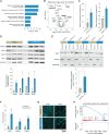
(A) Significantly enriched GO terms associated with response to hypoxia are highlighted for HUVEC_nSi when compared to control HUVEC. (B) A volcano plot showing DEGs for response to oxygen levels (GO: 0070482). Of these, AQP-1, ITGB4, and HMOX1 have been associated with nanoparticle-induced angiogenesis in HUVEC (Padj < 0.05 and absolute fold change > 0.5). (C) ROS levels were significantly (**P < 0.01, ***P < 0.001, n = 3) higher in HUVEC-nSi and HOMEC-nSi samples compared to untreated HUVECs and HOMECs. (D) Protein levels associated with Wnt signaling pathways were significantly higher (*P < 0.05, n = 3) in HUVECs treated with nanosilicates. (E) BCAT protein expression levels in the nuclear and cytoplasmic fractions of nSi-treated and untreated HUVECs showing significantly (*P < 0.05, n = 3) increased BCAT levels in nucleus of nSi-treated HUVECs. (F) Genes associated with hypoxic activation of angiogenesis showed significant (*P < 0.05, n = 3) up-regulation upon nanosilicate treatment. (G) Nanosilicate treatment showed initiation of tubular morphology in HUVECs which is an essential characteristic for proangiogenic behavior. (H) GSEA analysis showing up-regulation in the Reactome gene set aquaporin-mediated transport (NES = 1.49, P = 0.05). Statistical significance is denoted as follows: *P < 0.05, **P < 0.01, ***P < 0.001, and ****P < 0.0001.
Fig. 5.
Fig. 5.. Nanosilicates enhance microvessel formation and cellular infiltration in vivo.
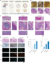
(A) Schematic representation of the subcutaneous mouse model to represent the influence of nanosilicate treatment in promoting angiogenesis. UV, ultraviolet. (B) Explant and histology images of the hydrogel postimplantation. Macroscopic presence of vessels around the hydrogel with increasing volume with nanosilicates and growth factor conjugated nanosilicates. (C) H&E images detailing the presence of microvessels and arterioles forming within, around, and on the periphery of the implanted hydrogels. (D) Histological analysis also shows improved collagen formation with nanosilicate integrated in hydrogels. Immunofluorescence analysis of endothelial markers like CD31, VEGF, and vWF shows increasing levels upon nanosilicate integration into the hydrogel. (E) Graphical representation of increase in endogenous cell infiltration and vessel coverage within the hydrogel incorporating nanosilicates and growth factor–conjugated nanosilicates (n = 4). Statistical significance is denoted as follows: **P < 0.01, ***P < 0.001, and ****P < 0.0001.

References

    1. Xing Y., Yerneni S. S., Wang W., Taylor R. E., Campbell P. G., Ren X., Engineering pro-angiogenic biomaterials via chemoselective extracellular vesicle immobilization. Biomaterials 281, 121357 (2022). - PubMed
    1. Briquez P. S., Clegg L. E., Martino M. M., Gabhann F. M., Hubbell J. A., Design principles for therapeutic angiogenic materials. Nat. Rev. Mater. 1, 15006 (2016).
    1. Gaharwar A. K., Singh I., Khademhosseini A., Engineered biomaterials for in situ tissue regeneration. Nat. Rev. Mater. 5, 686–705 (2020).
    1. Arlov Ø., Rütsche D., Asadi Korayem M., Öztürk E., Zenobi-Wong M., Engineered sulfated polysaccharides for biomedical applications. Adv. Funct. Mater. 31, 2010732 (2021).
    1. Chen S., Luo Y., He Y., Li M., Liu Y., Zhou X., Hou J., Zhou S., In-situ-sprayed therapeutic hydrogel for oxygen-actuated Janus regulation of postsurgical tumor recurrence/metastasis and wound healing. Nat. Commun. 15, 814 (2024). - PMC - PubMed

MeSH terms

LinkOut - more resources