. 2025 Sep:85:103746.
doi: 10.1016/j.redox.2025.103746. Epub 2025 Jun 28.

Anemoside B4 alleviates ulcerative colitis by attenuating intestinal oxidative stress and NLRP3 inflammasome via activating aryl hydrocarbon receptor through remodeling the gut microbiome and metabolites

Affiliations

Anemoside B4 alleviates ulcerative colitis by attenuating intestinal oxidative stress and NLRP3 inflammasome via activating aryl hydrocarbon receptor through remodeling the gut microbiome and metabolites

Hao Wu et al. Redox Biol. 2025 Sep.

Abstract

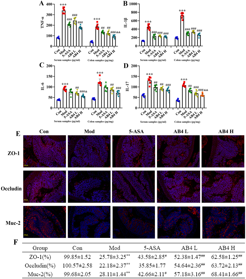
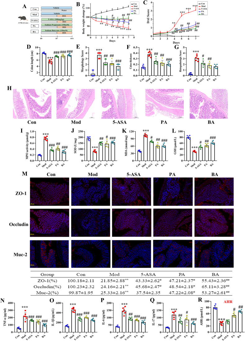
Ulcerative colitis (UC) is a chronic, non-specific inflammatory disease of the intestines with a significant increase in global incidence in recent years. Oxidative stress and inflammation are two hallmarks of UC pathogenesis. Anemoside B4 (AB4), a pentacyclic triterpenoid saponin, exhibits significant antioxidant and anti-inflammatory properties and shows potential for preventing UC. Here, an animal model induced by dextran sodium sulfate (DSS) was used to investigate the effect of AB4 on UC. The results demonstrated that AB4 significantly reduces intestinal oxidative stress and inflammation in UC mice, while also protecting intestinal barrier function. Furthermore, AB4 helps restore intestinal microbial balance primarily by modulating the abundance of Lactobacillus, which enhances the metabolism of short-chain fatty acids and upregulates the production of butyric acid (BA). Pseudogerm-free mice and fecal microbiota transplantation (FMT) demonstrated that AB4 significantly mitigated UC in a gut microbe-dependent manner. Both AB4 and BA markedly activate the aromatic hydrocarbon receptor (AhR). The intestinal organoid results suggest BA may activate the AhR to inhibit ROS production and activation of NLRP3 inflammasome, thereby protecting intestinal integrity. Administration of AhR antagonists abolished the protective effects, thus confirming the involvement of AhR in the underlying mechanism. Overall, these results indicate that AB4 is an effective agent against UC mainly by activating the AhR through gut microbial short-chain fatty acid metabolites to inhibit intestinal oxidative stress and inflammation.

Keywords: Anemoside B4; Aryl hydrocarbon receptor; Butyric acid; Gut microbiome; Oxidative stress; Ulcerative colitis.


Conflict of interest statement

Declaration of competing interest The authors declare that they have no known competing financial interests or personal relationships that could have appeared to influence the work reported in this paper.

Figures

Fig. 1
Fig. 1
AB4 alleviates DSS-induced mice colitis. (A) The flowchart of the part in vivo experiments. (B) The body weight of mice in each group. (C) The food intake. (D) The water intake. (E) DAI score. (F) Observation of colonic appearance in mice. (G) Colon length. (H) Morphology score. (I) Colon thickness. (J) Histopathological score. (K) Representative H&E images for colon tissues. Data are expressed as mean ± SEM for 8 mice in each group. ∗P < 0.05, ∗∗P < 0.01 and ∗∗∗P < 0.001 versus Con group, #P < 0.05, ##P < 0.01 and ###P < 0.001 versus Mod group.
Fig. 2
Fig. 2
AB4 suppressed inflammation and protected against intestinal barrier dysfunction to attenuate colitis in mice. (A–D) Inflammatory cytokines (TNF-α, IL-1β, IL-6, and IL-17) levels in serum and colonic tissues (n = 6). (E–F) Representative immunofluorescence images and the fluorescence intensities of ZO-1, Occludin, and Muc-2 in colonic tissues (n = 3). ∗∗∗P < 0.001 versus Con group, ##P < 0.01 and ###P < 0.001 versus Mod group, &P < 0.05 and &&P < 0.01 versus 5-ASA group.
Fig. 3
Fig. 3
AB4 improved UC mice by restoring gut microbial diversity. (A) OUT rank. (B) Pan analysis. (C) Core analysis. (D) Sobs index. (E) Shannon index. (F) Chao index. (G) Ace index. (H) PCA analysis. (I) PcoA analysis. (J) NMDS analysis. (K) NCM analysis. (L) PLS-DA analysis. (M) Microbial dysbiosis index. (N) Hierarchical clustering. Data are expressed as mean ± SEM for 6 mice in each group. ∗∗P < 0.01 and ∗∗∗P < 0.001 versus Con group, #P < 0.05 versus Mod group.
Fig. 4
Fig. 4
AB4 improved gut microbial composition in UC mice. (A) Venn diagram. (B) Community abundance analysis at the phylum level. (C) Community abundance analysis at the genus level. (D) Circos plots analysis at the phylum level. (E) Circos plots analysis at the genus level. (F) LEfSe multilevel species level tree. (G) Heat map. (H) LDA discriminant histogram. (I) PICRUSt2 functional prediction.
Fig. 5
Fig. 5
Effect of AB4 on comparative analysis of gut microbial metabolites in UC mice. (A–B) PCA analysis under positive and negative ions. (C–E) PLSDA analysis under positive ions. (F–H) PLSDA analysis under negative ions. (I–K) Volcano plots.
Fig. 6
Fig. 6
Effect of AB4 on gut microbial metabolites in DSS-induced UC mice. (A) Venn diagram. (B–C) VIP bar charts. (D) Differential metabolite cluster analysis. (E–F) KEGG enrichment analysis. (G-H)KEGG functional analysis.
Fig. 7
Fig. 7
AB4 inhibits oxidative stress and inflammation in UC mice. (A) The expression of butyric acid. (B) The expression of propionic acid. (C) Detection of ROS expression levels in the colon tissue by flow cytometry. (D) The mean fluorescence intensity of the colon tissue in each group. (E–H) The content of MPO, SOD, MDA and GSH in the colon tissue. (I) Western blot analysis for NLRP3 proteins in colonic tissues of colitis mice. (J) Box plot showing the densitometry analysis of NLRP3 normalized by β-Actin. (K–M) The expression level of ASC, Caspase-1 and IL-1β in colon tissue. Data are expressed as mean ± SEM for 6 mice in each group. ∗∗∗P < 0.001 versus Con group, #P < 0.05, ##P < 0.01 and ###P < 0.001 versus Mod group.
Fig. 8
Fig. 8
Effect of AB4 on DSS-induced UC mice after fecal microbiota transplantation. (A) The flowchart of the part in vivo experiments. (B) The body weight of mice in each group. (C) The food intake. (D) The water intake. (E) DAI score. (F) Colon length. (G) Morphology score. (H) Colon thickness. (I) Histopathological score. (J–M) Inflammatory cytokines (TNF-α, IL-1β, IL-6, and IL-17) levels in colonic tissues. ∗P < 0.05, ∗∗P < 0.01 and ∗∗∗P < 0.001 versus Con group, #P < 0.05, ##P < 0.01 and ###P < 0.001 versus Mod group.
Fig. 9
Fig. 9
AB4 attenuates UC in a gut microbiota short-chain fatty acid metabolites-dependent manner and activating AHR. (A) Representative H&E images for colonic tissues. (B) Representative immunofluorescence images of ZO-1, Occludin, and Muc-2 in colonic tissues (n = 3). (C) The expression of butyric acid. (D) The expression of propionic acid. (E) Detection of ROS expression levels in the colonic tissue by flow cytometry. (F) The mean fluorescence intensity of the colonic tissue in each group. (G–J) The content of MPO, SOD, MDA and GSH in the colonic tissue. (K) The expression of AHR in the colonic tissue. (L) Western blot analysis for NLRP3 proteins in colonic tissues of colitis mice. (M) Box plot showing the densitometry analysis of NLRP3 normalized by β-Actin. (N–P) The expression level of ASC, Caspase-1 and IL-1β in colon tissue. Data are expressed as mean ± SEM for 6 mice in each group. ∗∗∗P < 0.001 versus Con group, ##P < 0.01 and ###P < 0.001 versus Mod group.
Fig. 10
Fig. 10
The gut microbiota SCFAs metabolites of AB4 improved UC mice by activating AHR. (A) The flowchart of the part in vivo experiments. (B) The body weight of mice in each group. (C) DAI score. (D) Colon length. (E) Morphology score. (F) Colon thickness. (G) Histopathological score. (H) Representative H&E images for colonic tissues. (I–L) The content of MPO, SOD, MDA and GSH in the colon tissue. (M) Representative immunofluorescence images of ZO-1, Occludin, and Muc-2 in colonic tissues (n = 3). (N–Q) Inflammatory cytokines (TNF-α, IL-1β, IL-6, and IL-17) levels in colonic tissues. (R) The expression of AHR in the colonic tissue. Data are expressed as mean ± SEM for 6 mice in each group. ∗P < 0.05, ∗∗P < 0.01 and ∗∗∗P < 0.001 versus Con group, #P < 0.05, ##P < 0.01 and ###P < 0.001 versus Mod group.
Fig. 11
Fig. 11
Butyric acid protects intestinal organoids damaged by DSS on activating AHR. (A) The morphological change of intestinal organoids in DSS stimulation. (B–E) The levels of pro-inflammatory cytokines (IL-1β, IL-6, IL-17, and TNF-α) in the supernatant. (F) The expression of AHR. (G) Detection of ROS expression levels by flow cytometry. (H) The mean fluorescence intensity in each group. (I) Western blot analysis for NLRP3 proteins in intestinal organoids. (J) Box plot showing the densitometry analysis of NLRP3 normalized by β-Actin. (K–M) The expression level of ASC, Caspase-1 and IL-1β in intestinal organoids. ∗∗∗P < 0.001 versus Con group, ##P < 0.01 and ###P < 0.001 versus Mod group.
Fig. 12
Fig. 12
AhR is a key target that mediates the anti-UC effect of AB4. (A) The flowchart of the part in vivo experiments. (B) The body weight of mice in each group. (C) DAI score. (D) Colon length. (E) Morphology score. (F) Colon thickness. (G) Histopathological score. (H–K) The expression of MPO, SOD, MDA, and GSH. (L) Representative H&E images for colonic tissues. (M–P) Inflammatory cytokines (TNF-α, IL-1β, IL-6, and IL-17) levels in colonic tissues. (O) Representative immunofluorescence images of ZO-1, Occludin, and Muc-2 in colonic tissues (n = 3). Data are expressed as mean ± SEM for 6 mice in each group. ∗P < 0.05, ∗∗P < 0.01 and ∗∗∗P < 0.001 versus Con group, #P < 0.05, ##P < 0.01 and ###P < 0.001 versus Mod group.

References

    1. Ordás I., Eckmann L., Talamini M., Baumgart D.C., Sandborn W.J. Ulcerative colitis. Lancet. 2012 Nov 3;380(9853):1606–1619. - PubMed
    1. Segal J.P., LeBlanc J.F., Hart A.L. Ulcerative colitis: an update. Clin. Med. 2021 Mar;21(2):135–139. - PMC - PubMed
    1. Voelker R. What is ulcerative colitis? JAMA. 2024 Feb 27;331(8):716. - PubMed
    1. Eisenstein M. Ulcerative colitis: towards remission. Nature. 2018 Nov;563(7730):S33. - PubMed
    1. Wangchuk P., Yeshi K., Loukas A. Ulcerative colitis: clinical biomarkers, therapeutic targets, and emerging treatments. Trends Pharmacol. Sci. 2024 Oct;45(10):892–903. - PubMed

MeSH terms

LinkOut - more resources