. 2025 Jul 15;6(7):102213.
doi: 10.1016/j.xcrm.2025.102213. Epub 2025 Jul 1.

Overcoming multidrug resistance in gastrointestinal cancers with a CDH17-targeted ADC conjugated to a DNA topoisomerase inhibitor

Affiliations

Overcoming multidrug resistance in gastrointestinal cancers with a CDH17-targeted ADC conjugated to a DNA topoisomerase inhibitor

Rui Wang et al. Cell Rep Med. .

Abstract

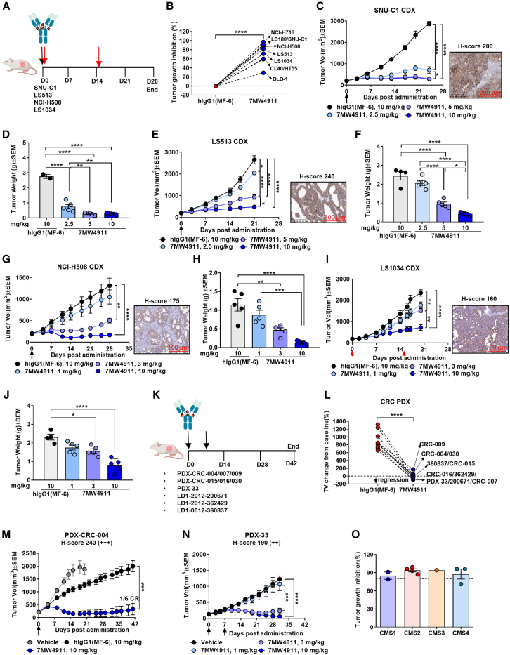
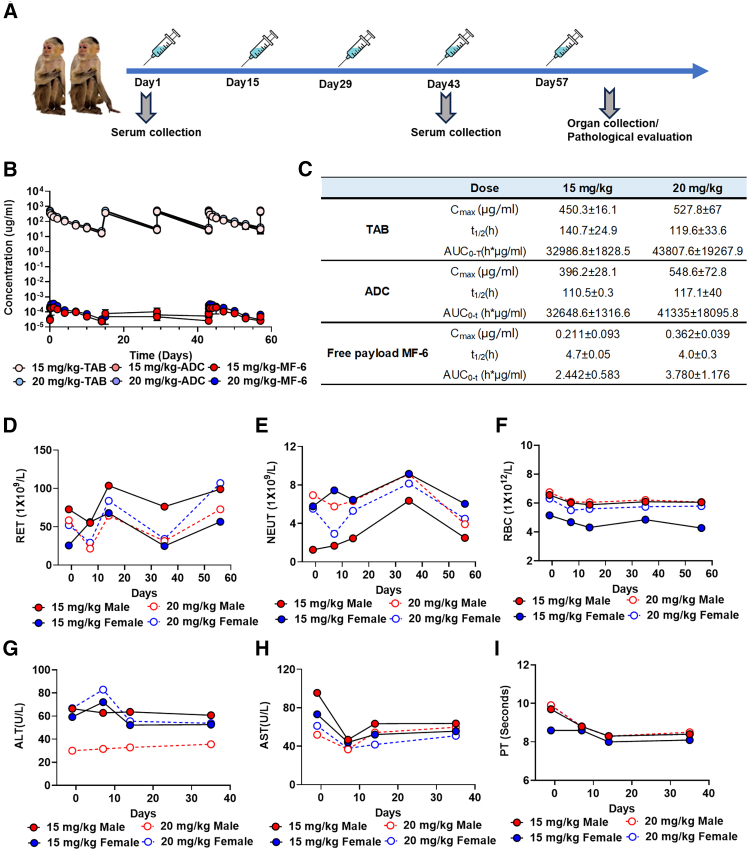
Cadherin 17 (CDH17) has emerged as a promising target for gastrointestinal (GI) cancers, which are often complicated by multidrug resistance (MDR) and recurrence. In this study, we developed 7MW4911, a CDH17-targeted antibody-drug conjugate (ADC) that incorporates a topoisomerase inhibitor MF-6 (Topi MF-6) payload linked via a cleavable linker, designed specifically to address MDR in GI cancers. 7MW4911 exhibited high specificity for CDH17-expressing cancer cells and potent cytotoxicity in vitro. In preclinical models, including patient-derived xenografts (PDXs) with distinct mutations, 7MW4911 achieved tumor growth inhibition ranging from 71% to 99%. Remarkably, 7MW4911 outperformed monomethyl auristatin E (MMAE)-based and Deruxtecan (DXd)-based ADCs in MDR models, highlighting its effectiveness against drug-resistant cancer phenotypes. Additionally, 7MW4911 showed favorable pharmacokinetics and a highest non-severely toxic dose (HNSTD) exceeding 20 mg/kg in cynomolgus monkeys, underscoring its promising safety profile. Together, these findings position 7MW4911 as a promising ADC candidate capable of enhancing therapeutic outcomes in GI cancers.

Keywords: ADC; CDH17; colorectal cancer; gastrointestinal cancers; multidrug resistance.


Conflict of interest statement

Declaration of interests All authors are employees of Mabwell (Shanghai) Bioscience Co., Ltd., or Jiangsu Mabwell Health Pharmaceutical R&D Co., Ltd., and may hold shares in Mabwell (Shanghai) Bioscience Co., Ltd. Rui Wang, P.F., W.Z., X.T., X.G., and D.L. are listed as inventors on a patent application for the anti-CDH17 ADC 7MW4911.

Figures

None
Graphical abstract
Figure 1
Figure 1
Identification of CDH17 as a potential therapeutic target for GI cancers (A) Schematic overview of the multi-omics strategy for identifying gastrointestinal cancer markers. Key steps included (I) selection of genes upregulated in tumors vs. normal tissues, (II) exclusion of genes highly expressed in normal organs (organ images from Figdraw), (III) removal of markers expressed on immune cells, and (IV) identification of tumor epithelium-specific genes. The top five colorectal cancer candidates and a representative simulated protein structure are shown (V). Statistical analyses were performed in R 4.2.2. (B) CDH17 expression analysis in gastrointestinal cancer tissue microarrays. H-scores and sample numbers are detailed in Table S1. Representative IHC images show variable staining intensities. Data are represented as mean ± SEM. (C) CDH17 surface expression in gastrointestinal cancer cell lines, as assessed by flow cytometry. Negative controls included primary endothelial cells and non-cancerous cell lines. Data are represented as mean ± SEM. (D and E) CDH17 mRNA (D) and protein (E) expression levels across CRC CMS1–4 subtypes using TCGA and clinical sample data, respectively. Data are presented as mean ± SEM. (F) Summary of CDH17 expression in CRC PDX models by molecular subtype. Data are represented as mean ± SEM. Statistical significance was determined by Kruskal-Wallis test in (D)–(F). ∗p < 0.05, ∗∗p < 0.01, ∗∗∗p < 0.001, and ∗∗∗∗p < 0.0001. See also Figures S1 and S2 and Table S1.
Figure 2
Figure 2
Generation and characterization of anti-CDH17 ADC 7MW4911 (A) Structure of 7MW4911: the ADC consists of the MF-6 payload conjugated to Mab0727 via a linker system composed of IDconnect and PEG-VA, enabling efficient payload release. (B and C) Physicochemical properties of 7MW4911 were measured by HIC-HPLC (B) and SEC-HPLC (C). (D and E) Binding affinities of 7MW4911 to human (D) and cynomolgus monkey (E) CDH17 were measured by Octet RED96 system. (F) Binding of 7MW4911 to CDH17-expressing cell lines (SK-CO-1, SNU-C1, SNU-16) was assessed by flow cytometry. (G) CDH17-mediated internalization of 7MW4911 was evaluated in CDH17-positive cell lines. (H) Cytotoxicity of 7MW4911 was measured in cell lines with varying CDH17 expression using CellTiter-Glo assays. (I–L) Stability of 7MW4911 in cynomolgus monkey plasma. Binding activity (I and J) and concentrations (K and L) of total antibody (TAB) and intact ADC were evaluated over time, as described in the STAR Methods. Data in (K) and (L) are normalized to baseline values. (M) Pharmacokinetics of 7MW4911 in BALB/c mice following a single intravenous dose on day 0. Serum levels of TAB and intact ADC were quantified over time. Data are represented as mean ± SEM (n = 4). (N) Bystander killing effect of 7MW4911 evaluated using a co-culture model. See also Figures S3–S6 and Table S2.
Figure 3
Figure 3
Potent anti-tumor activity of 7MW4911 in multiple CRC CDX and PDX models (A) Schematic of the CRC CDX treatment regimen. Tumor-bearing mice received intravenous 7MW4911 or control ADC on day 0 (black or red arrows). (B) TGI in CRC CDX models following 10 mg/kg 7MW4911 treatment. (C–J) In vivo anti-tumor efficacy of 7MW4911 was evaluated in various CRC CDX models following the treatment regimen. In vivo anti-tumor efficacy of 7MW4911 in various CRC CDX models. Tumor volume and weight were measured in SNU-C1 (C and D; n = 2–5), LS513 (E and F; n = 4–5), NCI-H508 (G and H; n = 5), and LS1034 (I and J; n = 4–5). Data are represented as mean ± SEM. Mice were euthanized early if tumors exceeded 3,000 mm3 (3 in SNU-C1 and 1 each in LS513 and LS1034). (K) Schematic of the CRC PDX treatment regimen. Mice received intravenous administration of 7MW4911 or control ADC on day 0. (L) Summary of tumor volume changes from baseline in CRC PDX models. (M and N) Tumor volume changes in PDX-CRC-004 (n = 6) and PDX-33-F4-1 (n = 6–8). Data are represented as mean ± SEM. (O) Efficacy summary of 7MW4911 across CMS1–4 subtypes in PDX models. CR, complete regression. Statistical significance was determined using one-way ANOVA for all models except (B) and (L), which were analyzed using paired t tests. ∗p < 0.05, ∗∗p < 0.01, ∗∗∗p < 0.001, and ∗∗∗∗p < 0.0001. See also Figures S7 and S8 and Tables S3 and S4.
Figure 4
Figure 4
Impressive anti-tumor activity of 7MW4911 in gastric and pancreatic cancer models (A) Schematic of treatment regimens in gastric and pancreatic cancer CDX models. Mice received 7MW4911 or control ADC on day 0 (black, red, or purple arrows). (B) Summary of TGI following 10 mg/kg 7MW4911 in gastric cancer CDX models and 3 mg/kg in the pancreatic cancer CDX model. (C–F) In vivo efficacy in gastric cancer CDX models: tumor volume and weight in SNU-16 (C and D; n = 6) and tumor volume and weight in 23132/87 (E and F; n = 5). Data are represented as mean ± SEM (n = 6). (G–J) Tumor volume changes and tumor weights in three gastric cancer PDX models. One mouse in each group of the PDX-STAD-050 model died on day 18, probably due to tumor-related factors. Data are represented as mean ± SEM (n = 3–5). STAD, stomach adenocarcinoma. (K and L) Efficacy in the AsPC-1 pancreatic cancer CDX model: tumor volume (n = 5) and weight (n = 4). Data are represented as mean ± SEM. Statistical analysis was performed using unpaired two-tailed t tests except for (B), which was analyzed using paired t tests. CR, complete regression. ∗p < 0.05, ∗∗p < 0.01, ∗∗∗p < 0.001, and ∗∗∗∗p < 0.0001. See also Table S3.
Figure 5
Figure 5
Strong anti-tumor efficacy of 7MW4911 in MDR models (A) Schematic of MMAE-resistant models (created with Figdraw). (B–D) Tumor volume changes in CDX models treated with 7MW4911 or comparators: LS513 (B), LS1034 (C), and AsPC-1 (D). Treatments included 7MW4911, Mab0727(MMAE), 07-0663-h7(MMAE), 07-0663-h7(MF-6), or controls. Data are represented as mean ± SEM (n = 5). Data were analyzed using one-way ANOVA. (E–G) Tumor volume changes in CRC PDX models treated with 7MW4911, 07-0663-h7(MMAE), or vehicle: PDX-CRC-015 (E), PDX-CRC-016 (F), and PDX-CRC-030 (G). Data are represented as mean ± SEM (n = 6). Data were analyzed using one-way ANOVA. (H and I) In vivo efficacy in MMAE-resistant LS513 (H) and NCI-H716 (I) CDX models. Mice were pretreated with 07-0663-h7(MMAE) or Mab0727(MMAE), then received 7MW4911 or continued MMAE treatment. Data are represented as mean ± SEM (n = 3–6). Data were analyzed using one-way ANOVA. (J and K) Tumor volume progression in NCI-H508 (J; 5 mg/kg) and NCI-H716 (K; 10 mg/kg) CDX models following 7MW4911, Mab0727-GGFG-DXd (DAR 4), or vehicle. Data are represented as mean ± SEM (n = 4–5). Statistical analysis was performed using one-way ANOVA. CR, complete regression. ns, not significant; ∗p < 0.05, ∗∗p < 0.01, ∗∗∗p < 0.001, and ∗∗∗∗p < 0.0001. See also Figures S8–S10.
Figure 6
Figure 6
Nonclinical safety and PK assessment of 7MW4911 in cynomolgus monkeys (A) Dosing regimen: cynomolgus monkeys (n = 2/group; 1 male and 1 female) received 15 or 20 mg/kg 7MW4911 intravenously every 2 weeks for 8 weeks (days 1, 15, 29, 43, and 57). Clinical observations, pathology, and blood sampling for pharmacokinetic (PK) and clinical assessments were conducted throughout. Necropsy was performed 1 week post-final dose. (B) PK profiles of TAB, intact ADC, and MF-6 were evaluated over time. Data are represented as mean ± SEM. (C) Toxicokinetic analysis following repeat dosing assessed systemic exposure and drug accumulation. (D–F) Hematology parameters. Hematological assessments, including red and white blood cell counts and reticulocyte levels, were monitored for hematologic toxicity. (G and H) Serum chemistry: liver and kidney function markers were analyzed for organ toxicity. (I) Coagulation: prothrombin time (PT) and related parameters were assessed for effects on hemostasis. See also Figure S11.

References

    1. Bray F., Laversanne M., Sung H., Ferlay J., Siegel R.L., Soerjomataram I., Jemal A. Global cancer statistics 2022: GLOBOCAN estimates of incidence and mortality worldwide for 36 cancers in 185 countries. CA Cancer J. Clin. 2024;74:229–263. doi: 10.3322/caac.21834. - DOI - PubMed
    1. Baraniskin A., Van Laethem J.-L., Wyrwicz L., Guller U., Wasan H.S., Matysiak-Budnik T., Gruenberger T., Ducreux M., Carneiro F., Van Cutsem E., et al. Clinical relevance of molecular diagnostics in gastrointestinal (GI) cancer: European Society of Digestive Oncology (ESDO) expert discussion and recommendations from the 17th European Society for Medical Oncology (ESMO)/World Congress on Gastrointestinal Cancer, Barcelona. Eur. J. Cancer. 2017;86:305–317. doi: 10.1016/j.ejca.2017.09.021. - DOI - PubMed
    1. Sonnenberg W.R. Gastrointestinal Malignancies. Prim. Care. 2017;44:721–732. doi: 10.1016/j.pop.2017.07.013. - DOI - PubMed
    1. Diaz L.A., Le D.T. PD-1 Blockade in Tumors with Mismatch-Repair Deficiency. N. Engl. J. Med. 2015;373:1979. doi: 10.1056/NEJMc1510353. - DOI - PubMed
    1. Baah S., Laws M., Rahman K.M. Antibody–Drug Conjugates—A Tutorial Review. Molecules (Basel) 2021;26:2943. doi: 10.3390/molecules26102943. - DOI - PMC - PubMed

MeSH terms