. 2025 Jul 3;10(1):209.
doi: 10.1038/s41392-025-02295-8.

Oleic acid restores the impaired antitumor immunity of γδ-T cells induced by palmitic acid

Affiliations

Oleic acid restores the impaired antitumor immunity of γδ-T cells induced by palmitic acid

Yanmei Zhang et al. Signal Transduct Target Ther. .

Abstract

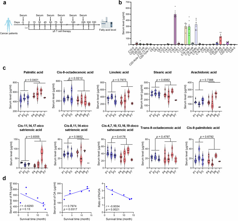
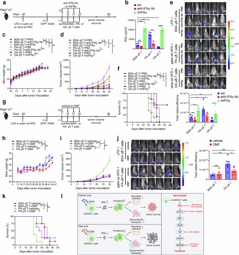
Dietary fatty acids (FAs) are associated with the therapeutic intervention under various health conditions. Human γδ-T cells are indispensable for immunosurveillance toward malignant cells. However, their impact on γδ-T cell metabolism and function remains poorly unexplored. Here, we applied targeted metabolomics analysis to serum FAs among cancer patients undergoing γδ-T cell therapy and discovered that palmitic acid (PA) or oleic acid (OA) levels were associated with the efficacy of Vγ9Vδ2-T cell therapy. We further elucidated that PA suppresses the antitumor activity of Vγ9Vδ2-T cells by disrupting metabolic processes and inhibiting the secretion of lytic granules, whereas OA restores the impaired antitumor activity of Vγ9Vδ2-T cells. Mechanistically, we surprisingly found that PA stimulates Vγ9Vδ2-T cells to secrete excessive IFNγ, which in turn induces cell pyroptosis, ultimately resulting in decreased antitumor activity. In contrast, OA reduces IFNγ secretion and mitigates cell pyroptosis, thereby restoring their antitumor activity. Alternatively, direct blockade of IFNγ by anti-IFNγ mAb or inhibition of pyroptosis by dimethyl fumarate (DMF) also restores their antitumor activity. This study highlights a novel mechanism whereby dietary FAs modulate γδ-T cell function through regulating IFNγ-mediated pyroptosis. Additionally, it offers proof-of-concept for an innovative approach by targeting IFNγ-mediated pyroptosis or dietary OA supplementation to strengthen the antitumor immunity of γδ-T cells against cancers.


Conflict of interest statement

Competing interests: The authors declare no competing interests.

Figures

Fig. 1
Fig. 1
The level of PA or OA is associated with the efficacy of Vγ9Vδ2-T cell-based anticancer therapy in patients. a Diagram showing the protocol for collecting serum of HCC patients treated with Vγ9Vδ2-T cells. b Mass spectrometric analysis of FAs in serum of cancer patients before Vγ9Vδ2-T cell therapy (n = 7). c Serum FFA levels of seven cancer patients who received Vγ9Vδ2-T cell treatment are displayed in violin plots. FA levels were measured at baseline and before each Vγ9Vδ2-T cell infusion, corresponding to all timepoints in (a), for responders and non-responders until therapy cessation. Patients with complete response to Vγ9Vδ2-T cell therapy are represented by blue violin plots, and patients with progressive disease under Vγ9Vδ2-T cell therapy are represented by red violin plots. d Correlation between average serum level of PA, OA, or ratio of PA/OA and survival time of patients. The data are shown as the mean ± SEM
Fig. 2
Fig. 2
OA restores the reduced antitumor activity of Vγ9Vδ2-T cells induced by PA. a The apoptotic K562 cells were determined by flow cytometry after coculture with Vγ9Vδ2-T cells for 6 h (n = 4). b The percentages of apoptotic tumor cells after coculture with Vγ9Vδ2-T cells for 6 h (n = 4). c Diagram of the protocol in (dg). Rag2−/−γc−/− mice fed on the LFD, palm oil, olive oil or palm and olive oil HFDs for 30 days were subcutaneously (s.c.) injected with GFP+ A549 tumor cells. Expanded BSA-, PA-, OA- or PA + OA-Vγ9Vδ2-T cells were intravenously (i.v.) transferred into the mice at the indicated time (n = 6 mice per group). d Main components of control LFD, palm oil, olive oil, and palm and olive oil HFDs. e Tumor volumes were obtained at the indicated time. f On day 26 following GFP+ A549 tumor cells inoculation, whole-body fluorescence images (left) and total radiant efficiency of fluorescence intensity (right) of the mice are shown after Vγ9Vδ2-T cells treatment. g Survival curves were acquired at the specified time. Quantitative data are shown as the mean ± SEM. ns not significant; *p < 0.05; **p < 0.01; ***p < 0.001
Fig. 3
Fig. 3
OA rescued the reduced lytic granule secretion, glycolysis and OXPHOS in Vγ9Vδ2-T cells induced by PA. a The secretions of granzyme A/B, perforin, granulysin, sFas and sFasL collected from the supernatant of the co-culturing system between BSA-, PA-, OA-, or PA + OA-treated Vγ9Vδ2-T cells and K562 tumor cells were measured (n = 6). b, c Real-time analysis of aerobic glycolysis (ECAR) and OXPHOS (OCR) in BSA-, PA-, OA-, PA + OA-Vγ9Vδ2-T cells were performed. b In the presence of oligomycin and 2-DG, ECAR curves were evaluated. Comparisons of glycolysis, glycolytic capacity, and glycolytic reserve are shown (n = 4). c After the supplement of oligomycin, FCCP, and rotenone/antimycin A, OCR curves were obtained. Comparisons of maximum respiration, ATP production, and spare respiration capacity, are shown (n = 4). The data are shown as the mean ± SEM. ns not significant; *p < 0.05; **p < 0.01; ***p < 0.001
Fig. 4
Fig. 4
OA restores Vγ9Vδ2-T cell antitumor activity by preventing PA-induced cell pyroptosis. Vγ9Vδ2-T cells were expanded from PBMCs of healthy donors using PAM and IL-2 with the supplement with BSA, PA, OA, or a mixture of PA and OA for 14 days. The absolute cell numbers (a) and the percentages (b) of Vγ9Vδ2-T cells were examined (n = 6). c The level of LDH release in the culture medium of BSA-, PA-, OA-, or PA + OA-Vγ9Vδ2-T cells after culturing for 14 days was measured (n = 4). d Representative immunoblot (left) and quantification (right) of GSDMD, cleaved GSDMD, caspase-1, and cleaved caspase-1 in BSA-, PA-, OA-, or PA + OA -Vγ9Vδ2-T cells (n = 4). eg Expanded BSA-, or PA- Vγ9Vδ2-T cells were treated with DMF to block pyroptosis. e The level of LDH release in the culture medium of BSA-, or PA -Vγ9Vδ2-T cells after DMF treatment overnight was measured (n = 4). f Representative immunoblot (left) and quantification (right) of GSDMD, cleaved GSDMD, caspase-1, and cleaved caspase-1 in BSA-, or PA -Vγ9Vδ2-T cells after DMF treatment overnight (n = 4). g The percentages of apoptotic tumor cells after coculture with BSA-, or PA-Vγ9Vδ2-T cells with or without DMF treatment were examined (n = 4). h The secretions of granzyme A/B, perforin, and granulysin collected from the supernatant of the co-culturing system after coculture BSA-, or PA -Vγ9Vδ2-T cells with tumor cells were examined (n = 4). ik Expanded BSA-, or OA- Vγ9Vδ2-T cells were treated with nigericin to induce pyroptosis. i The level of LDH release in the culture medium of BSA-, or OA -Vγ9Vδ2-T cells after nigericin treatment overnight was measured (n = 5). j Representative immunoblot (left) and quantification (right) of GSDMD, and cleaved GSDMD in BSA-, or OA -Vγ9Vδ2-T cells after nigericin treatment overnight (n = 4). k The percentages of apoptotic tumor cells after coculture with BSA-, or OA-Vγ9Vδ2-T cells with or without nigericin treatment were examined (n = 5). The data are shown as the mean ± SEM. ns not significant; *p < 0.05; **p < 0.01; ***p < 0.001; ****p < 0.0001
Fig. 5
Fig. 5
IFNγ mediates PA-induced pyroptosis in Vγ9Vδ2-T cells. a Proteomics analysis of cellular proteins from BSA-, PA-, or OA-Vγ9Vδ2-T cells after culturing for 14 days (n = 3 per group). Then pathway analysis of the distinguished proteins was performed in PA- and OA-treated Vγ9Vδ2-T cells compared to BSA-treated Vγ9Vδ2-T cells. b IFNα and IFNγ secretions collected from the supernatant of BSA-, PA-, OA-, or PA + OA-Vγ9Vδ2-T cells were measured by ELISA (n = 6). cg Expanded BSA-, PA- or OA-Vγ9Vδ2-T cells were treated with or without anti-IFNγ Ab or rhIFNγ for 3 days. c The secretion of IFNγ collected from the supernatant of Vγ9Vδ2-T cells was measured by ELISA (n = 4). The absolute cell numbers (d) and the percentages (e) of Vγ9Vδ2-T cells were examined (n = 4). f The level of LDH release in the culture medium of Vγ9Vδ2-T cells was measured (n = 4). g Representative immunoblot (left) and quantification (right) of GSDMD, cleaved GSDMD, caspase-1 and cleaved caspase-1 in Vγ9Vδ2-T cells (n = 4). hk Expanded BSA-, or OA- Vγ9Vδ2-T cells were treated with rhIFNγ for 3 days, and then DMF was used to block pyroptosis. h Representative immunoblot (left) and quantification (right) of GSDMD, cleaved GSDMD, caspase-1 and cleaved caspase-1 in BSA-, or OA-Vγ9Vδ2-T cells (n = 4). i The level of LDH release in the culture medium of BSA-, or OA-Vγ9Vδ2-T cells was measured (n = 4). j The percentages of apoptotic tumor cells after coculture with BSA-, or OA-Vγ9Vδ2-T cells were examined (n = 4). k The secretions of granzyme A/B, perforin, and granulysin collected from the supernatant of the co-culturing system after coculture BSA-, or OA -Vγ9Vδ2-T cells with tumor cells were examined (n = 4). The data are shown as the mean ± SEM. ns not significant; *p < 0.05; **p < 0.01; ***p < 0.001; ****p < 0.0001
Fig. 6
Fig. 6
pSTAT1-IRF1-iNOS pathway mediates IFNγ-induced pyroptosis in Vγ9Vδ2-T cells. a, b Vγ9Vδ2-T cells were cultured with BSA, PA, OA, or a mixture of PA and OA for 14 days. a pSTAT1, IRF1, iNOS expression on Vγ9Vδ2-T cells, and NO level in their supernatants were examined (n = 5). b The NO level in Vγ9Vδ2-T cells was quantified using DAF-FM DA probe under confocal microscopy. Representative confocal images and quantification of average fluorescence intensity are shown (n = 5). The scale bar represents 20 µm. c, d Expanded BSA-, PA- or OA-Vγ9Vδ2-T cells were treated with or without anti-IFNγ Ab or rhIFNγ. c pSTAT1, IRF1, and iNOS expression on Vγ9Vδ2-T cells and NO level in their supernatants were examined (n = 5). d The NO level in Vγ9Vδ2-T cells was quantified using DAF-FM DA probe under confocal microscopy. Representative confocal images and quantification of average fluorescence intensity are shown (n = 5). The scale bar represents 20 µm. The level of LDH release in the culture medium of BSA-, or PA-Vγ9Vδ2-T cells after fludarabine (e) or 1400 W (f) treatment was measured (n = 5). g Representative immunoblot of GSDMD, cleaved GSDMD, caspase-1, and cleaved caspase-1 in BSA-, or PA-Vγ9Vδ2-T cells after fludarabine (left) or 1400 W (right) treatment. hj Expanded BSA-, or OA- Vγ9Vδ2-T cells were treated with fludarabine or 1400 W, then rhIFNγ was used. h, i The level of LDH release in the culture medium of BSA-, or OA-Vγ9Vδ2-T cells was measured (n = 6). j Representative immunoblot of GSDMD, cleaved GSDMD, caspase-1 and cleaved caspase-1 in BSA-, or OA-Vγ9Vδ2-T cells after fludarabine (left) or 1400 W (right) treatment. The percentages of apoptotic tumor cells after coculture with BSA-, or PA-Vγ9Vδ2-T cells with or without fludarabine (k) or 1400 W (l) treatment were examined (n = 4). The percentages of apoptotic tumor cells after coculture with BSA-, or OA-Vγ9Vδ2-T cells in the presence of rhIFNγ with or without fludarabine (m) or 1400 W (n) treatment were examined (n = 4). The data are shown as the mean ± SEM. ns not significant; *p < 0.05; **p < 0.01; ***p < 0.001; ****p < 0.0001
Fig. 7
Fig. 7
IFNγ mediates PA-induced functional and metabolic defects in Vγ9Vδ2-T cells. a The antitumor activity of expanded BSA-, PA- or OA-Vγ9Vδ2-T cells with or without anti-IFNγ Ab or rhIFNγ was detected by co-culturing Vγ9Vδ2-T cells with K562 tumor cells at an effector/target cells of 10:1 for 6 h, and then the apoptotic K562 cells were determined by flow cytometry. b Quantitative comparisons of apoptotic K562 cells are shown (n = 5). c The secretions of granzyme A/B, perforin and granulysin collected from the supernatant of the co-culturing system between expanded BSA-, PA- or OA-Vγ9Vδ2-T cells with or without anti-IFNγ Ab or rhIFNγ and K562 tumor cells were measured (n = 6). dg Real-time analysis of aerobic glycolysis (ECAR) and OXPHOS (OCR) in expanded BSA-, PA- or OA-Vγ9Vδ2-T cells with or without anti-IFNγ Ab or rhIFNγ was performed. d In the presence of oligomycin and 2-DG, ECAR curves were assessed. e Quantitative comparisons of glycolysis, glycolytic capacity, and glycolytic reserve, are shown (n = 4). f After the supplement of oligomycin, FCCP, and rotenone/antimycin A, OCR curves were obtained. g Quantitative comparisons of maximum respiration, ATP production, and spare respiration capacity, are shown (n = 4). The data are shown as the mean ± SEM. ns not significant; *p < 0.05; **p < 0.01; ***p < 0.001; ****p < 0.0001
Fig. 8
Fig. 8
Blockade of IFNγ and pyroptosis restores PA-impaired antitumor activity of Vγ9Vδ2-T cells in vivo. a Diagram of the protocol in (bf). Rag2−/−γc−/− mice fed on the LFD, palm oil, or olive oil HFDs for 30 days were subcutaneously (s.c.) injected with GFP+ A549 tumor cells. Then anti-IFNγ Ab or rhIFNγ was intraperitoneally (i.p.) injected into mice. Expanded BSA-, PA-, or OA-Vγ9Vδ2-T cells were intravenously (i.v.) transferred into the mice at the indicated time (n = 5 mice per group). b The level of IFNγ from mice serum collected on the 2nd day after injection of anti-IFNγ Ab or rhIFNγ was measured. c Mouse weight was monitored for 2 months. d Tumor volumes were obtained at the indicated time. e On day 29 following GFP+ A549 tumor cells inoculation, whole-body fluorescence images (upper) and total radiant efficiency of fluorescence intensity (lower) of the mice are shown after Vγ9Vδ2-T cell treatment. f Survival curves were obtained at the indicated time. g Diagram of the protocol in (hk). Rag2−/−γc−/− mice fed on the LFD, or palm oil HFD for 30 days were subcutaneously (s.c.) injected with GFP+ A549 tumor cells. Then vehicle or DMF was intragastrically (i.g.) injected into mice. Expanded BSA-, or PA-Vγ9Vδ2-T cells were intravenously (i.v.) transferred into the mice at the indicated time (n = 6 mice per group). h Mouse weight was monitored at the indicated time. i Tumor volumes were obtained at the indicated time. j On day 29 following GFP+ A549 tumor cells inoculation, whole-body fluorescence images (upper) and total radiant efficiency of fluorescence intensity (lower) of the mice were assessed after Vγ9Vδ2-T cell treatment. k Survival curves were acquired at the specified time. l Schematic overview of the effect of PA and OA on the antitumor activity and the mechanism of IFNγ-induced pyroptosis in Vγ9Vδ2-T cells. The schematic was made using Biorender and included a publication license (Zhang (2025) https://BioRender.com/h90t444). Quantitative data are shown as the mean ± SEM. ns not significant; *p < 0.05; **p < 0.01; ***p < 0.001; ****p < 0.0001

References

    1. Mozaffarian, D. Dietary and policy priorities for cardiovascular disease, diabetes, and obesity: a comprehensive review. Circulation133, 187–225 (2016). - PMC - PubMed
    1. Nava Lauson, C. B. et al. Linoleic acid potentiates CD8(+) T cell metabolic fitness and antitumor immunity. Cell Metab.35, 633–650.e639 (2023). - PubMed
    1. Lai, Y. et al. Dietary elaidic acid boosts tumoral antigen presentation and cancer immunity via ACSL5. Cell Metab.36, 822–838.e828 (2024). - PubMed
    1. Fan, H. et al. Trans-vaccenic acid reprograms CD8(+) T cells and anti-tumour immunity. Nature623, 1034–1043 (2023). - PMC - PubMed
    1. de Vries, N. L. et al. gammadelta T cells are effectors of immunotherapy in cancers with HLA class I defects. Nature613, 743–750 (2023). - PMC - PubMed