. 2025 Aug;32(8):870-883.
doi: 10.1038/s41417-025-00928-2. Epub 2025 Jul 2.

SLC34A2 inhibits tumorigenesis and progression via upregulating LRRK2/TTF-1/SELENBP1 axis in lung adenocarcinoma

Affiliations

SLC34A2 inhibits tumorigenesis and progression via upregulating LRRK2/TTF-1/SELENBP1 axis in lung adenocarcinoma

Ying Zhu et al. Cancer Gene Ther. 2025 Aug.

Abstract

Alveolar type II epithelial (AT2) cells have the properties of stem cells, abnormal AT2 cells serve as one of the original cells in lung adenocarcinoma (LUAD). However, the abnormal genes expression of AT2 cells during their malignant transformation into LUAD cells remain poorly understood. Importantly, SLC34A2 is a specific gene in AT2 cells of the lung. Our previous researches have reported that overexpression of SLC34A2 significantly inhibited proliferation, migration and invasion of LUAD cells. But, the underlying mechanisms of SLC34A2 in LUAD are largely unknown until now. Here, the present study discovered that the protein expression of Napi2b (SLC34A2), SELENBP1, TTF-1 and LRRK2 were all located in human AT2 cells of adjacent non-tumor tissues. However, the expression level of SLC34A2, SELENBP1, TTF-1 and LRRK2 were significantly decreased in LUAD tissues, and the expression of SLC34A2 was obviously positive correlation with the expression of SELENBP1, TTF-1 and LRRK2, respectively. Mechanistically, our study elucidated that overexpression of SLC34A2 could inhibit the activation of MEK/ERK signaling pathway through up-regulating the expression of LRRK2, and subsequently suppressed the expression of p-TTF-1(Ser327), which upregulated the expression of SELENBP1 by enhancing TTF-1 transcriptional activity. Ultimately, overexpression of SLC34A2 depressed the activation of PI3K/AKT/mTOR signaling pathway via up-regulating the expression of SELENBP1, which significantly inhibited the malignant characteristics of LUAD. In summary, our current research revealed a novel SLC34A2/LRRK2/TTF-1/SELENBP1 axis and its involvement in inhibiting the malignant characteristics of LUAD cells for the first time, which made contribution to further exploring the clinical application of SLC34A2. Furthermore, it also might offer novel insights into understanding how AT2 cells undergo malignant transformation into LUAD cells in the future.


Conflict of interest statement

Competing interests: The authors declare no competing interests. Figure 7 was drawn by Figdraw ( www.figdraw.com ). Ethics approval and consent to participate: All methods were performed in accordance with the relevant guidelines and regulations. The clinical samples study was approved by the West China Hospital of Sichuan University Biomedical Research Ethics Committee and the Medical Ethics Committee, Sichuan Academy of Medical Sciences, Sichuan Provincial People’s Hospital (NO.2023-138). This study was performed in accordance with the Declaration of Helsinki. Informed consent was obtained from each patient. The animal experiments and all procedures involving the handling and treatment of mice during this study were approved by Experimental Animal Ethics Committee of West China Hospital (NO.20220721002), Sichuan University (Chengdu, China). All the experiments were performed according to the guidelines of the National Institutes of Health Guide for the Care and Use of Laboratory Animals.

Figures

Fig. 1
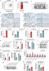
Fig. 1. SLC34A2 was downregulated in LUAD and its abnormal low expression was associated with the tumorigenesis and progression of LUAD.
A The expression of SLC34A2 in unpaired (left) and paired (right) LUAD relative to adjacent non-tumor tissues in TCGA database. N, adjacent non-tumor tissues; T, tumor tissues. B The expression of SLC34A2 in 72 collected LUAD and adjacent tissues. N, adjacent non-tumor tissues; T, tumor tissues. C A representative immunohistochemical staining (IHC) image and IHC score of Napi2b in collected human LUAD tissue and paired adjacent non-tumor tissues (n = 11). D Western blot analysis and statistical diagram of Napi2b levels in collected human LUAD tissues and matched adjacent non-tumor tissues (n = 6). The relationship between the expression of SLC34A2 and clinical stage in patients with LUAD in TCGA database (E) and collected LUAD clinical tissues (F). G The relationship between the expression of SLC34A2 and differentiation in collected LUAD tissues. Kaplan-Meier analysis of the overall survival of LUAD patients based on SLC34A2 levels from TCGA (H) and GEO database (I). J The relationship between the expression of SLC34A2 and lymph node metastasis in collected LUAD tissues. K ROC curve analysis of SLC34A2 expression in collected LUAD tissues (n = 72) and adjacent normal tissues. Statistical analysis in (A, C, J) were performed by using unpaired Student’s t test, and (B, D) were obtained by paired t test. (F, G) were conducted by One-way ANOVA. *P < 0.05, ns no significance.
Fig. 2
Fig. 2. The expression of SLC34A2 was positive correlation with SELENBP1.
The correlations of transcript levels between SLC34A2 and SELENBP1 in human LUAD tissues in TCGA (A), GEO database GSE166720 (B) and GSE140343 (C). D The expression of SELENBP1 in 45 LUADs with low SLC34A2 expression related to adjacent non-tumor tissues. E Western blot analysis of Napi2b and SELENBP1 protein levels in collected human LUAD tissues and matched adjacent non-tumor tissues (n = 10). F A representative IHC image of Napi2b and SELENBP1 in collected human LUAD tissue and paired adjacent non-tumor tissues. Red arrows indicated colocalization of Napi2b and SELENBP1. G The mRNA levels of SLC34A2 and SELENBP1 were measured by qRT-PCR. Western blot analysis (H) and statistical diagrams (I) of Napi2b and SELENBP1 protein levels in human LUAD cells. Data are presented as mean ± SD. The P values were performed by spearman correlation analysis (B, C) or two-tailed unpaired t test (G, I). *P < 0.05, **P < 0.01, ****P < 0.0001.
Fig. 3
Fig. 3. Overexpression of SLC34A2 suppressed malignant characteristics of LUAD cells via increasing the expression levels of SELENBP1 in vitro.
qRT-PCR (A) and western blot (B) analysis confirmed the downregulation of SELENBP1 mediated by siRNA targeting SELENBP1 in A549-SLC34A2 and H1299-SLC34A2 cells. Cell proliferation curves (CF) and cell colony-forming ability (G) of SLC34A2 stable overexpression LUAD cells after knockdown of SELENBP1. H Microscopic observations were recorded at 0, 24 and 48 h after scratching the surface of a confluent layer of the indicated LUAD cells. Scale bar, 200 μm. The cell migration ability (I) and cell invasion ability (J) of SLC34A2 stable overexpression LUAD cell lines after knockdown of SELENBP1. Scale bar, 50 μm. Western blot analysis (K) and statistical diagrams (L) of PI3K/AKT/mTOR pathway key markers in indicated cells. Levels of β-actin were used as a loading control. All experiments were repeated at least three times. Data were presented as mean ± SD. The P values were performed by two-tailed unpaired t test (A) or One-way ANOVA (CJ, L). *P < 0.05, **P < 0.01, ***P < 0.001, ****P < 0.0001, ns no significance.
Fig. 4
Fig. 4. Overexpression of SLC34A2 elevated the transcriptional activity of TTF-1 by inhibition of MEK/ERK signaling pathway in LUAD cells in vitro.
A The correlations of transcript levels between SLC34A2 and TTF-1 in human LUAD tissues in TCGA database. B The expression of TTF-1 in 35 collected clinical LUADs with low SLC34A2 and SELENBP1 expression related to adjacent non-tumor tissues. C Western blot analysis of Napi2b and TTF-1 protein levels in collected human LUAD tissues and matched adjacent non-tumor tissues (n = 6). D A representative IHC image of Napi2b, TTF-1 and SELENBP1 in collected human LUAD tissue and paired adjacent non-tumor tissues. Red boxes, tumor tissues. Black boxes, adjacent non-tumor tissues. E The colocalization of Napi2b, TTF-1 and SELENBP1 in serial sections of adjacent non-tumor tissues. Red arrows indicated representative images of colocalization. F The mRNA levels of TTF-1 was measured by qRT-PCR. Western blot analysis (G) and statistical diagrams (H) of TTF-1 protein levels in human LUAD cells. I The mRNA levels of TTF-1 and SELENBP1 were measured by qRT-PCR. Western blot analysis (J) and statistical diagram (K) of TTF-1 and SELENBP1 protein levels after knockdown of TTF-1 in human LUAD cells. L CUT&TAG assay showing the fold enrichment of TTF-1 in the promotor region of SELENBP1 and TTF-1 in human LUAD cells. TTF-1 antibody enrichment was quantified relative to the amount of input DNA spike-in (n = 3). Western blot analysis (M) and statistical diagrams (N) of p-TTF-1(Ser327) protein levels in indicated human LUAD cells. Data were presented as mean ± SD. The P values were performed two-tailed unpaired t test (F, H, I, K, L) or One-way ANOVA (N). *P < 0.05, **P < 0.01, ***P < 0.001, ns no significance.
Fig. 5
Fig. 5. Overexpression of SLC34A2 inactivated the MEK/ERK signaling pathway via upregulating LRRK2 in LUAD cells in vitro.
The correlations of transcript levels between SLC34A2 and LRRK2 in human in LUAD tissues in TCGA (A) and GEO (B) database. C The expression of LRRK2 in 29 collected clinical LUADs with low SLC34A2 expression related to adjacent non-tumor tissues. D Western blot analysis of Napi2b and LRRK2 protein levels in collected human LUAD tissues and matched adjacent non-tumor tissues (n = 6). E The colocalization and expression of Napi2b and LRRK2 in collected human LUAD tissue and paired adjacent non-tumor tissues. Red arrows indicated representative images of colocalization. F The mRNA levels of LRRK2 were measured by qRT-PCR. Western blot analysis (G) and statistical diagrams (H) of LRRK2 protein levels in human LUAD cells. I qRT-PCR analysis confirmed the downregulation of LRRK2 mediated by siRNA targeting LRRK2 in A549-SLC34A2 and H1299-SLC34A2 cells. Western blot analysis (J) and statistical diagrams (K) of MEK/ERK pathway key markers after konckdown of LRRK2 in indicated cells. Levels of β-actin were used as a loading control. L Western blot analysis of p-LRRK2(T1503) protein levels in human LUAD cells. Data were presented as mean ± SD. The P values were performed by spearman correlation analysis (B), two-tailed unpaired t test (F, H, I) or One-way ANOVA (K). *P < 0.05, **P < 0.01, ***P < 0.001.
Fig. 6
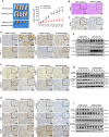
Fig. 6. Overexpression of SLC34A2 inhibiting growth of LUAD cells was associated with the inactivation of MEK/ERK and PI3K/AKT/mTOR signaling pathway in vivo.
Establishment of stable SLC34A2 overexpressing A549 cells and control cells tumor Xenograft Model. Athymic nude mice (4-week-old male, n = 5 /point/group) were subcutaneously injected with A549-SLC34A2 cells or A549-control cells, respectively. A The images and tumor sizes of subcutaneous xenografts were presented. Scale bar, 20 mm. B Representative images of organization structure in retrieved subcutaneous tumor samples detected by H&E staining. Scale bar, 50 μm. C Representative images of Ki-67 in retrieved subcutaneous tumor samples detected by IHC staining. Scale bar, 20 μm. Representative IHC staining images (D) and western blot analysis (E) of Napi2b, SELENBP1, TTF-1, and LRRK2 (n = 5). Scale bar, 20 μm. Representative IHC staining images (F) and western blot analysis (G) of MEK/ERK signaling key markers in retrieved subcutaneous tumor samples (n = 5). Scale bar, 20 μm. Representative IHC staining images (H) and western blot analysis (I) of PI3K/AKT/mTOR signaling key markers in retrieved subcutaneous tumor samples (n = 5). Scale bar, 20 μm. Data were presented as mean ± SD. The P values were obtained by two-tailed unpaired t test (A). **P < 0.01, ****P < 0.0001.
Fig. 7
Fig. 7. The molecular model of SLC34A2 inhibiting the tumorigenesis and progression of LUAD.
Overexpression of SLC34A2 could inhibit the activation of MEK/ERK signaling pathway through up-regulating the expression of LRRK2, and subsequently suppressed the expression of p-TTF-1(Ser327), which upregulated the expression of SELENBP1 by regulating TTF-1 transcriptional activity. Ultimately, overexpression of SLC34A2 depressed the activation of PI3K/AKT/mTOR signaling pathway via up-regulating the expression of SELENBP1, which significantly inhibited the malignant characteristics of LUAD.

Similar articles

Cited by

References

    1. Bray F, Laversanne M, Sung H, Ferlay J, Siegel RL, Soerjomataram I, et al. Global cancer statistics 2022: GLOBOCAN estimates of incidence and mortality worldwide for 36 cancers in 185 countries. CA Cancer J Clin. 2024;74:229–63. - PubMed
    1. Fang H, Sun Q, Zhou J, Zhang H, Song Q, Zhang H, et al. m(6)A methylation reader IGF2BP2 activates endothelial cells to promote angiogenesis and metastasis of lung adenocarcinoma. Mol Cancer. 2023;22:99. - PMC - PubMed
    1. Miller KD, Nogueira L, Devasia T, Mariotto AB, Yabroff KR, Jemal A, et al. Cancer treatment and survivorship statistics, 2022. CA Cancer J Clin. 2022;72:409–36. - PubMed
    1. Guillot L, Nathan N, Tabary O, Thouvenin G, Le Rouzic P, Corvol H, et al. Alveolar epithelial cells: master regulators of lung homeostasis. Int J Biochem cell Biol. 2013;45:2568–73. - PubMed
    1. Hill W, Lim EL, Weeden CE, Lee C, Augustine M, Chen K, et al. Lung adenocarcinoma promotion by air pollutants. Nature. 2023;616:159–67. - PMC - PubMed

MeSH terms

Substances