Review
. 2024 Aug 2;2(1):23.
doi: 10.1038/s44303-024-00027-1.

New tactics in the design of theranostic radiotracers

Affiliations
Review

New tactics in the design of theranostic radiotracers

Cesare Berton et al. Npj Imaging. .

Abstract

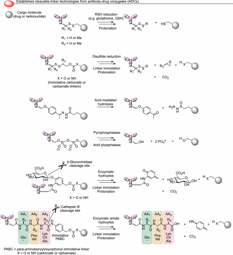
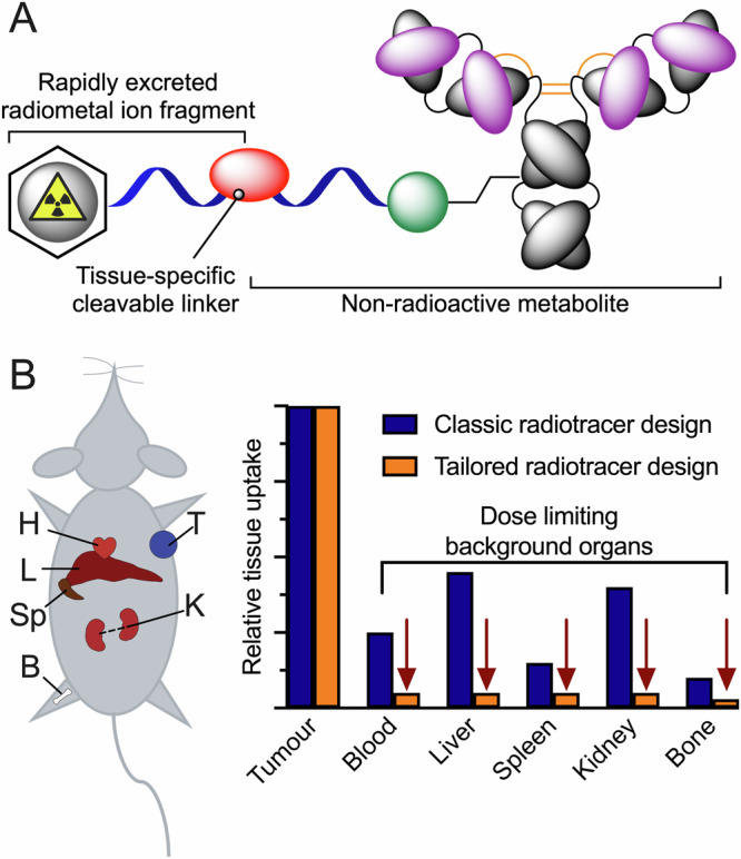
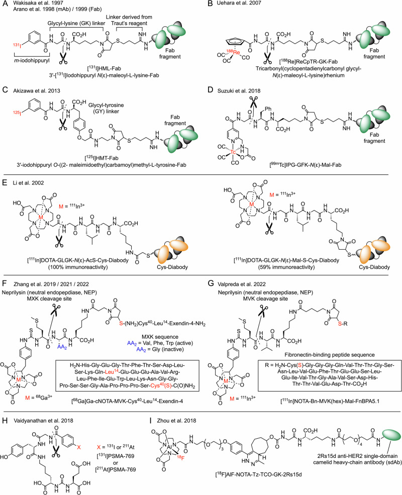
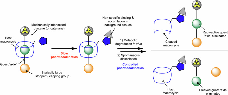
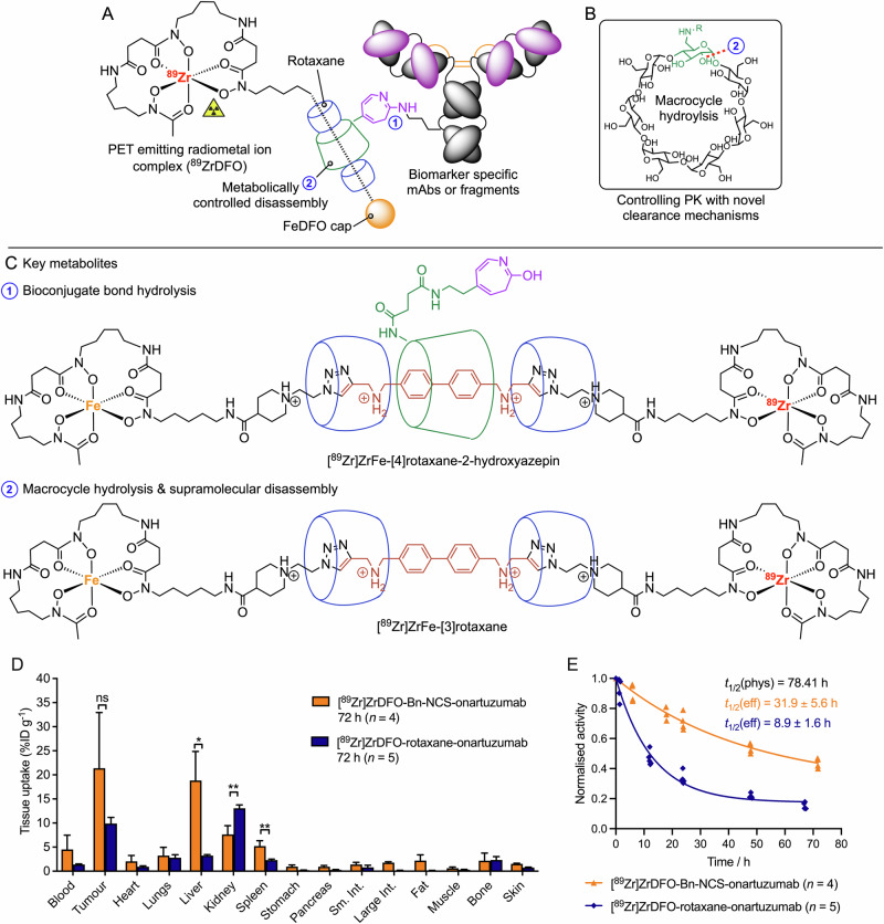
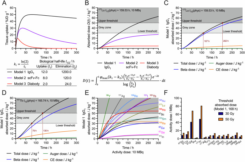
In the context of molecularly targeted radiotherapy, dosimetry concerns in off-target tissues are a major limitation to the more wide-spread application of radiopharmaceuticals to treat diseases like cancer. Reducing off-target accumulation of radionuclides in background tissues, whilst maintaining high and specific uptake in disease sites and improving the therapeutic window, requires rethinking common radiotracer design concepts. This article explores ways in which innovative radiotracer chemistry (the making and breaking of bonds) is used to modify interactions with the host organism to control excretion profiles and dosimetry at the tissue-specific level.


Conflict of interest statement

Competing interests: The authors declare no competing interests.

Figures

Fig. 1
Fig. 1. The founding father of radiotracer science.
A Photograph of Prof. George de Hevesy circa 1950 (https://arkiv.dk/en/vis/5941286) who received the Nobel Prize in Chemistry in 1943 for developing the ‘radiotracer principle’. B An excerpt from a manuscript by Chiewitz and de Hevesy in 1935, reporting one of the earliest examples of a ‘biodistribution study’ using a radionuclide to track the distribution and metabolic fate of [32P]NaPO4 in rats after ingestion in the food.
Fig. 2
Fig. 2. Classic examples of the bifunctional chelate concept in radiotracer design.
A Schematic of the classic antibody-based radiotracer design employing a covalent modification of the protein structure via bioconjugate bond formation (green), a linker or spacer group (blue), and a multidentate chelate (black) that provides thermodynamically and kinetically stable complexation of a specific radiometal ion (red) for applications in diagnostic imaging or radioimmunotherapy (RIT). B Two examples of the conventional radiopharmaceutical design showing (top) the structure of a monoclonal IgG1 antibody modified with a thiourea bond formed at a surface-exposed lysine residue radiolabelled with 89Zr-desferrioxamine B (DFO) complex, and (bottom) a 177Lu-radiolabelled mAb employing an octadentate macrocyclic chelate (DOTAGA) and a tris-polyethylene glycol (PEG3) linker which has been demonstrated to enhance the metabolism and clearance of the radionuclide complex compared with non-PEGylated designs,. This Fast-ClearTM linker technology is under development at Fusion Pharmaceuticals Inc, Canada (United States Patents: US10093741 and US11191854).
Fig. 3
Fig. 3. Modulating the global pharmacokinetics of radioimmunoconjugates through protein engineering of antibody size.
A Schematic structures of conventional monoclonal antibody fragments. B Representative plot showing the tumour-associated activity (in percentage injected dose per gram of tissue, %ID g−1) versus time (in hours) post-radiotracer administration (figure composed based on the work of Wu and Senter referring to experimental data acquired from radioiodinated monoclonal antibody fragments).
Fig. 4
Fig. 4. Five conceptually distinct mechanisms that use chemistry to modulate radionuclide dosimetry in vivo.
A Pretargeting involving pre-administration of a non-radioactive component with slow pharmacokinetics followed by administration of a rapid-action small-molecule radiotracer after a suitable time delay that allows blood-pool and non-specific background clearance for the first agent. Note that mechanisms (BE) focus on eliminating radioactivity from non-target tissues. B Chelation therapy (or radioactive chase experiments) whereby administration of a classic radiopharmaceutical with slow pharmacokinetics is followed by administration of a competitive binding agent that can (in theory) extract and eliminate the radionuclide. C Controlled metabolic cleavage which harnesses endogenous catabolism in vivo to break a bond and release a small-molecule radiolabelled fragment which is eliminated rapidly from the body. D Stimulated or chemically-triggered radionuclide (or fragment) release whereby after initial radiotracer delivery, administration of a non-radioactive bioorthogonal partner induces bond cleavage and immolative release of the radionuclide complex. E Controlled supramolecular disassembly of a molecular inclusion complex or mechanical bond.
Fig. 5
Fig. 5. Summary of selected classes of cleavable linkers, used in antibody-drug conjugate technologies that could potentially be applied to cleavable radiotracer design via changing the cargo molecule.
(Top-to-bottom) A reducible disulfide linker, a disulfide combined with a chemically labile carbonate or carbamate, an acid labile hydrazide linker, an enzymatically-labile pyrophosphate linker, a glycoside linker cleavable by β-glucuronidase followed by linker immolation, and a di- or tripeptide linker hydrolysed by cathepsin B followed by PABC degradation leading to cargo release.
Fig. 6
Fig. 6. A new design principle for molecularly targeted radio(immuno)therapy agents that draws inspiration from the cleavable linkers used in antibody-drug conjugates (ADCs).
A Installation of a metabolically cleavable group (red) and/or the use of alternative bioconjugation chemistries (green) that facilitate tissue-specific metabolism in background organs, and rapid (ideally renal) excretion of a stable, radiolabelled small-molecule fragment. B Graphical illustration of the objective of retaining high tumour uptake and specificity whilst reducing the dosimetry burden experienced by dose limiting background organs including, but not restricted to: K = kidneys, L = liver, H = heart/blood pool, Sp = spleen, B = bone/marrow.
Fig. 7
Fig. 7. Prominent examples of radiolabelled compounds whose structure includes a cleavable linker designed for hydrolysis in the kidney by brush border enzymes like carboxypeptidase M and neprilysin (neutral endopeptidase, NEP).
A An early example of a radioiodinated meta-iodohippuryl-glycyl-lysine linker–,. B 188Re-radiometal ion complexes featuring the Gly-Lys cleavable linker,. C Installation of a cleavable Gly-Tyr linker to a radioiodinated-Fab radiotracer. D A 99mTc(I)-tricarbonyl-Fab radiotracer for SPECT imaging. E An 111In-radiolabelled diabody with a cleavable GLGK tetrapeptide. F A 68Ga-radiolabeled Exendin-4 radiotracer with a cleavable MXK peptide for tuneable hydrolysis by NEP. G A fibronectin-binding peptide radiolabelled with 111In3+ ions via a cleavable MVK peptide. H A cleavable radiohalogenated (131I or 211At) derivative of a PSMA-targeting vector with a hydrolysable glycine linker. I An Al18F-radiotracer with a tetrazine-TCO conjugation, a cleavable GK linker and classic amide functionalisation on a bioactive single-domain antibody (sdAb). For further examples see the references.
Fig. 8
Fig. 8. A supramolecular approach for controlling tissue-specific processing of radiotracers.
This method is a combination of both metabolically induced bond cleavage and supramolecular disassembly (see Fig. 4D, E, respectively). The schematic radiotracer is composed of a mechanically interlocked rotaxane (or alternatively, a catenane) featuring non-covalent interactions between the macrocyclic host (blue rings) and the guest ‘axle’ molecules. Bulky axle capping agents which act as stoppers to prevent disassembly of the mechanical bond can be tune to include a combination of radiometal complexes, fluorophores, cytotoxic drugs. Metabolic cleavage of a covalent bond in either the host macrocycle (top right) or axle (bottom right) breaks the mechanical bond leading to the release and rapid renal elimination of a small-molecule radiolabelled fragment.
Fig. 9
Fig. 9. Examples of supramolecular radiotracers constructed using molecularly-interlocked rotaxanes.
Head-to-head comparison of the supramolecular rotaxane-based radiotracer [89Zr]ZrDFO-rotaxane-onartuzumab versus conventional [89Zr]ZrDFO-Bn-NCS-onartuzumab synthesised via the standard protocol by Vosjan et al. which is used in the clinical preparation of many 89Zr-mAbs (see Fig. 2B for the molecular structure of the control compound). A Schematic representation of the rotaxane-based radiotracer featuring a non-covalent mechanically-interlocked bond between the radiolabelled guest axle and the host macrocycle which is attached via a covalent bond to the mAb or mAb-fragment using our photoradiosynthesis methods. B A metabolically labile 1,4-glycosidic bond in the functionalised host β-CD macrocycle, hydrolysis of which breaks the mechanical bond of the rotaxane. C Potential hydrolysis of (1) the novel bioconjugate bond leading to rapid renal excretion of a supramolecular [4]rotaxane metabolite or (2) supramolecular disassembly to eliminate a radiolabelled fragment. D Comparison of the biodistribution of the supramolecular [Zr]ZrDFO-rotaxane-onartuzumab versus conventional [89Zr]ZrDFO-Bn-NCS-onartuzumab at 72 h post-intravenous administration in female athymic nude mice bearing subcutaneous MKN-45 gastrointestinal adenocarcinoma tumours, and (E) the experimentally measured effective half-life of the two radiotracers showing a clear difference in the distribution and excretion rates.
Fig. 10
Fig. 10. Modelling the pharmacokinetic profile and tissue absorbed dose (in J kg−1) for protein-based radiotracers.
A Modelled tissue (for example, tumour) time-activity curves based on the experimental studies from Wu and co-workers in rodents (see Fig. 3B) showing profiles generated from typical uptake and elimination biological half-lives associated with Model 1: a full-length IgG1 (~150 kDa, black), Model 2: an scFv-Fc fragment (~100 kDa, purple) and Model 3: a diabody (~50 kDa, red). DoseItRight© web-application: https://doseitright.streamlit.app/. B Plot of the calculated absorbed dose for 177Lu using pharmacokinetic models 1–3. C Deconvolution of the contributions of different particle emissions to the total absorbed dose for the example using 177Lu-Model 1. D An equivalent plot of the adsorbed dose for Model 1 using 161Tb. E An overlay showing the total absorbed dose calculated for Model 1 using 14 different therapeutic radionuclide. F Bar chart showing the activity required to achieve threshold absorbed dose of 30 Gy (blue bars) and 50 Gy (yellow bars) at an arbitrary time point of 168 h post-radiotracer administration for 14 different radionuclides.

Similar articles

References

    1. Sgouros, G., Bodei, L., McDevitt, M. R. & Nedrow, J. R. Radiopharmaceutical therapy in cancer: clinical advances and challenges. Nat. Rev. Drug Discov.19, 589–608 (2020). - PMC - PubMed
    1. Strosberg, J. et al. Phase 3 Trial of 177Lu-Dotatate for midgut neuroendocrine tumors. N. Engl. J. Med.376, 125–135 (2017). - PMC - PubMed
    1. Sartor, O. et al. Lutetium-177-PSMA-617 for metastatic castration-resistant prostate cancer. N. Engl. J. Med.385, 1091–1103 (2021). - PMC - PubMed
    1. Research, P. Nuclear Medicine Market. https://www.precedenceresearch.com/nuclear-medicine-market/ ‘The global nuclear medicine market, forecast period 2024 to 2033.’ (2024).
    1. Holland, J. P., Williamson, M. J. & Lewis, J. S. Unconventional nuclides for radiopharmaceuticals. Mol. Imaging9, 1–20 (2010). - PMC - PubMed

LinkOut - more resources