. 2025 Jul 2;15(1):23171.
doi: 10.1038/s41598-025-05134-6.

A lentiviral vector targeting a KRAS neoepitope for cancer immunotherapy

Affiliations

A lentiviral vector targeting a KRAS neoepitope for cancer immunotherapy

Anastasia Goloudina et al. Sci Rep. .

Abstract

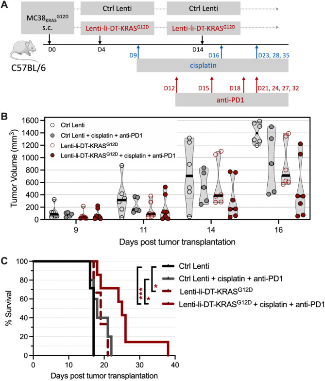
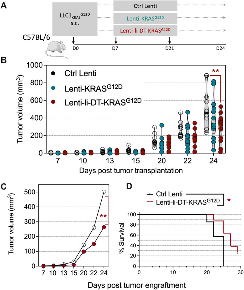
Mutated oncogenic Kirsten rat sarcoma virus (KRAS) antigen is expressed in a large variety of cancers, including pancreatic, colorectal, and pulmonary cancers. The oncogenic KRAS mutations cause malignancies and are usually ubiquitously expressed by all cells of a tumor. The KRAS amino acid substitutions at the positions 12 or 13 are among the most frequent mutations in human cancers. Here, we developed immuno-oncotherapeutic non-integrative lentiviral vectors encoding a segment encompassing KRASG12D, either alone, or associated with antigen carriers. These carriers can improve the intracellular antigen routing to major histocompatibility complex presentation machineries or provide universal helper CD4+ epitopes. Immunotherapy with one of these vectors resulted in significant immune control of tumor growth in colorectal or pulmonary preclinical cancer models, in several murine genetic backgrounds. The antitumor effect was correlated with increased proportions of intra-tumoral hematopoietic cells and notably CD8+ T cells. Although this effect was partial, it was robust, reproducible and advantageously combinable with conventional chemotherapies and immunotherapies to improve antitumor protection. Therefore, this approach shows promise as an immuno-oncotherapy against KRAS-mediated malignant transformation.

Keywords: Cancer immunotherapy; Inhibition of tumor growth; KRAS oncogenic mutations; KRASG12D; Lentiviral vectors.


Conflict of interest statement

Declarations. Competing interests: PC is the founder and CSO of TheraVectys. AG, FLC, PA, KN, IF, FM, BV and AN are employees of TheraVectys. LM has a consultancy activity for TheraVectys. AG, FLC, IF, AN, CB, PC and LM are inventors of a pending patent directed to the potential of lentiviral vectors encoding KRAS mutations in immuno-oncotherapy. Other authors declare no competing interests.

Figures

Fig. 1
Fig. 1
Antigen designs of KRASG12D as encoded by non-integrative lentiviral vectors. A Scheme of KRAS1-23G12D protein segment: alone (top), fused at its N-terminal extremity with full length li to facilitate its routing through the MHC-II pathway (middle), or fused at its N-terminal extremity with li-DT (bottom). B Protein sequences of the KRASG12D (25 aa), li-KRASG12D (244 aa) and li-DT-KRASG12D (425 aa) antigens. The lower-case characters indicate linkers inserted between the sequences to avoid generation of irrelevant junctional T cell epitopes. KRAS1-23G12D substitution is indicated in red bold. The KRASG12D sequence is in black, li sequence in grey and DT sequence in blue.
Fig. 2
Fig. 2
Therapeutic vaccination of mice bearing CT26 colorectal tumors with lentiviral vectors coding for various KRASG12D antigen designs. A Timeline of CT26 tumor inoculation and vaccination. BALB/c mice (n = 8 for Ctrl Lenti, Lenti-li-KRASG12D and Lenti-li-DT-KRASG12D groups, and n = 9 for Lenti-KRASG12D group) were engrafted s.c. on the right flank with 2 × 105 CT26 cells. Seven days later, when tumors became palpable, mice were randomized and injected i.m. with 1 × 109 TU of Ctrl Lenti, Lenti-KRASG12D, Lenti-li-KRASG12D or Lenti-li-DT-KRASG12D. B Plots of tumor size in individual mice over time. Statistical significance was determined by two-way ANOVA test by Log-rank Mantel-Cox test (*p < 0.05). C Survival curve of the animals. Statistical significance was determined by Log-rank Mantel-Cox tests (*p < 0.05). Mice were sacrificed when the tumor volume reached 1500 mm3, defined as humane endpoints. The experiment shown is representative of two independent experiments.
Fig. 3
Fig. 3
Therapeutic vaccination of mice bearing MC38KRASG12D tumors with Lenti-li-DT-KRASG12D. A Timeline of tumor engraftment and vaccination. C57BL/6 mice were engrafted s.c. on the right flank with 2 × 105 MC38KRASG12D cells. At day 6 post tumor engraftment, mice were randomized and immunized i.m. with 1 × 109 TU of Ctrl Lenti or Lenti-li-DT-KRASG12D. B-C Plots of tumor size in individual mice over time in two independent experiments (n = 7 or 12, respectively for Ctrl Lenti or Lenti-li-DT-KRASG12D group in C, and n = 7 or 6 for Ctrl Lenti or Lenti-li-DT-KRASG12D group in D). Statistical significance was determined by two-way ANOVA test by Log-rank Mantel-Cox tests (*p < 0.05). Mice were sacrificed when the tumor volume reached 1500 mm3, defined as humane endpoints. D-E TILs in Ctrl Lenti- or Lenti-li-DT-KRASG12D-treatted mice. C57BL/6 mice were engrafted with 2 × 105 MC38KRASG12D cells as detailed in B and injected on day 6 with 1 × 109 TU Lenti Ctrl or Lenti-li-DT-KRASG12D (n = 5 or 4 for Ctrl Lenti-or Lenti-li-DT-KRASG12D group). Tumors were studied on day 11 post-vaccination. D Gating strategy for flow cytometry analysis and representative blots of tumor infiltrating T cells. E The percentage of each subset was compared between the two groups and statistical significance determined using two-tailed unpaired t tests (*p ≤ 0.05). The experiment shown is representative of two independent experiments.
Fig. 4
Fig. 4
Presence of anti-KRAS CD8+ T cell effectors within tumor infiltrates. C57BL/6 mice were engrafted with 1 × 105 MC38KRASG12D cells and were primed (day 2) and boosted (day 9) i.m. with 1 × 109 TU/mouse of Ctrl Lenti or Lenti-li-DT-KRASG12D. The tumor infiltrates were studied at day 20 after enzymatical digestion of tumors, enrichment of cell suspensions in lymphocytes on Ficoll, and overnight co-culture with syngeneic bone-marrow-derived dendritic cells loaded with KRAS1-20G12D (MTEYKLVVVGADGVGKSALT), KRAS1-20WT (MTEYKLVVVGAGGVGKSALT) or a negative control peptide. A Cytometric gating strategy. B Typical results of intracellular IFN-γ- and TNF-α detection in CD8+ T cell infiltrates. C Percentages of CD4+ and CD8+ within the CD45+ cells in the tumor infiltrates of Ctrl Lenti- or Lenti-li-DT-KRASG12D-treated mice after enrichment on Ficoll and in vitro culture. D Percentages of IFN-γ+ TNF-α+ CD8+ within the CD45+ cells in the tumors after antigenic stimulation as detailed above. To obtain sufficient cells to perform these cytofluorometric assays, tumor samples had to be pooled in pairs from 6 mice per group for A-D. E Cytometric analysis of the tumor infiltrates after enzymatical digestion yet without enrichment on Ficoll. F Percentages of CD45+ or tumor cells, identified as large CD45- cells, within the whole cells in Ctrl Lenti- or Lenti-li-DT-KRASG12D-treated mice. The percentage of each subset was compared between the two groups and statistical significance determined using two-tailed unpaired t tests (*p ≤ 0.05).
Fig. 5
Fig. 5
Phenotype of antitumor effector T cells generated by Lenti-li-DT-KRASG12D therapy. A Timeline of tumor engraftment, vaccination and anti-CD4, or anti-CD8 mAb treatments in C57BL/6 mice (n = 8 for Ctrl Lenti, Lenti-li-DT-KRASG12D + Ctrl Ig and Lenti-li-DT-KRASG12D + anti-CD8 groups, and n = 7 for Lenti-li-DT-KRASG12D + anti-CD4 group). Mice received 8 i.p. at days 2, 4, 7, 10 and 14 of 250 µg anti-CD4 (clone GK1.5), anti-CD8 (clone H35.17.2) or an irrelevant control Ig. B Plots of tumor size in individual mice over time. C Mean tumor size in each group. Statistical significance was determined by two-way ANOVA test by Log-rank Mantel-Cox tests (*p ≤ 0.05). Mice were sacrificed when the tumor volume reached 1500 mm3, defined as humane endpoints. D Efficacy of T subset depletion in anti-CD4 or anti-CD8 mAb-treated mice, assessed at day 6 on the peripheral blood leukocytes from one representative mouse/group, by anti-CD3, anti-CD4 and anti-CD8 mAb staining and cytometry study.
Fig. 6
Fig. 6
Beneficial antitumor effect of a combination of Lenti-li-DT-KRASG12D with cisplatin and anti-PD1 treatment. A Timeline of MC38KRASG12D tumor engraftment, Lenti-li-DT-KRASG12D prime-boost, cisplatin chemotherapy and anti-PD1 mAb therapy. C57BL/6 mice (n = 6 for Ctrl Lenti, n = 5 for Ctrl Lenti + cisplatin + anti-PD1, n = 6 for Lenti-li-DT-KRASG12D, and n = 7 for Lenti-li-DT-KRASG12D + cisplatin + anti-PD1 groups) were engrafted s.c. with 1 × 105 MC38KRASG12D cells on d0. The prime (d4) and boost (d14) immunization were performed i.m. with 1 × 109 TU of Lenti-li-DT-KRASG12D. The combination of cisplatin and anti-PD1 consisted of i.p. injections of 4 mg/kg of cisplatin on d9, d16, d20, d23, d28 and d35 and 200 µg of anti-PD1 mAb on d12, d15, d18, d21, d24, d27 and d32. B Evolution of tumor size in individual mice over time. Significance was determined by two-way ANOVA test by Log-rank Mantel-Cox tests (*p < 0.05). C Survival curve of the animals. Statistical significance was determined by Log-rank Mantel-Cox tests (*p < 0.01, ***p < 0.0001). Mice were sacrificed when the tumor volume reached 1500 mm3, defined as humane endpoints. The experiment shown is representative of two independent experiments.
Fig. 7
Fig. 7
Therapeutic vaccination of mice bearing LLC1KRASG12D tumors with lentiviral vectors coding for various of KRASG12D antigen designs. A Timeline of LLC1KRASG12D tumor engraftment and vaccination. C57BL/6 mice (n = 11 for Ctrl Lenti, and n = 13 for Lenti-KRASG12D and Lenti-li-DT-KRASG12D groups) were challenged s.c. on the right flank with 2 × 105 LLC1KRASG12D cells. Seven days later mice were randomized and primed i.m. with 1 × 109 TU of Ctrl Lenti or Lenti-KRASG12D or Lenti-li-DT-KRASG12D. Mice were boosted at day 21. B Plots of tumor size in individual mice over time. Mice were sacrificed when the tumor volume reached 1500 mm3, defined as humane endpoints. Statistical significance was determined by 2-way ANOVA (ns: not significant, **p ≤ 0.01). C Mean tumor size in each group. Statistical significance was determined by two-way ANOVA test by Log-rank Mantel-Cox tests (**p ≤ 0.01). D Survival curve of the animals. Statistical significance was determined by Log-rank Mantel-Cox tests (*p < 0.01). The experiment shown is representative of two independent experiments.

References

    1. Hanahan, D. & Weinberg, R. A. Hallmarks of cancer: The next generation. Cell144, 646–674 (2011). - PubMed
    1. Borden, E. S., Buetow, K. H., Wilson, M. A. & Hastings, K. T. Cancer neoantigens: Challenges and future directions for prediction, prioritization, and validation. Front Oncol12, 836821 (2022). - PMC - PubMed
    1. Schumacher, T. N. & Schreiber, R. D. Neoantigens in cancer immunotherapy. Science348, 69–74 (2015). - PubMed
    1. Ward, J. P., Gubin, M. M. & Schreiber, R. D. The role of neoantigens in naturally occurring and therapeutically induced immune responses to cancer. Adv Immunol130, 25–74 (2016). - PMC - PubMed
    1. Rojas, L. A. et al. Personalized RNA neoantigen vaccines stimulate T cells in pancreatic cancer. Nature618, 144–150 (2023). - PMC - PubMed

MeSH terms