. 2025 Jul 2;15(1):23307.
doi: 10.1038/s41598-025-05383-5.

Volatile dimethyl disulphide emission from Burkholderia cepacia LS-044 suppresses metabolism and budding in caspofungin-resistant Nakaseomyces glabratus NT2

Affiliations

Volatile dimethyl disulphide emission from Burkholderia cepacia LS-044 suppresses metabolism and budding in caspofungin-resistant Nakaseomyces glabratus NT2

Nishmat Abdul Kadar Rahmath et al. Sci Rep. .

Abstract

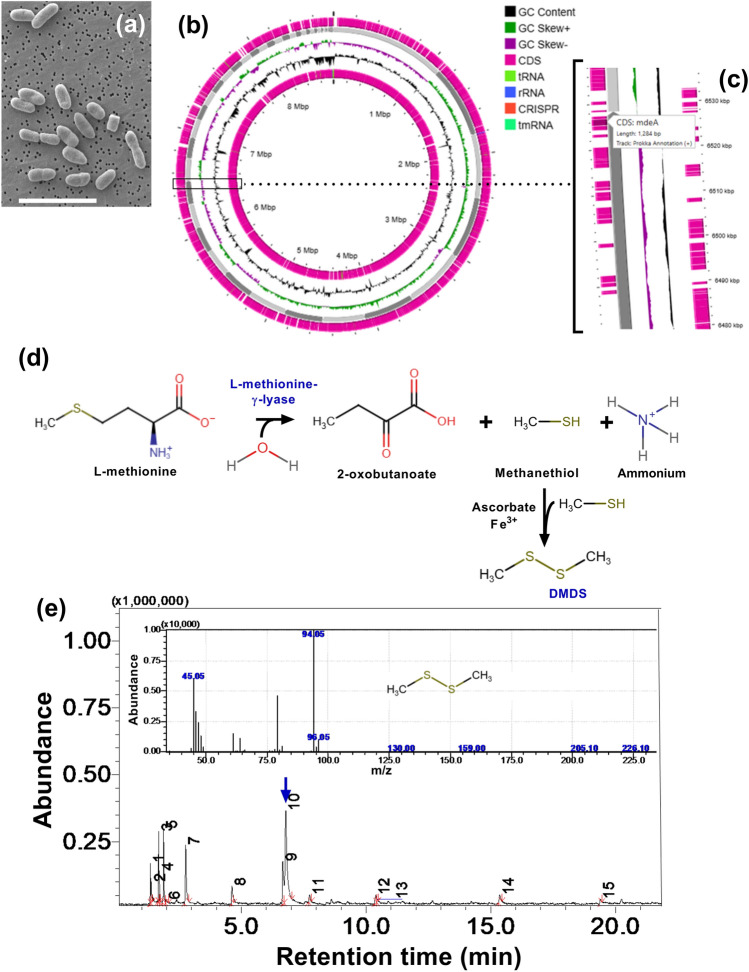
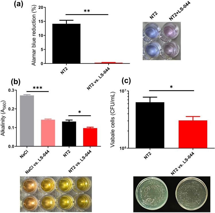
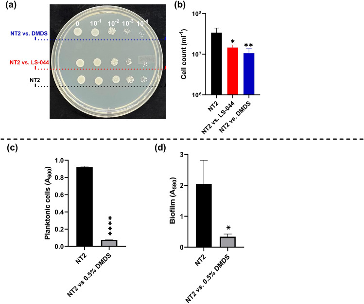
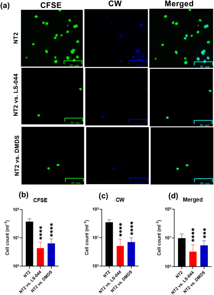
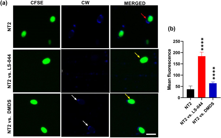
Some bacteria emit dimethyl disulphide (DMDS), one of the bioactive volatile sulphurous compounds (VSCs) of environmental and ecological significance, yet unexplored in combating drug-resistant yeasts. Here, we show the anti-budding and fungicidal activity of volatile DMDS emitted from Burkholderia cepacia LS-044 on the caspofungin-resistant yeast Nakaseomyces glabratus NT2. We identified a gene encoding L-methionine-γ-lyase (mdeA) catalysing DMDS formation in LS-044 and detected volatile DMDS as one of the VSCs (12% peak area) emitted by LS-044 through solid-phase microextraction followed by gas chromatographic-mass spectrometry. Exposure to volatiles of LS-044 resulted in a significant decline in metabolism (~ 98%), media alkalinity (~ 26%), and viable cell count (52‒95%) of NT2. Confocal microscopy of carboxyfluorescein succinimidyl ester- and calcofluor white stained cells revealed significantly high mean fluorescence in NT2 exposed to the volatiles of LS-044 (~ fivefold) and standard DMDS vapour (1.7-fold), suggesting a significant thickening of the cell wall. The surface area-to-volume ratio decreased significantly in NT2 cells exposed to volatiles of LS-044 and DMDS versus unexposed NT cells (6.5‒10.2 vs. 8.3‒13.2). DMDS exhibited a minimum inhibitory concentration of 0.5% (v/v) on NT2 cells in liquid broth dilution assay, and displayed fractional inhibitory concentration index of 0.95 with commercial antifungal clotrimazole reflecting lack of synergy or antagonism during the present combination therapy. The 26S rRNA gene sequence-based phylogeny revealed a tight phylogenetic association between NT2 and N. glabratus of environmental and clinical origins. Our study provided novel mechanistic insights into DMDS-driven bacterial antagonism on drug-resistant budding yeast N. glabratus NT2 that could be exploited in the ecological engineering and therapeutics of drug-resistant fungal infection.

Keywords: Candida glabrata; Antagonism; Fungicide; Volatile organic compounds (VOCs); Volatile sulphurous compounds (VSCs); Volatilome.


Conflict of interest statement

Declaration. Competing interests: The authors declare no competing interests.

Figures

Fig. 1
Fig. 1
Identification of dimethyl disulphide (DMDS) biosynthetic pathway and gas chromatographic evidence for the emission of DMDS in Burkholderia cepacia LS-044. Scanning electron microscopic view of LS-044 (a), circular plot of its genome (b), enlarged view of the circular genome showing localization of mdeA (c), which encodes L-methionine-γ-lyase involved in the methionine degradation (d); subsequent non-enzymatic reactions results in the formation of DMDS that emits from bacterial biomass as evidenced through SPME GC–MS (blue arrow) (e). The MS fingerprint of DMDS is shown in the inset.
Fig. 2
Fig. 2
Bipartition plate assay results revealed the lethal impact of the volatiles of Burkholderia cepacia LS-044 on the budding yeast Nakaseomyces glabratus NT2. The impact of volatiles of LS-044 on the metabolic activity (a), alkalinity (b) and viable cell count of N. glabratus NT2 (c) are shown. Statistical significance (*p < 0.1, **p < 0.05, ***p < 0.01) was determined by t-test using GraphPad Prism, ns, non-significant.
Fig. 3
Fig. 3
Identification of DMDS as one of the bioactive volatiles of Burkholderia cepacia LS-044, suppressing growth and biofilm formation in Nakaseomyces glabratus NT2. A PDA plate showing the colonies of control NT2 without volatile exposure and NT2 pre-exposed to volatiles of LS-044 and standard DMDS vapour (a). The corresponding viable cells are shown in the bar chart (b). The optical cell density (c) and biofilm formation (d) when exposed to 0.5% DMDS in liquid cultures. Statistical significance (*p < 0.1 and **p < 0.05) in (b) was determined using One-way ANOVA. Statistical significance (*p < 0.05 and ****p < 0.0001) in (c) and (d) was determined using the t-test.
Fig. 4
Fig. 4
Epifluorescence microscopy revealing the susceptibility of Nakaseomyces glabratus NT2 to the volatilome of Burkholderia cepacia LS-044 and standard dimethyl disulphide (DMDS) vapour. Differential cell wall staining of control NT2 cells without exposure to bacterial volatile (uppermost panel) and NT2 exposed to the volatiles of LS-044 (middle panel) and the standard DMDS (lowermost panel) (a). Cells were labeled with carboxyfluorescein succinimidyl ester (CFSE) and calcofluor white (CW) and subjected to excitation at green (491 nm) and blue (405 nm) lasers, respectively. The images were captured with Zoe. Scale bar, 25 μm. The relative abundance of cells strained with CFSE (b), CW (c) and undergone dual straining (d) when exposed to the volatiles of LS-044 and DMDS standard as compared to control NT2 (without exposure to volatiles) are shown. Statistical significance was determined using One-way ANOVA; ***p < 0.01, ****p < 0.0001; ns, non-significant.
Fig. 5
Fig. 5
Confocal microscopy revealing the impacts of the volatilome of Burkholderia cepacia LS-044 and standard dimethyl disulphide (DMDS) vapour on budding and cell wall peptidoglycans of Nakaseomyces glabratus NT2. Differential cellular morphology of control NT2 cells without exposure to bacterial volatiles (uppermost panel) and when exposed to the volatiles of LS-044 (middle panel) and the vapour of standard DMDS (lowermost panel) are shown (a). Cells were labeled with carboxyfluorescein succinimidyl ester (CFSE) and calcofluor white (CW) and subjected to excitation at green (491 nm) and blue (405 nm) lasers, respectively, and observed under a Zeiss confocal microscope. Budding cells, thickened luminous cell wall and scars are highlighted in red, yellow and white arrows, respectively. Scale bar, 5 μm. Differential fluorescence of yeast cells strained with CFSE when exposed to the volatiles of LS-044 and DMDS standard, as compared to control NT2 (without exposure to volatiles), is shown (b). Error bar, mean ± SD (n = 15). Statistical significance was determined using One-way ANOVA. ****p < 0.0001.
Fig. 6
Fig. 6
Unrooted maximum likelihood tree depicting the phylogenetic position of the strains of Nakaseomyces glabratus (NT2 and NT5, red triangles; accession No. PQ846718 and PQ846721, respectively) used in this study and related strains based on the Internal Transcribed Spacer sequencing targeted to the gene encoding larger subunit of ribosomal RNA (26S rRNA gene D1/D2 domain). Isolation source and/or origin of the isolates are indicated after the accession number. Bootstrap values of > 70% after 1,000 bootstrap replicates are shown at the branch points. Reference sequences were retrieved from GenBank under the accession numbers given in parentheses. Type strains are marked with strain number followed by ‘T’. Bar, 0.05 substitutions per site.

Similar articles

References

    1. European Food Safety, A. et al. Peer review of the pesticide risk assessment of the active substance dimethyl disulfide. EFSA J17 e05905 10.2903/j.efsa.2019.5905 (2019). - PMC - PubMed
    1. Fang, N. et al. Dissipation and residues of dimethyl disulfide in tomatoes and soil under greenhouse and open field conditions. J. Environ. Sci. Health B.55, 566–573. 10.1080/03601234.2020.1740531 (2020). - PubMed
    1. Yan, D. et al. Dimethyl disulfide (DMDS) as an effective soil fumigant against nematodes in China. PLoS ONE14, e0224456. 10.1371/journal.pone.0224456 (2019). - PMC - PubMed
    1. Yu, J., Land, C. J., Vallad, G. E. & Boyd, N. S. Tomato tolerance and pest control following fumigation with different ratios of dimethyl disulfide and chloropicrin. Pest. Manag. Sci.75, 1416–1424. 10.1002/ps.5262 (2019). - PubMed
    1. Yu, J., Sharpe, S. M., Vallad, G. E. & Boyd, N. S. Pest control with drip-applied dimethyl disulfide and chloropicrin in plastic-mulched tomato (Solanum lycopersicum L.). Pest Manag. Sci.76, 1569–1577. 10.1002/ps.5678 (2020). - PubMed

MeSH terms

LinkOut - more resources