

Glans penis electric stimulation modulates cerebral activity and functional connectivity in lifelong premature ejaculation revealed by functional MRI
- PMID: 40603932
- PMCID: PMC12222508
- DOI: 10.1038/s41598-025-03994-6
Glans penis electric stimulation modulates cerebral activity and functional connectivity in lifelong premature ejaculation revealed by functional MRI
Abstract
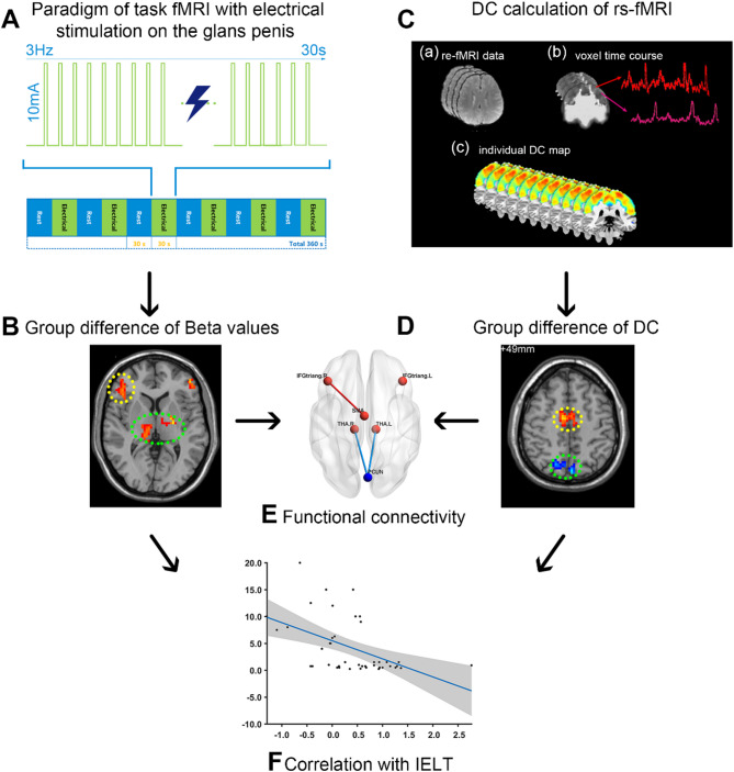
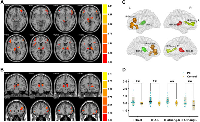
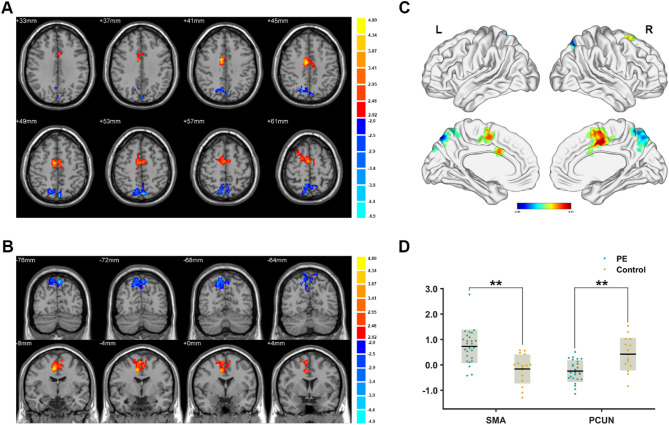
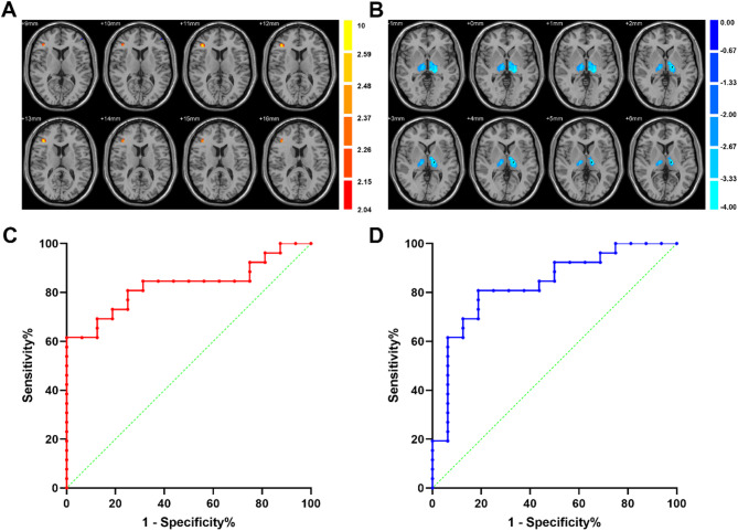
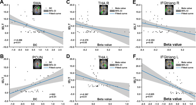
To compare brain activation in the dopaminergic reward system between 26 LPE patients and 16 normal controls (NCs) via glans penis electric stimulation task-fMRI and resting-state fMRI (rs-fMRI). The beta value, degree centrality (DC), and functional connectivity (FC) were calculated. The Pearson correlation was used to analyze the correlation between the fMRI measurements and disease severity. After task-fMRI, PE patients had significantly higher beta values in the dopaminergic reward system, including the bilateral thalamus and inferior frontal gyrus than NCs. In the rs-fMRI, higher DC values in the bilateral supplementary motor area (SMA) and lower DC values in the bilateral precuneus were found. Furthermore, our results showed enhanced FC between the right inferior frontal gyrus and the bilateral SMA and decreased FC between the bilateral precuneus and bilateral thalamus after electrical stimulation. The sensitivity was 80.77%, the specificity was 81.25%, and the AUC was 0.83 (p < 0.001) when differentiating the PE and NC using the FC between the inferior frontal gyrus and SMA. The sensitivity was 73.08%, the specificity was 75.00%, and the AUC was 0.82 (P = 0.002) when differentiating the two groups using the FC between the precuneus and thalamus.
Keywords: Degree centrality; Electrical stimulation; Premature ejaculation; Reward system; fMRI.
© 2025. The Author(s).
Conflict of interest statement
Declarations. Competing interests: The authors declare no competing interests.
Figures
Similar articles
-
Alteration in functional connectivity of SC_thalamus with primary trigeminal neuralgia.BMC Neurol. 2025 Jul 5;25(1):278. doi: 10.1186/s12883-025-04289-z. BMC Neurol. 2025. PMID: 40618014 Free PMC article.
-
Abnormal intrinsic functional hubs and connectivity in nurses with occupational burnout: a resting-state fMRI study.Front Public Health. 2025 Jun 16;13:1595550. doi: 10.3389/fpubh.2025.1595550. eCollection 2025. Front Public Health. 2025. PMID: 40589802 Free PMC article.
-
Exploring the changes in functional connectivity of the limbic system in Patients with amnestic mild cognitive impairment treated by acupuncture based on fMRI.Front Neurol. 2025 Jun 13;16:1506367. doi: 10.3389/fneur.2025.1506367. eCollection 2025. Front Neurol. 2025. PMID: 40584525 Free PMC article.
-
Altered brain connectivity in hyperkinetic movement disorders: A review of resting-state fMRI.Neuroimage Clin. 2023;37:103302. doi: 10.1016/j.nicl.2022.103302. Epub 2022 Dec 24. Neuroimage Clin. 2023. PMID: 36669351 Free PMC article.
-
The effects of noninvasive brain stimulation on cognitive function in patients with mild cognitive impairment and Alzheimer's disease using resting-state functional magnetic resonance imaging: A systematic review and meta-analysis.CNS Neurosci Ther. 2023 Nov;29(11):3160-3172. doi: 10.1111/cns.14314. Epub 2023 Jun 22. CNS Neurosci Ther. 2023. PMID: 37349974 Free PMC article.
References
-
- Hatzimouratidis, K. et al. Guidelines on male sexual dysfunction: Erectile dysfunction and premature ejaculation. Eur. Urol.57, 804–814 (2010). - PubMed
-
- Shindel, A. W. et al. Disorders of ejaculation: An AUA/SMSNA guideline. J. Urol.207, 504–512 (2022). - PubMed
-
- Serefoglu, E. C. et al. An evidence-based unified definition of lifelong and acquired premature ejaculation: Report of the second international society for sexual medicine Ad Hoc committee for the definition of premature ejaculation. J. Sex. Med.11, 1423–1441 (2014). - PubMed
-
- Corona, G. et al. Different testosterone levels are associated with ejaculatory dysfunction. J. Sex Med.5, 1991–1998 (2008). - PubMed
MeSH terms
Grants and funding
LinkOut - more resources
Full Text Sources
Medical
