

Dual functions of the ΔNp63-miR-141-3p-YAP1 regulatory axis in cervical cancer progression are dependent on histological subtype
- PMID: 40603978
- PMCID: PMC12222551
- DOI: 10.1038/s41598-025-07237-6
Dual functions of the ΔNp63-miR-141-3p-YAP1 regulatory axis in cervical cancer progression are dependent on histological subtype
Abstract
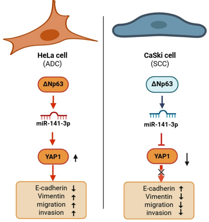
Cervical cancer (CC) poses a significant global health challenge, necessitating the development of novel therapeutic strategies. The interplay between the p63 isoform (ΔNp63), microRNA-141-3p (miR-141-3p), and Yes-associated protein 1 (YAP1) has emerged as a potential area of interest in cancer progression. This study aimed to investigate the functional relationship between the between the p63 isoform (ΔNp63), miR-141-3p and YAP1 in modulating migration, invasion, and epithelial-mesenchymal transition (EMT) in two CC cell lines, CaSki, and HeLa, which are human cervical squamous cell carcinoma (SCC) and adenocarcinoma (ADC) cells, respectively. The Gene Expression Omnibus (GEO) datasets, were utilized to assess the expression profiles of TP63 and YAP1 in the cervical SCC and ADC samples by using the GEO2R tool. The prediction software (miRDIP, miRmap, and TargetScan), and RT-qPCR were used to determine the relationship between genes. Different assays were performed for proliferative, migratory and invasive abilities of cells. The results were confirmed by western blot analysis. The ΔNp63-miR-141-3p-YAP1 axis exhibited distinct, cell-line-specific functions. In HeLa cells, this axis promoted a prometastatic phenotype by upregulating YAP1, leading to increased proliferation, migration, invasion, and EMT. Conversely, in CaSki cells, the same axis demonstrated an antimetastatic function by downregulating YAP1. YAP1 expression was significantly higher in HeLa cells compared to CaSki cells. In HeLa cells, YAP1 expression appeared to be regulated not solely by the upstream ΔNp63-miR-141-3p axis, suggesting its role as a key oncogene in HeLa cell progression. MiR-141-3p demonstrated context-dependent effects, exhibiting both pro- and anti-metastatic activities depending on the specific cell line. These findings highlight the complex and subtype-specific functions of the ΔNp63-miR-141-3p-YAP1 regulatory network in cervical cancer progression. In summary, our data highlights the different functions of ΔNp63-miR-141-3p-YAP1 axis in regulating proliferation, migration and invasion as well as EMT of different cervical cancer cells.
Keywords: Cervical cancer; EMT; YAP1; miR-141-3p; ΔNp63.
© 2025. The Author(s).
Conflict of interest statement
Declarations. Ethics approval and consent to participate: Not applicable. Consent for publication: Not applicable. Competing interests: The authors declare no competing interests.
Figures


Similar articles
-
LncRNA SNHG15 targets miR-200a-3p affects the proliferation, apoptosis, migration, and invasion of cervical cancer cells.BMC Cancer. 2025 Aug 7;25(1):1279. doi: 10.1186/s12885-025-14600-3. BMC Cancer. 2025. PMID: 40775293 Free PMC article.
-
MIR155HG promotes metastasis and cisplatin resistance of cervical cancer cells by regulating the miR-409-3p and ZEB1 axis.Sci Rep. 2025 Jul 1;15(1):20541. doi: 10.1038/s41598-025-08727-3. Sci Rep. 2025. PMID: 40593322 Free PMC article.
-
Blocking YAP1-Liprin-β2 interaction impedes metastasis and promotes tumor suppression in head and neck squamous carcinoma.Sci Rep. 2025 Jul 24;15(1):26968. doi: 10.1038/s41598-025-11652-0. Sci Rep. 2025. PMID: 40707583 Free PMC article.
-
The Protective Role of miR-125b in Hepatocellular Carcinoma: Unraveling Tumor-Suppressive Mechanisms.Curr Mol Med. 2025;25(6):663-671. doi: 10.2174/0115665240304247240529074123. Curr Mol Med. 2025. PMID: 38859784 Review.
-
Cost-effectiveness of using prognostic information to select women with breast cancer for adjuvant systemic therapy.Health Technol Assess. 2006 Sep;10(34):iii-iv, ix-xi, 1-204. doi: 10.3310/hta10340. Health Technol Assess. 2006. PMID: 16959170
References
-
- Siegel, RL, Miller, KD & Jemal, A. Cancer statistics, 2018. Cancer J. Clin.68 (1), 7–30 (2018). - PubMed
-
- Jemal, A. et al. Global cancer statistics. Cancer J. Clin.61 (2), 69–90 (2011). - PubMed
-
- Galic, V. et al. Prognostic significance of adenocarcinoma histology in women with cervical cancer. Gynecol. Oncol.125 (2), 287–291 (2012). - PubMed
-
- Pastushenko, I. & Blanpain, C. EMT transition States during tumor progression and metastasis. Trends Cell Biol.29 (3), 212–226 (2019). - PubMed
