. 2025 Jul 2;15(1):23467.
doi: 10.1038/s41598-025-07723-x.

FAM72A promotes UNG2 degradation and mutagenesis in human cancer cells

Affiliations

FAM72A promotes UNG2 degradation and mutagenesis in human cancer cells

Yuqing Feng et al. Sci Rep. .

Abstract

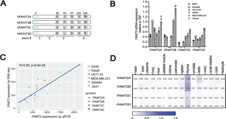
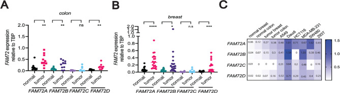
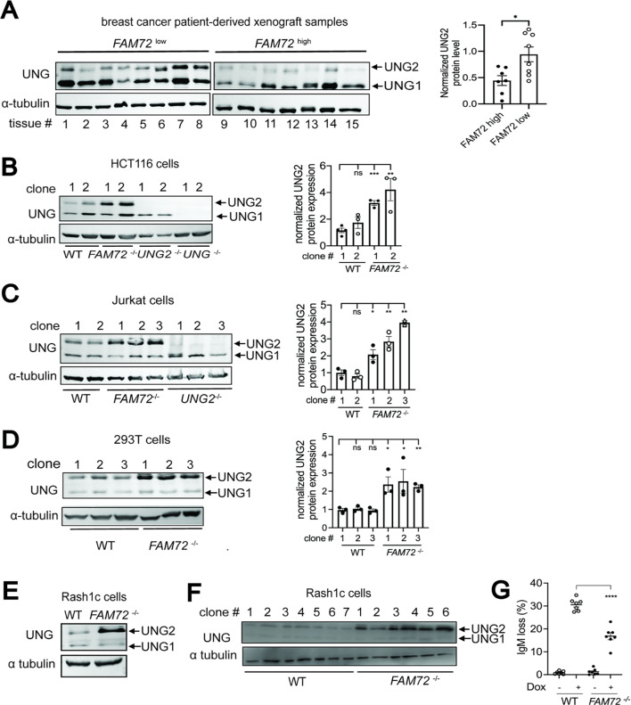
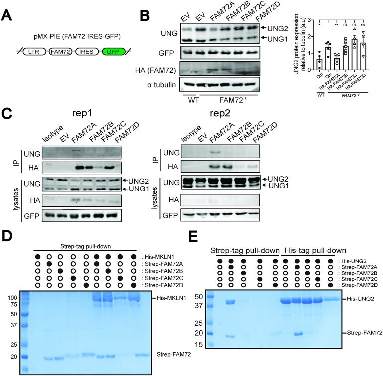
Genetic lesions drive cancer development and progression, and understanding their origins will reveal the mechanisms of carcinogenesis. We showed that murine FAM72A promotes mutagenic DNA repair during antibody maturation by acting as a substrate adaptor of the CTLHMKLN1 E3 ligase to induce the proteasome degradation of Uracil DNA glycosylase 2 (UNG2), a pivotal enzyme of the base excision repair. In humans, the FAM72 gene has expanded to include four paralogues named FAM72A-D. Bioinformatic studies suggested that the human FAM72 genes are overexpressed in a broad range of cancers. However, the functional roles of FAM72A-D in human biology and cancer are unknown. Here, we show that FAM72 family members are minimally expressed in most healthy tissues except for thymus, and that FAM72A, B and D are overexpressed in primary tumorigenic tissues. Human FAM72 expression inversely correlates with UNG2 protein level in human cell lines and primary tumorigenic tissues suggesting that human FAM72 promotes UNG2 degradation. However, only FAM72A is able to bind to and induce UNG2 degradation in human cells. Our results suggest that the ability of FAM72A to induce UNG2 degradation contributes to neoplasia in a variety of cancer types by promoting mutagenic repair of genomic dUs.

Keywords: Base excision repair; Cancer; Cytosine deamination; FAM72; Mutation; Uracil DNA glycosylase 2.


Conflict of interest statement

Declarations. Competing interests: The authors declare no competing interests. Conflict of interest: F.S is a founder and consultant for Repare Therapeutics and Induxion Therapeutics.

Figures

Fig. 1
Fig. 1
Expression profile of human FAM72 gene family in cell lines and healthy tissues. (A) Schematic of the human FAM72 family members demonstrating that they differ at the amino acid positions 82, 94, 99, 122, and 125. Of note, FAM72 paralogues differ at six nucleotide positions. The substitutions at nucleotide position 6 (amino acid position 2 shown in blue) in FAM72C and FAM72D results in silent mutations. Hence, only five out of six nucleotide substitutions result in changes at the protein level. (B) Assessment of FAM72 expression in multiple human cell lines by RT-qPCR. A549: human lung carcinoma; HET293T: human embryonic kidney; MDA-MB-231: human breast adenocarcinoma; HCT116: human colorectal carcinoma; Daudi: human Burkitt’s lymphoma; SW480: human colorectal adenocarcinoma. FAM72 mRNA expression relative to TBP was calculated using the 2^(delta Ct) method. (C) Correlation of FAM72 expression in human cell lines assessed by RNA-seq versus qPCR. (D) FAM72 mRNA levels in heathy human tissues as assessed from commercially available multiple tissue cDNA panels. FAM72 transcript levels were compared to housekeeping gene TBP. The color scheme provides quantitative information, as the full range of purple (low expression) to red (high expression) is used to as a surrogate measurement of the expression data. Numbers inside each box represent the relative fold change of FAM72 expression in comparison to TBP calculated using 2^ (delta Ct) method. Three replicates were done for each condition, and the averaged values were shown in the box.
Fig. 2
Fig. 2
Elevated expression of human FAM72 genes in primary cancers. (A) FAM72 mRNA expression level in primary colon cancer tissue specimen paired with normal flanking tissue obtained from Ontario Institute for Cancer Research (OICR) as assessed by RT-qPCR using FAM72 paralogue-specific primers. The relative FAM72 transcript expression was compared to TBP using 2^(delta Ct) method. (B) Same as (A), except FAM72 mRNA expression levels in primary breast cancer tissue specimen are shown. (C) Expression of FAM72 genes in normal and cancer tissues in comparison to human cell lines as shown in Fig. 1B. Purple (low expression) to red (high expression) is used to as a surrogate measurement of expression data. Two-tailed unpaired Student’s t-test was used for statistically analysis. *, P < 0.05; **, P < 0.01; ***, P < 0.001 ****; P < 0.0001.
Fig. 3
Fig. 3
FAM72 expression promotes UNG2 degradation and mutagenesis. (A) Western blotting for UNG protein in 15 human xenograft breast cancer tissues that express either high or low level of FAM72. Graph on the right shows the expression of UNG2 protein relative to a-tubulin. (B) Western blotting of whole cell lysates to assess UNG level in different clones of WT and FAM72−/− Cas9-HCT116 cells. Alpha-tubulin was used as a loading control for all immunoblots. Densitometric analysis shown on right was carried out on 3 independent Western blots. (C) Same as B, except that UNG expression was analyzed in WT and FAM72-deficient Jurkat cells. (D) Same as B, except that UNG expression was analyzed in WT and FAM72-deficient 293 T cells. (E) Western blotting of whole cell lysates to assess UNG levels in WT and FAM72−/− Cas9-expressing RASH1c cells. (F) Western blotting of whole cell lysates to assess UNG level in seven independent subclones derived from WT and FAM72−/− RASH1c cells. (G) Quantification of IgM loss in WT and FAM72−/− RASH1c cells treated with or without doxycycline (dox) for 4 days. Two-tailed unpaired Student’s t-test was used for statistically analysis. *, P < 0.05; **, P < 0.01; ***, P < 0.001 ****; P < 0.0001.
Fig. 4
Fig. 4
FAM72A, but not FAM72B-D, binds to and induces UNG2 degradation in human cells. (A) Schematic showing the pMX-Pie-HA-FAM72A-GFP expression vector that was used to transduce FAM72−/− HCT116 cells. (B) Western blotting of whole cell lysates to examine HA-FAM72 and UNG level in FAM72−/− HCT116 cells expressing pMX-PIE empty vector (EV), or pMX-PIE HA-tagged FAM72A/B/C/D. Cells were sorted based on GFP expression after viral transduction, and GFP level was assessed based on Western blot. Alpha-tubulin was used as a loading control. Densitometric analysis was done with 3 independent experiments. Two-tailed unpaired Student’s t-test was used for statistically analysis. (C) Immunoprecipitation of HA-FAM72 in MG132-treated HCT116 cell transduced with the indicated constructs. Two independent experiments were shown. (D) Strep-Tag II pull-down from Sf9 lysates expressing the indicated His6x-tagged MKLN1 and Strep-Tag II FAM72A/B/C/D proteins, followed by visualization by Coomassie-stained SDS-PAGE. (E) His6x-tag and Strep-Tag II pull-downs from Sf9 lysates expressing the indicated Strep-Tag II FAM72A/B/C/D and His6x-tagged UNG2 proteins, followed by visualization by Coomassie-stained SDS-PAGE. Data is presented as mean ± s.d of a minimum of 3 biological replicates. ns, not significant; *, P < 0.05; **, P < 0.01.

Similar articles

References

    1. Feng, Y., Seija, N., Di Noia, J. M. & Martin, A. AID in antibody diversification: there and back again. Trends Immunol.41, 586–600. 10.1016/j.it.2020.04.009 (2020). - PMC - PubMed
    1. Methot, S. P. & Di Noia, J. M. Molecular mechanisms of somatic hypermutation and class switch recombination. Adv. Immunol.133, 37–87. 10.1016/bs.ai.2016.11.002 (2017). - PubMed
    1. Cascalho, M., Wong, J., Steinberg, C. & Wabl, M. Mismatch repair co-opted by hypermutation. Science279, 1207–1210 (1998). - PubMed
    1. Martin, A. & Scharff, M. D. AID and mismatch repair in antibody diversification. Nat. Rev. Immunol.2, 605–614 (2002). - PubMed
    1. Phung, Q. H. et al. Increased hypermutation at G and C nucleotides in Immunoglobulin variable genes from mice deficient in the MSH2 mismatch repair protein. J. Exp. Med.187, 1745–1751 (1998). - PMC - PubMed

Substances

LinkOut - more resources