Comparative Study
. 2025 Jul 2;15(1):23575.
doi: 10.1038/s41598-025-08979-z.

Clinical efficacy analysis of arthroscopically assisted orthcord suture fixation in the treatment of tibial intercondylar eminence fractures: a retrospective comparative cohort study

Affiliations
Comparative Study

Clinical efficacy analysis of arthroscopically assisted orthcord suture fixation in the treatment of tibial intercondylar eminence fractures: a retrospective comparative cohort study

Rongfang Zhang et al. Sci Rep. .

Abstract

To explore the efficacy of arthroscopically assisted fixation of type II and type III tibial intercondylar eminence fractures with Orthcord sutures. A retrospective analysis was performed on 80 patients with intercondylar eminence fractures admitted to our hospital from April 2020 to March 2023. According to different surgical methods, the patients were divided into special suture fixation group (n = 30), cannulated screw fixation group (n = 24), and wire fixation group (n = 26). The suture group used arthroscopic Orthcord sutures to fix tibial intercondylar eminence fractures, and the cannulated screw group used cannulated compression screws for fixation. Patients in the wire group underwent arthroscopic wire fixation. The basic information of all patients was collected and followed up for 1 year. The Lysholm score and Range of motion of the knee joint and was performed at 3 months and 1 year after surgery. The patients' general data, surgical conditions, operation time, blood loss, hospitalization costs, postoperative recovery (Lysholm score and Range of motion of knee joint and at 3 months and 1 year after surgery) and other data were analyzed by variance analysis. P < 0.05 was considered statistically significant. There was no statistical difference in the general data of all patients. One-year follow-up showed that all patients had achieved bone healing without displacement, or bone malformation. The hospitalization time in the wire group was (11 ± 1.02) days, the screw group was (11.58 ± 1.61) days, and the Orthcord suture group was shortened to (10.03 ± 1.07) days. The differences among the three groups were statistically significant (P < 0.05). At the same time, the cost of Orthcord suture surgery (1310.7 ± 0.29) $ was significantly lower than that of the other two groups (P<0.05). The operation time of the suture group (68.13 ± 1.11 min) was significantly shorter than that of the wire group (76.76 ± 11.57 min) and the screw group (90.62 ± 1.99 min) (P<0.05). In the follow-up, the score of Orthcord suture 3 months after operation (94.07 ± 2.72 points) was better than that of the wire group (90.23 ± 5.23 points) and the screw group (90.37 ± 5.41 points); the difference was statistically significant (P<0.05).Three months after surgery, the range of motion of the knee joint in the Orthcord suture group (124.8°±7.2°) was significantly better than that in the screw group (105.7°±9.3°) and the wire group (112.4°±8.6°) (P<0.05). However, there was no statistically significant difference in the Lysholm score of the three groups of patients 1 year after operation (96.26 ± 1.89, 96.33 ± 2.44, 97.3 ± 1.70) (P>0.05).Similarly, there was no significant difference in the range of knee motion among the three groups of patients 1 year after surgery (135.1°±4.2°), (134.6°±4.8°), and (136.3°±3.5°) (P>0.05).Late fixation fracture and chronic pain complications occurred in both the wire and screw groups, but not in the suture group. (P<0.05). The use of Orthcord sutures in the arthroscopically assisted treatment of intercondylar ridge fractures can shorten the length of hospital stay and surgery, while greatly reducing hospitalization costs. It can achieve better short-term (3 months) recovery effects while avoiding second surgery, and ultimately show no weaker fixation effect than conventional screws and wires when full weight-bearing is restored.

Keywords: Arthroscopy; Eminence fracture; Internal fixation; Orthcord suture.


Conflict of interest statement

Declarations. Competing interests: The authors declare no competing interests. Ethics approval and consent to participate: The studies involving human participants were reviewed and approved by the Ethics Committee of Nanyang Hospital of Traditional Chinese Medicine (Nanyang Orthopedic Hospital; NO.202003). The patients/participants provided written informed consent to participate in this study.

Figures

Fig. 1
Fig. 1
A female patient was hospitalized for tram injury. Her knee joint was obviously swollen. X-ray examination of the knee joint showed a fracture of the lower insertion point of the anterior cruciate ligament and a raised fracture fragment (the red arrows in the figure show the intercondylar ridge fracture a and b).
Fig. 2
Fig. 2
In addition to X-rays, intra-articular fractures usually require CT three-dimensional reconstruction to further clarify the fracture situation. Three-dimensional reconstruction can show displacement of the lower insertion point of the fracture.
Fig. 3
Fig. 3
As shown in Figure a, an arthroscopic approach is established to clear the blood in the joint to expose the fracture fragment and thoroughly clean the bone bed. b, an anteromedial and anterolateral bone tunnels are established under the guidance of the locator. Figures c and d show the introduction of the suture through the right-angle clamp and the fixation of the fracture fragment in the shape of"8"after bypassing the anterior cruciate ligament.
Fig. 3
Fig. 3
As shown in Figure a, an arthroscopic approach is established to clear the blood in the joint to expose the fracture fragment and thoroughly clean the bone bed. b, an anteromedial and anterolateral bone tunnels are established under the guidance of the locator. Figures c and d show the introduction of the suture through the right-angle clamp and the fixation of the fracture fragment in the shape of"8"after bypassing the anterior cruciate ligament.
Fig. 4
Fig. 4
Cleaning the blood clots around the fracture, the ligament-connected fracture fragment was seen to be lifted up (a and b), which was consistent with the preoperative MRI examination results. (The red arrow in c shows that the anterior cruciate ligament is loose, the fracture fragment is lifted up, and the ligament loses tension) During the operation, Kirschner wires were used to temporarily fix the fracture fragments (d) to maintain a stable position, and an anterior cruciate ligament locator was used to create a tibial bone tunnel (e). After the bone tunnel was made, Orthcord sutures were introduced and cross-fixed. The tension of the anterior cruciate ligament was restored (f). The wound was minimally invasive and beautiful. The first dressing change after the operation was minimally invasive and did not require suture removal. It was beautiful and did not require suture removal (g).
Fig. 4
Fig. 4
Cleaning the blood clots around the fracture, the ligament-connected fracture fragment was seen to be lifted up (a and b), which was consistent with the preoperative MRI examination results. (The red arrow in c shows that the anterior cruciate ligament is loose, the fracture fragment is lifted up, and the ligament loses tension) During the operation, Kirschner wires were used to temporarily fix the fracture fragments (d) to maintain a stable position, and an anterior cruciate ligament locator was used to create a tibial bone tunnel (e). After the bone tunnel was made, Orthcord sutures were introduced and cross-fixed. The tension of the anterior cruciate ligament was restored (f). The wound was minimally invasive and beautiful. The first dressing change after the operation was minimally invasive and did not require suture removal. It was beautiful and did not require suture removal (g).
Fig. 5
Fig. 5
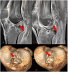
Postoperative knee joint MRI (a and b) was performed. MRI showed that the tension of the anterior cruciate ligament was significantly restored compared with that before the operation (indicated by the red arrow: a shows that the anterior cruciate ligament lost tension, and b shows that the ligament tension was restored after fixation) Postoperative review of the knee joint CT three-dimensional reconstruction showed that the fracture was well reduced c was the preoperative CT, and postoperative d showed that the fracture was well reduced.

Similar articles

References

    1. Tuca, M., Bernal, N., Luderowski, E. & Green, D. W. Tibial spine avulsion fractures: treatment update. Curr. Opin. Pediatr.31 (1), 103–111. 10.1097/MOP.0000000000000719 (2019). - PubMed
    1. Rudran, B., Little, C., Wiik, A. & Logishetty, K. Tibial plateau fracture: anatomy, diagnosis and management. Br. J. Hosp. Med. (Lond). 81 (10), 1–9. 10.12968/hmed.2020.0339 (2020). - PubMed
    1. Meyers, M. H. & McKeever, F. M. Fracture of the intercondylar eminence of the tibia. J. Bone Joint Surg. Am.52 (8), 1677–1684 (1970). - PubMed
    1. Zaricznyj, B. Avulsion fracture of the tibial eminence: treatment by open reduction and pinning. J. Bone Joint Surg. Am.59, 1111–1114 (1977). - PubMed
    1. Kobayashi, S. et al. Arthroscopic treatment of tibial eminence avulsion fracture with suture tensioning technique. Arthrosc. Tech.7 (3), e251–e256. 10.1016/j.eats.2017.08.078 (2018). Published 2018 Feb 19. - PMC - PubMed

Publication types